Loading
Archives of Clinical Ophthalmology
ISSN: 2771-7925
All Articles
Comment on “Retinitis Pigmentosa and Molar Tooth Sign Caused by Novel AHI1 Compound Heterozygote Pathogenic Variants: A Case Report”
Qing Lv, Ailian Du
Joubert syndrome (JS) is a rare congenital neurodevelopmental disease which is basically a primary Ciliopathy. It’s characteristic manifestation on imaging is so called ‘molar tooth sign’ in the brainstem and cerebellum. JS can involve multiple organs, mainly including retina, kidney, bone and liver. Clinical signs of early onset JS include hypotonia, developmental delay, breathing abnormalities, and ocular motor apraxia.
Arch Clin Ophthalmol, 2022, Volume Volume 2, Issue Issue 1, p1-2 | DOI: 10.33696/Ophthalmology.2.005
Uhthoff ’s Phenomenon as Presentation of COVID-19 Infection
Pieter Gouws, Alexander Gouws
This is the first reported case of COVID-19 associated optic neuritis (ON) presenting with classic Uhtoff’s phenomenon typically associated with multiple sclerosis (MS). Uhthoff phenomenon, also known as Uhthoff sign or syndrome, is a transient worsening of neurological function lasting less than 24 hours that can occur in multiple sclerosis patients due to increases in core body temperature.
Arch Clin Ophthalmol, 2023, Volume Volume 3, Issue Issue 1, p1-2 | DOI: 10.33696/Ophthalmology.3.008
Iris Applications in Forensic Ophthalmology: Promise, Limitations, and the Future Directions
Sushil Bhatt, Jagmahender Singh Sehrawat, Vishali Gupta, Savita Kumari
Human iris, with its highly complex, stable, and unique patterns, has gained increasing attention as a reliable biometric modality. In forensic ophthalmology, iris recognition presents significant promise for secure identification of suspects, victims, and missing persons, as well as in disaster victim identification (DVI). Unlike fingerprints or DNA, which may be degraded or unavailable, iris-based methods provide a non-invasive and highly accurate alternative with strong potential for forensic application.
Arch Clin Ophthalmol, Volume 5, Issue 1, p1-5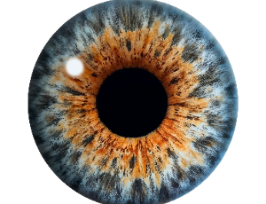
Macular Microcirculation after Rhegmatogenous Retinal Detachment Repair Evaluated by OCT-Angiography
Evita Evangelia Christou, Maria Stefaniotou
In the process of rhegmatogenous retinal detachment (RRD), retinal homeostasis may be adversely affected with resultant modifications in retinal and choroidal tissue. Hypoxia and nutrient deprivation along with inflammation at the detached retina may lead to morphological and microvascularity alterations. These changes imply that the functional status of the macula may not be entirely restored despite anatomical repair.
Arch Clin Ophthalmol, 2021, Volume Volume 1, Issue Issue 1, p1-7 | DOI: 10.33696/Ophthalmology.1.001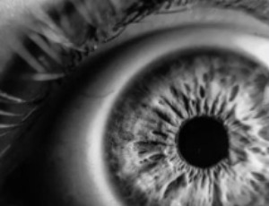
Repeatability of Scheimpflug Corneal Tomography in Patients with Keratoconus and Different Body Mass Indices
James S Lewis, Lize Angelo, Akilesh Gokul, Charles NJ McGhee, Mohammed Ziaei
To evaluate the repeatability of corneal tomographic parameters in keratoconus patients across different body mass index (BMI) categories. This prospective study was conducted at the University of Auckland, New Zealand, from June 2021 to June 2022. A total of 243 eyes from keratoconus patients aged 18-45 years were categorized into normal (BMI ≤24.9; n=55), overweight (BMI 25.0-29.9; n=58), and obese (BMI ≥30.0; n=130) groups. Patients underwent three consecutive scans using the Pentacam AXL.
Arch Clin Ophthalmol, 2025, Volume Volume 4, Issue Issue 1, p1-11 | DOI: 10.33696/Ophthalmology.4.015
- Abstract |
- Full Text |
- Cite |
- Supplementary File
Assessment of Visual Function for Education: A Commentary on ‘VEP Visual Acuity in Children with Cortical Visual Impairment’
Alison M Mackay
Last year’s article In the International Journal of Clinical and Experimental Ophthalmology [1] highlighted that Cortical Visual Impairment (CVI) is now the leading cause of visual impairment in the developed world [2]. It also provided a definition of CVI [3,4], and summarized its functional deficits, and methods of assessment.
Arch Clin Ophthalmol, 2023, Volume Volume 3, Issue Issue 1, p3-4 | DOI: 10.33696/Ophthalmology.3.009
Stroke and Visual Loss in a Young Girl with Dengue Fever – Report of a Case and a Mini Review
Sweety Trivedi, Ambar Chakravarty
The case of a young girl with Dengue fever presenting with seizures and bilateral visual loss is presented. At the time of presentation, she had right hemiplegia and dysarthria but was not dysphasic. Fundoscopy revealed presence of macular and disc oedema in the right eye and vitreous haemorrhage in the left eye.
Arch Clin Ophthalmol, 2022, Volume Volume 2, Issue Issue 1, p3-8 | DOI: 10.33696/Ophthalmology.2.006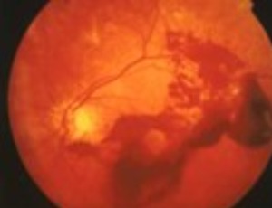
Are Women with COVID-19 and Migraine Prone to AMN Type 2?
Maxime Depasse, Marcel PM ten Tusscher, Vincent De La Porte, Robert W Kuijpers
Acute macular neuroretinopathy (AMN), first described by Bos and Deutman in 1975 [1], is an infrequent yet increasingly diagnosed retinal condition, characterized by the acute onset of persisting paracentral scotomata with capricious vision loss in one or both eyes, associated with (peri)foveal petaloid (wedge-shaped) outer retinal lesions generally accepted to be caused by microvascular damage to the retinal capillary network of the macula [2,3].
Arch Clin Ophthalmol, 2023, Volume Volume 3, Issue Issue 1, p5-12 | DOI: 10.33696/Ophthalmology.3.010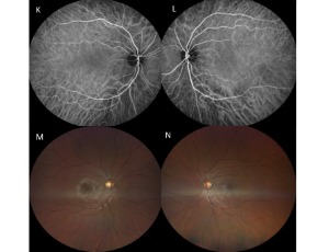
Rapid-onset Bilateral Cataracts after Hyperbaric Oxygen Therapy: A Case Report
Natalie Marie Lane, David Lane
Background: Hyperbaric oxygen therapy (HBOT) is used to manage various medical conditions and has been associated with the gradual formation of cataracts after prolonged exposure. However, rapid-onset cataract development remains uncommon. This case describes an unusual presentation of rapid bilateral cataract development following HBOT, occurring over a much shorter timeframe than previously reported in literature.
Arch Clin Ophthalmol, Volume 5, Issue 1, p6-9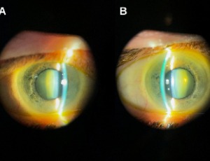
Focal Aggregates of Normal or Near Normal Uveal Melanocytes (FANNUMs) in the Choroid. A Practical Clinical Category of Small Ophthalmoscopically Evident Discrete Melanocytic Choroidal Lesions
James J. Augsburger
Focal aggregate of normal or near normal uveal melanocytes (FANNUM) of the choroid is a term the author has proposed to categorize small melanocytic choroidal lesions that are not detectably thicker than surrounding normal choroid by B-scan ocular ultrasonography. In this article, the author describes the clinical features of small melanotic choroidal lesions he categorizes clinically as FANNUMs and discusses the presumed compositional spectrum of such lesions.
Arch Clin Ophthalmol, 2021, Volume Volume 1, Issue Issue 1, p8-19 | DOI: 10.33696/Ophthalmology.1.002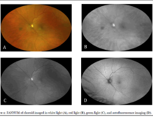
Lens Clarity and Visual Fatigue in Children: The Role of Eyewear Hygiene and Healthcare Professionals
Mirela Tushe
Visual fatigue, also known as digital eye strain (DES), has become a growing concern among children due to increased exposure to digital screens, especially after the global shift to remote learning during the COVID-19 pandemic. Although prolonged screen time and poor ergonomics are well-recognized contributors, the influence of eyewear hygiene-specifically lens cleanliness-on visual discomfort is less explored.
Arch Clin Ophthalmol, 2025, Volume Volume 4, Issue Issue 1, p12-16 | DOI: 10.33696/Ophthalmology.4.016
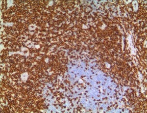
Orbital Lymphoproliferative Disorders (OLPDs) in a 3-year-old Child: Case Report and Review of Literature
Caiping Shi, Jia Feng, Sujuan Zhao, Jiao Zhan, Yanhong Ren, Weizhong Gu, Xiaoyu Zheng
Orbital lymphoproliferative disorders (OLPDs) consist of a spectrum of diseases ranging from benign to malignant lesions including reactive lymphoid hyperplasia, atypical lymphoid hyperplasia, and lymphoma. OLPDs rarely present as an orbital mass lesion in children. Accurate discrimination of OLPDs is crucial for treatment planning. We report a case to investigate the clinical and pathological
features of OLPDs in children.
Gabapentin for Ocular Surface Disorders: Bridging Molecular Mechanisms to Therapeutic Innovation
Caterina Gagliano, Alessandro Avitabile, Dario Rusciano
This commentary critically evaluates Rusciano's (2024) comprehensive review on gabapentin (GBP) as a multifaceted therapy for ocular surface diseases, emphasizing its transition from systemic to topical applications. We highlight the review's synthesis of GBP's polypharmacology—spanning calcium channel modulation, anti-inflammatory effects, and neuroprotection—and its innovative integration with nanotechnology (e.g., nanoceria platforms) to overcome corneal delivery challenges while potentially reducing systemic side effects associated with oral administration.
Arch Clin Ophthalmol, 2025, Volume Volume 4, Issue Issue 1, p17-25 | DOI: 10.33696/Ophthalmology.4.017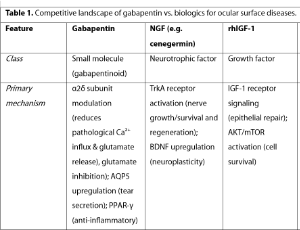
Mega-Dose Dietary Riboflavin in Treatment in Keratoconus, Post-Refractive Cornea Ectasia and Migraine. Has Its Time Arrived?
John Steven Jarstad
Recently, several studies and investigators have shown the beneficial effects of high dose dietary riboflavin (vitamin B2) in the treatment of keratoconus, post-refractive (LASIK, PRK & Radial Keratotomy) ectasia (with sunlight exposure) and patients treated with our own protocol (NIH Clinical Study – www.clinicaltrials.gov - # NCT 03095235) discovered significant relief for intractable migraine headaches and/or ophthalmic migraine (classic migraine visual symptoms without headache).
Arch Clin Ophthalmol, 2021, Volume Volume 1, Issue Issue 1, p20-20 | DOI: 10.33696/Ophthalmology.1.003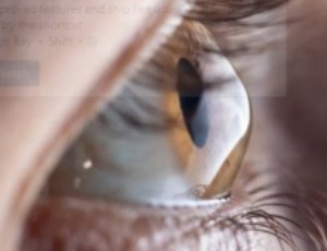
A Rare Case of Vitreous Cyst
Yogesh Kumar, Jatinder Singh Bhalla, Ridhima Sakhuja, Neha Yadav
Vitreous cysts are regarded as ‘ocular curiosities’ as they are rarely seen and reported. They can be either congenital or acquired. Congenital vitreous cysts are usually isolated clinical findings and acquired cysts are observed in conditions like high myopia, ocular trauma, degenerative pathologies of the retina & choroid, and infections like Cysticercosis and Toxoplasmosis.
Arch Clin Ophthalmol, 2023, Volume Volume 3, Issue Issue 1, p20-22 | DOI: 10.33696/Ophthalmology.3.012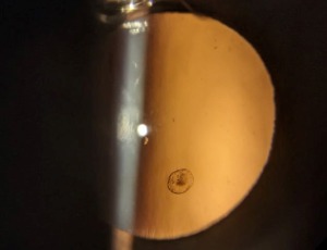
Multidisciplinary Acute Care of Central Retinal Artery Occlusion with a Stroke Paradigm: A Call to Action
Stacey Q. Wolfe, Stephanie A. Coffman, Mark Perez, Katriel Lee, Bartlett H.Hayes, Tamra Ranasinghe, Patrick A. Brown, Kyle M. Fargen
Central retinal artery occlusion (CRAO) is an ophthalmologic emergency that can result in permanent vision loss. Over 25% of CRAO are associated with acute cerebral ischemia, and there are many parallels between CRAO and acute ischemic stroke. There are no definitive treatment algorithms for CRAO, however there may be opportunities to treat CRAO as an “eye stroke”. Given the similarities to acute ischemic stroke, multidisciplinary involvement and stroke algorithms should be considered and tested for this disease.
Arch Clin Ophthalmol, 2021, Volume Volume 1, Issue Issue 1, p20-26 | DOI: 10.33696/Ophthalmology.1.004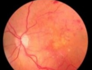
An Unusual Case of an Open Globe due to Airbag Injury and Scleral Lens Use
Mary K. Wilson, Lee P. Bowman, Jonathan K. Hu, Charles R. Blake
A 30-year-old woman who wears scleral contact lenses for keratoconus was brought to the emergency room after a motor vehicle collision where a deployed airbag hit her left eye. The patient was taken to the operating room for globe exploration and found to have two large scleral lacerations (one superiorly and one temporally) with significant uveal prolapse.
Arch Clin Ophthalmol, 2023, Volume Volume 3, Issue Issue 1, p23-26 | DOI: 10.33696/Ophthalmology.3.013
Generating Awareness and a Planned Multidisciplinary Treatment Approach Can Save Both the Sight and Life in Retinoblastoma in Developing Countries
Soma Rani Roy
While rare, retinoblastoma is the most common (1:16000 – 18000 live births) intraocular and life threatening tumor of childhood. According to the World Health Organization (WHO), 66% of children present with symptoms before 2 years of age and 95% before 5 years of age. About 8000 new cases are detected annually with the highest incidence in Africa and India. In fact, more than 1400 cases each year are from India. According to Mukesh et al., 43% of the global burden lives in 6 countries of Asia (India, China, Indonesia, Pakistan, Bangladesh & Philippines).
Arch Clin Ophthalmol, 2021, Volume Volume 1, Issue Issue 1, p27-29 | DOI: 10.33696/Ophthalmology.1.005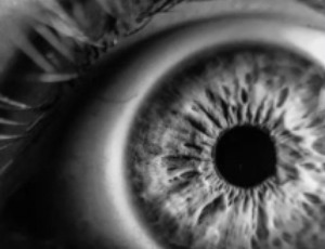
Treatment Burden Associated with Intravitreal Injections: A Cross-sectional Study at a Tertiary Eye Centre in Ireland
Ann Marie O’Leary, Daniel Coakley, Eamonn O’Connell
Treatment burden significantly impacts patient adherence and quality of life when it comes to chronic conditions requiring frequent medical interventions. Intravitreal injections are administered to over 20 million patients globally annually, yet the associated treatment burden remains poorly quantified in Irish healthcare settings. This study aimed to assess treatment burden and identify key predictors among patients receiving intravitreal injections for retinal conditions at a tertiary eye center in Ireland.
Arch Clin Ophthalmol, 2025, Volume Volume 4, Issue Issue 1, p31-37 | DOI: 10.33696/Ophthalmology.4.019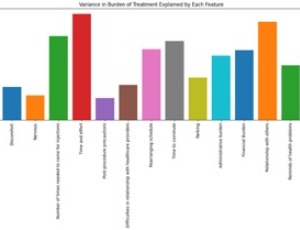
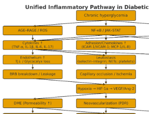
Advances in Inflammatory Biomarkers for Diabetic Retinopathy
Honghong Dong, Ying Xie
Diabetic retinopathy is a leading cause of blindness in diabetic patients, and its onset and progression are influenced by inflammation. This article provides an overview of local inflammatory biomarker research in diabetic retinopathy, covering serum, aqueous humor, vitreous inflammatory factors, and retinal inflammation markers. Through a systematic review and analysis, we found that inflammatory biomarkers play a crucial role in the prediction, diagnosis, and treatment of diabetic retinopathy, as well as in understanding its pathological mechanisms and improving clinical diagnostic and therapeutic strategies.
Arch Clin Ophthalmol, 2025, Volume Volume 4, Issue Issue 1, p38-46 | DOI: 10.33696/Ophthalmology.4.020