

Abstract
Cohort studies involving more than 500,000 transfers have shown that having an endometrial lining less than 7 or 8 mm at the time of IVF trigger or cryo-defrost progesterone cycle start is associated with having a lower clinical pregnancy and live birth rate than for women with cycles having thicker endometrial linings. The use of platelet-rich plasma has been proposed as a therapy to compensate for having a thin endometrial lining. Its use is purported to increase the thickness of the endometrial linings of affected women or otherwise increase their endometrial receptivity.
Casper hypothesized that the diminished endometrial receptivity seen in women with thin endometrial linings was due to their having a higher oxygen concentration in the lumen of their uterus than they would have had with a thicker endometrial lining. This hypothesis is examined in the context of uterine anatomical arguments, accepted chemical/physiological principles, and mechanistic biochemical data.
Platelet-rich plasma (PRP) may enhance endometrial growth and receptivity through vascular endothelial growth factor (VEGF) and other platelet-derived growth factors. PRP can deliver supraphysiologic concentrations of these factors directly to the basal endometrium, augmenting angiogenesis and indirectly promoting glandular development during endometrial repair. Increased glandular development and the associated more hypoxic oxygen environment may further stimulate endogenous VEGF and other growth factors, potentially altering midcycle endometrial luminal secretions and improving the environment for implantation and early embryonic development. PRP will be most effective when the epithelial surface is disrupted, permitting greater access of platelet-derived factors to underlying tissues before degradation or clearance.
Keywords
Casper's hypothesis, Endometrial lining, Gynecology, Implantation, Infertility and medicine, Platelet Rich Plasma (PRP), Receptivity, Reproductive medicine and endocrinology, Thin endometrial lining, Vascular Endothelial Growth Factor (VEGF)
Introduction
Most frequently, a thin endometrial lining is defined as having a late proliferative phase or “pre-trigger” measurement of less than 7 or 8 mm in the thickest transverse part of the endometrium as seen on transvaginal ultrasound [1–4]. A thin endometrial lining is an established cause of ART inefficiency and treatment failure in that endometrial lining thickness positively correlates to the clinical pregnancy rate (CPR) and the live birth rate (LBR) in cohort series involving a total of more than 540,000 cycles [1–4]. These observational reports demonstrated that the incidence of having a thin endometrial lining less than 7 mm was between 3.4 and 5% [1,3,4] and having a thin endometrial lining less than 8 mm was between 13.2% and15% [1–4]. These cohort studies were large enough to isolate many of the variables (age, type of stimulation, number of oocytes collected, stage of embryo transferred, donor or autologous, fresh or cryo-thaw, use of PGD, infertility diagnosis, endometrial pattern) relevant to the practicing clinician and arrive at the same general conclusion. Namely, cycles with a thicker endometrial lining had better ART outcomes than with a thinner endometrial lining even in subgroups identified by the variables listed above. For example, Liu et al. found that for each one mm less than an 8 mm endometrial lining thickness in women, there was a decrease in the LBR per transfer [1]. Specifically, the LBR for fresh blastocyst transfer was 40.6% for endometrial linings ≥8 mm, 33.3% for endometrial linings 7 mm–7.9 mm, 31.7% for endometrial linings 6 mm–6.9 mm, 16.9% for endometrial linings 5 mm–5.9 mm and 0% for endometrial linings <5 mm [1]. Gingold et al., using the SART CORS United States national registry of IVF transfers from 2016–2018, found a linear increase in the LBR from 31.2% for an endometrial lining thickness of <6 mm to 45% for an endometrial lining thickness of 8–12 mm, with the LBR leveling off (but not decreasing) for endometrial linings greater than 12 mm [3].
Many approaches, including increasing the dose or duration of estrogen exposure, low dose aspirin, sildenafil, vitamin E, pentoxifylline, and granulocyte colony stimulating factor, have been used to treat this condition [5,6]. Although most of these approaches have been shown to be effective in a few publications, none of these approaches have been shown to be sufficiently effective to be considered the standard of care. Clinicians often choose to use multiple approaches simultaneously, with more hope than confidence, in the highly motivated patient.
A regenerative medicine approach using platelet-rich plasma (PRP) is progressively being used to treat various problems in sundry areas of medicine including orthopedics, dentistry, plastic surgery, and general surgery. Presumptively, these approaches are effective because platelets release supraphysiologic quantities of many of the growth factors that enable standard wound healing to take place. The use of PRP is currently an active area of research and publications in reproductive endocrinology including treatment of the thin endometrial lining. Study design has ranged from case series, cohort series, prospective cohort series, to randomized trials. Generally, these have shown a benefit of treatment. This paper’s intention was not to formally review this literature, in part, because many such reviews have already been published [7,8]. Rather, the focus of this paper was to examine relevant physiology of the menstrual cycle together with some established facts about key growth factors and submit that this article be viewed as a part of the foundation on which the clinician can best build an effective treatment for the thin endometrial lining using PRP.
Endometrial Receptivity of the Thin Endometrial Lining
Although there are gynecological conditions that are completely refractive to embryo implantation, most conditions that are believed to impair the ability of an embryo to implant are not absolute. Endometrial thickness provides a good example in that women with a 6 mm thick endometrial lining have a lower ability for an embryo to implant than average women, but an embryo still has a positive probability of implanting in a thin endometrium. DNA microarray technology has been used to characterize the genes that are either up or down regulated during all phases of the menstrual cycle. The fundamental importance of transcription in natural biological function led to the hypothesis that evaluating gene expression at the time of implantation might largely explain implantation failure of euploid embryos. This transcriptomic approach to endometrial receptivity is clinically available as the Endometrial Receptivity Assay (ERA) and is one measure of endometrial receptivity. However, it does not appear to capture the specific pathology seen with the thin endometrial lining in that 77% of a sample of women with a thin endometrial lining had normal ERA testing [9]. These findings suggest that dysfunctional transcription at the presumed window of potential implantation does not appear to be an etiology for the reduced endometrial receptivity seen in women having a thin endometrial lining. This may reflect the inherently dynamic nature of implantation, where successful outcome depends not only on transcriptional activity but also on downstream processes such as protein synthesis, molecular signaling, and embryo-endometrial interactions. Consequently, transcriptional profiling alone may not adequately capture important subtilties such as quantity of functional end products, actions induced by key low concentration molecules such as cytokines, immune dysfunction, or the many transcriptional events triggered by early embryo attachment.
The Casper Hypothesis
Casper proposed a theory as to the mechanism of diminished endometrial receptivity in women with a thin endometrial lining. He posited that embryos transferred in closer proximity to the endometrial blood supply are exposed to higher levels of oxygen tension and therefore increased levels of reactive oxygen species [10].
Using mass spectrometry, Kigawa found the human uterine intraluminal partial pressure of O2 to be about 22 mm Hg, with pO2 lower in the proliferative compared to the luteal phase [11]. Yedwab et al. used a microelectrode inserted into their subjects’ uteri, measured pO2 on a blood gas machine (radiometer PHM–76), and found the pO2 to be about 14 mm Hg throughout the menstrual cycle [12]. Using a third methodology, Ottosen et al. measured oxygen tension in 21 women immediately before an intrauterine insemination and found the mean pO2 to be 18.9 mmHg. The pO2 range of the four women who achieved pregnancy was 4–15 mmHg compared to a range of 4 to 27 mmHg for those who did not [13]. By way of comparison, the common 5% O2 concentration used in IVF culture is equivalent to a partial pressure of 38 mmHg [14]. A blastocyst transferred into the uterus experiences the dissolved oxygen concentration of the luminal uterine secretions. Although it has never been directly measured in women with different thickness endometria, Fick’s law suggests that as the distance increases from the circulating blood supply, less oxygen will be transported to the luminal uterine secretions [14]. As illustrated in Figure 1, the thickness of the post-menstrual functional endometrium will largely determine the oxygen content experienced by the blastocyst. If a higher oxygen exposure in women with thin endometrial lining is a significant contributor to their lower pregnancy rates, it also helps explain why some women with very thin endometrial lining still get pregnant. Human IVF can be done in various oxygen concentrations, but lower oxygen concentrations have proved to provide a better culture environment. If Casper’s theory is correct, then an effective therapy to treat diminished receptivity in women with a thin endometrial lining would be to thicken their lining.
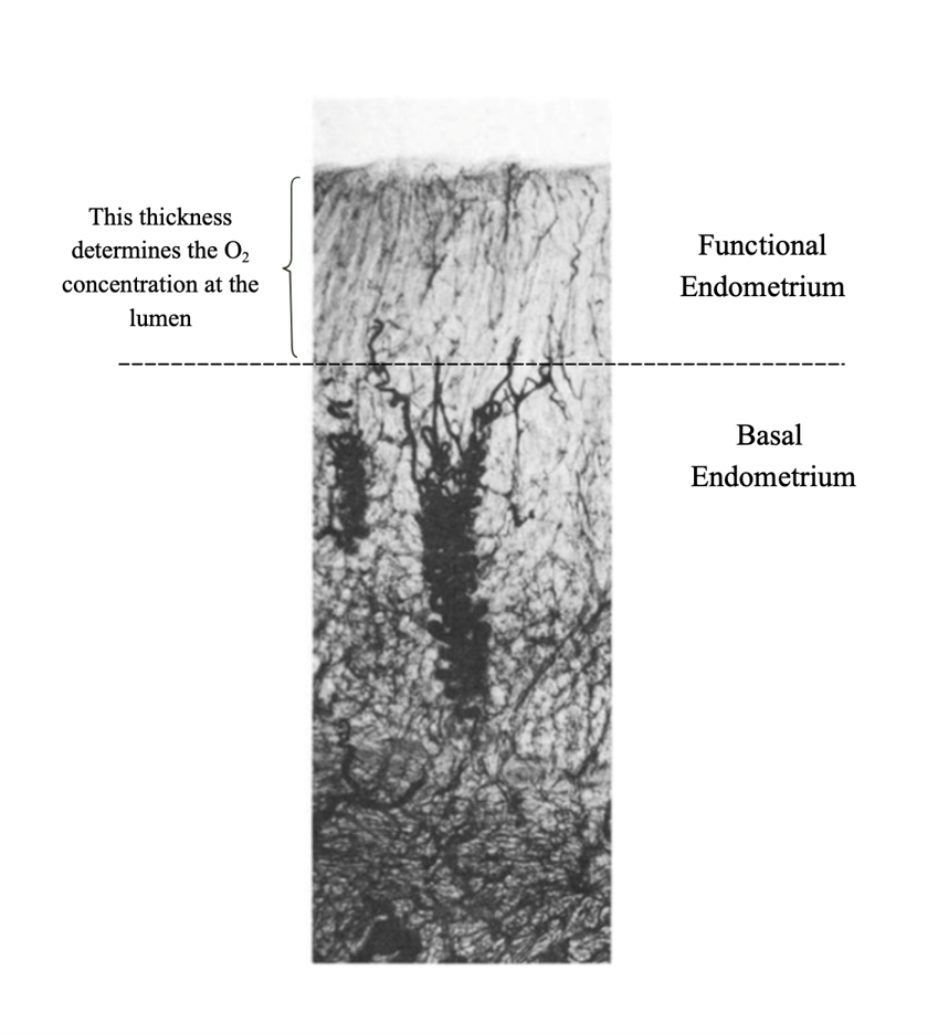
Uterine vascular anatomy is sufficiently well understood that one can use existing data to better understand the differences in oxygen concentrations in uterine luminal fluid for women with different thickness endometrial linings. The spiral arteries (branching off the uterine, arcuate, radial, and basal arteries) are numerous arterioles that each give rise to a capillary plexus supplying a small region of functional endometrium located directly above that spiral artery [15,16]. Although the spiral arteries become more prominently coiled during the luteal phase of the menstrual cycle, the vascular portion of the endometrium remains relatively unchanged throughout the menstrual cycle [15]. Operationally, the spiral arteries carry oxygen to the functional layer of the endometrium, but the capillary plexus distributes it. The capillary plexus is newly regenerated each cycle.
Alveolar air has a pO2 of about 104 mmHg. As oxygen is transported by the arterial system, the oxygen content of blood drops by diffusion through arterial walls and as the arteriole ends, blood has a pO2 level about 85–90 mmHg. Oxygen then diffuses through the capillary walls which supplies oxygen to tissues for metabolism. These capillary webs originating from different spiral arterioles will distribute oxygen on the microscopic level in a complex minimally overlapping pattern to the endometrial tissue related to tissue demand. If the functional endometrium were biologically inert, Fick’s law would suggest the pO2 level at a 3 mm depth of endometrium (6 mm lining) would be twice as high as at the lumen of a 6 mm of endometrium (in a 12 mm lining). Based on the studies cited above, this would suggest that a lumen has an oxygen concentration of about 2–2.5%, but 3 mm above the basal endometrium the oxygen concentration would be about 4–5%. However, applying a Krogh-type modification of Fisk’s law, oxygen release from hemoglobin in capillaries is designed to meet the tissue’s oxygen requirements. Fewer vessels require greater oxygen release per capillary. This flattens the oxygen loss curve from the spiral artery to the terminal endometrium.
Nevertheless, oxygen levels are still higher in the endometrium at a 3 mm depth compared to 6 mm depth. Oxygen transport within capillaries is affected by the metabolic requirements of passing through more tissue and the farther that red blood cells travel within capillaries, the less oxygen they carry before entering the venous system. This has been shown using models more sophisticated than Krogh which strongly correlate with direct measurements in rodent brains. In the rodent brain setting of high metabolic demand, pO2 drops by about 9 mmHg over a 0.05 mm distance [17]. Classical histological studies (for example, Bartelmez [16]) have shown a vascular architecture in late proliferative endometrium where a spiral arteriole gives rise to a long capillary plexus spanning the entire functional layer to supply a small, localized area of luminal epithelium with oxygen. With less capillary overlap, this could further increase metabolic demands from each capillary. Oxygen release into the uterine lumen (and proportional reactive oxygen species) will depend on oxygen availability in the pre-venule aspect of the capillaries.
Once blastocyst attachment begins, the endometrial environment becomes even more precisely regulated with respect to oxygen availability. Failure to achieve the appropriate degree of hypoxia is associated with an increased likelihood of pregnancy loss. Reduced oxygen tension stabilizes hypoxia-inducible factors (HIFs), key transcriptional regulators that coordinate the cellular response to low oxygen conditions. Among their many functions, HIFs promote trophoblast proliferation and differentiation, processes essential for early placental development [18,19]. HIFs increase transcription of vascular endothelial growth factor (VEGF). VEGF is an important actor in promoting angiogenesis. It is also a chemo attractant to monocytes and macrophages, particularly under hypoxic conditions, which are important immune contributors to a viable pregnancy. Relative hypoxia promotes conversion of macrophages to their M2 phenotype which help create a permissive, repair-oriented, and immune tolerant endometrial environment [20].
HIFs also contribute to maintaining this relatively hypoxic environment in early implantation by promoting the transient plugging of spiral arteries by trophoblasts. This plugging limits maternal blood flow during early pregnancy until the vessels are adequately remodeled to support the higher perfusion demands of later gestation [13,18,20]. Consistent with this mechanism, comparative studies of decidual tissue have shown that the proportion of spiral arteries occluded by trophoblasts is approximately sixfold higher in elective abortion specimens than in spontaneous abortions, indicating impaired vascular adaptation in the latter [19].
Hypoxia also plays a critical role in regulating immune function at the time of formation of the maternal–fetal interface. Macrophages preferentially accumulate in hypoxic environments and are integral to the coordinated processes that establish and sustain early pregnancy. Under low-oxygen conditions, these plastic cells adopt an anti-inflammatory phenotype, induce immune tolerance, and facilitate tissue remodeling. Inadequate hypoxia may disrupt these processes, leading to reduced macrophage recruitment, a heightened inflammatory response, and impaired differentiation and function, including defective remodeling of spiral arteries [20].
Estrogen increases the production of VEGF by stromal cells acting through its cognate receptors [21]. HIFs, induced by relative hypoxia, increase VEGF production by direct activation of the VEGF gene in endothelial and other cell types [22]. This relative hypoxia could be especially important for adequate ongoing angiogenesis throughout the course of the proliferative phase in those patients with reduced or abnormal estrogen receptors.
Pathophysiology of the Thin Endometrial Lining
Although the finding of having a thin endometrial lining is a clinical diagnosis, it has an identified underlying pathophysiology that could be more directly addressed in its treatment. Late proliferative phase biopsies of women with thin endometrial lining have 1/3 the number of cells found in fertile controls [23,24]. The number of epithelial glands in the endometrium in the mid-luteal phase is decreased in women with thin endometrial linings compared to usual endometrial linings [23–25]. Since stromal thickening of endometrial tissue is primarily driven by estrogen, a long-standing component of treatment has been to increase the dose and duration of estrogen exposure. However, both late proliferative phase and mid-luteal phase endometrial biopsies of women with thin endometrial lining were found to have decreased estrogen receptors compared to infertility patients with average thickness endometrial lining and fertile women [23,25]. Women with thin endometrial linings also have more estrogen receptor polymorphisms than women with thicker endometrial linings. Some alleles found in women with thin endometrial lining have been found to be associated with a decreased CPR after transfer of a euploid embryos compared to women not carrying them [26].
There is decreased blood flow to the endometrial lining in women with thin endometrial linings as measured by the resistance index (RI) in both the radial and uterine arteries supplying the endometrium throughout the menstrual cycle [24]. There is also a lower density of blood vessels and decreased cellularity suggestive of a diminished microcirculation in the endometrium of women with thin compared to a standard endometrial lining [24] (Figure 2).
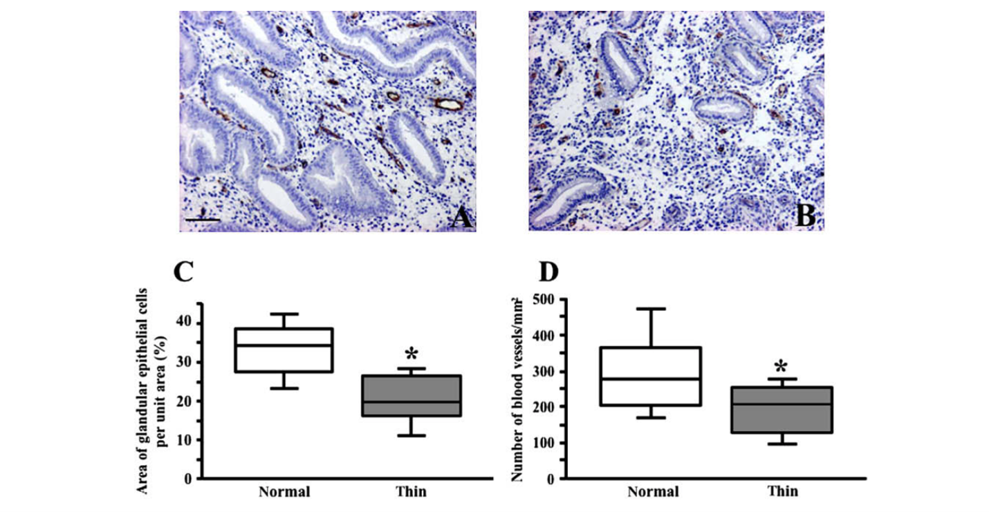
If establishing a sufficiently hypoxic endometrial environment is an important component for implantation, then both the decreased blood flow in the uterine and radial arteries and the less rich capillary plexus could be viewed as positive adaptations compensating for a thin endometrial lining rather than pathophysiology. The lower blood flow could lower oxygen availability from the spiral arteries and the metabolic demands on the diminished capillary plexus will cause greater oxygen utilization from it.
Decreased circulation could play a role in fewer available constituents for forming a normally thick endometrial lining. Circulating endothelial progenitor cells and mesenchymal stem cells may contribute to populating the recovering endometrium during menstrual repair. One source suggests that 48% of endothelial cells and 52% of stromal cells were derived from these circulation stem cells [27], which is likely an overestimate. Cells in both from both the circulation as well as cells resident in the basal layer of the endometrium are likely the genesis of the new endometrium [27,28].
Vascular Endothelial Growth Factor
There are decreased levels of VEGF in late proliferative phase and mid-luteal phase biopsies taken from women with thin endometrial lining compared to typical women, especially in the endometrial glands [23,24]. Much is known about the essential role that VEGF plays in healing the menstrual endometrium and creating a new capillary plexus [29]. Epithelial progenitor cells are recruited to sites for neoangiogeneis by areas of increased concentrations of VEGF [30]. VEGF is also plentiful in the mature endometrium, produced by stromal and glandular epithelial cells during the proliferative phase. VEGF is secreted into the uterine luminal fluid from the apical surfaces of the glandular epithelium. The uterine glands are plentiful by the late proliferative phase and often run for some distance parallel to the luminal surface.
The action of VEGF within the endometrium varies depending on the stage of the endometrial development in which VEGF is acting. Transgenic and knockout experiments have shown that VEGF is essential for the proliferation and migration of endothelial cells in forming new capillaries to support a new endometrium. VEGF blockade in rhesus and mouse models completely inhibits endometrial neovascularization without affecting existing vessels [31]. VEGF also appears essential for blastocyst implantation [32]. For example, immunization of rhesus monkeys with monoclonal anti-VEGF significantly reduces the establishment of pregnancy [33]. The importance of VEGF availability further supports Casper’s hypothesis in that VEGF secretion by epithelial cells is enhanced by hypoxia [31,34].
The endometrial glands also secrete other growth factors into the uterine fluid. TGF-β1 is also produced in large quantities [35]. In addition to modulating immunotolerance, TGF-β1 modulates other molecules known to be important for implantation including VEGF [36]. After implantation, TGF-β1 availability plays an expansive role in the processes required for live birth. Prior to implantation, the availability of TGF-β1 in the uterine cavity depends upon glandular secretion. After implantation, the endometrial glands continue to transform likely in response to estrogen and progesterone and play a significant role in support of the early pregnancy [37,38].
Growth Factors and Healing from Post Menstrual Shedding
After shedding, the endometrial epithelium is rapidly restored, typically reforming within 4 to 8 days after the start of menstruation [39]. The disordered piecemeal breakdown of the functionalis contains patches of completely shed endometrium adjacent to endometrium in a range of healing states [40]. Full surface breakdown also takes place over a number of days as endometrial cells can be found in menstrual blood during the entire course of flow. Prior to complete re-epithelialization, residual patches of intact epithelium are commonly observed in the cornual regions, central fundus, and around the cervical canal [41,42]. Surface re-epithelialization is thought to generally arise from patches of intact epithelium and from the stumps of epithelial glands located on the basal surface. Within these retained basal endometrium, small populations of epithelial progenitor cells are present and are believed to clonally expand to generate the surface epithelium. However, it is likely that a subset of these progenitor epithelial cells also arises from circulating mesenchymal stem cells/progenitor cells [27,28].
Endometrial gland regeneration was traditionally believed to occur through proliferation of glandular epithelial cells within the residual glandular stumps retained in the basal layer following menstruation. However, the limited mitotic activity observed in these structures led Garry et al. to propose that glandular regeneration is driven primarily by the differentiation of progenitor or stem cells residing within the basalis [42,43]. Newly formed glands undergo rapid morphological remodeling, initially appearing as elongated, narrow structures that extend horizontally parallel to the basalis and frequently aligned with newly developing capillaries [43]. This close spatial relationship suggests that angiogenesis may contribute to gland formation and organization. Once established, glandular growth and maturation are primarily regulated by estrogen.
Recent studies have demonstrated that endometrial fibroblasts (stromal cells) can arise from mesenchymal stem/progenitor cells located within the basal endometrium. These stem cells constitute approximately 1.3% of stromal cells (and 0.2% of epithelial cells) within this compartment [28]. Mesenchymal stem cells have been identified in the circulation and in menstrual fluid, where they are also likely to contribute to regeneration of the functional endometrium, although the extent of this contribution has not yet been quantified [28]. Capillary pericytes, which are thought to be of vascular origin and possess mesenchymal stem cell-like properties, may likewise participate in endometrial regeneration.
During the regenerative phase of menstruation, the menstrual wound environment is enriched with blood-derived components that come into direct contact with repairing epithelial, stromal, glandular, and vascular tissues. Immediately preceding menstruation, leukocytes are the predominant blood-derived cell population in the endometrium, comprising up to 50% of the total cellular content [40]. These immune cells secrete a diverse array of bioactive mediators, including growth factors, cytokines, and chemokines. Growth factors present in menstrual blood are therefore thought to play a pivotal role in coordinating tissue repair and regeneration, (Table 1).
|
Phase |
Major GFs |
Comments |
|
Hemostasis |
PDGF, VEGF, TGF-β |
|
|
Epithelial regeneration |
EFG, IGF-1 |
Migration and proliferation |
|
Angiogenesis |
VEGF, FGF |
New capillaries and repair |
|
Stromal remodeling |
PGDF, TGF-β |
Fibroblast proliferation Extracellular matrix deposition |
|
Functional regeneration |
IGF-1 (primarily estrogen) |
Gland elongation Stromal thickening |
Once the surface epithelium completely covers the uterine cavity, it forms a tight barrier to large molecules and thus large growth factors cannot passively diffuse through the intact endometrium. For example, VEGF is at least 38 kDA or 32,000 times as large as a water molecule. The smallest growth factor discussed here, EGF, is still about 333 times the size of a water molecule (Table 2). If these paracrine-acting growth factors from PRP do improve endometrial stromal or glandular growth and if treatment is initiated after the epithelium has healed over the entire uterine surface, then that substrate/receptor mediated response requires a breach in the uterine epithelial surface or there is some, presently unknown, growth factor transport system within the endometrial epithelium. However, such a transport system seems unlikely. Copious growth factors are being secreted outward from the endometrial glands. Once menstrual repair is complete, in the late proliferative phase, TGF-β and FGF signaling are attenuated in the stromal cells beneath the epithelial surface. During the late proliferative-to-secretory transition, endometrial epithelial cells express LEFTY, an endogenous antagonist of TGF-β superfamily signaling [44].
Platelet-Rich Plasma
Many published clinical studies utilize a PRP infusion to treat the problem of a thin endometrial lining in the late proliferative phase over an intact surface epithelium. PRP contains many growth factors including VEGF. With PRP administered in this way, growth factors are unlikely to play a role in changing the underlying endometrium. However, because activated PRP also contains more than 1,500 bioactive molecules [45], such an infusion during the late proliferative phase could still confer benefits for endometrial receptivity. In addition to the previously described growth factors, other bioactive components may influence the endometrial environment, either by penetrating the restored endothelial barrier or by otherwise modulating local cellular signaling pathways. Furthermore, when PRP is administered close to the time of implantation, an increased concentration of growth factors within the endometrial luminal fluid could enhance implantation potential. However, despite accumulating clinical interest and proposed general mechanisms, the currently available hypotheses remain limited and heterogeneous. As a result, it is difficult to definitively identify a dominant pathway underlying therapeutic effects of PRP infusion administered in the late proliferative phase.
PRP and Its Preparation
More than 40 different preparations of PRP have been described in the medical literature for the diverse applications of PRP [45]. Many reproductive endocrinology programs likely use a commercially available kit which employs the predominant double centrifugation approach. Based on in vitro published studies, this technique concentrates platelets 3–5-fold over blood levels [46]. Blood is usually drawn into a collecting device with sodium citrate adequate to bind all free calcium and prevent coagulation and platelet activation. For PRP therapy to work, platelets need to be activated (to empty the granules that they carry to store these substances). This can be done endogenously (by contact with thrombin, basement membrane, etc.) or exogenously by adding, for example, calcium chloride which is typical in most PRP preparations for infusion.
Activated platelets release their granules within the PRP fluid. Most VEGF from platelets is of the form VEGF-A, isoform 165. This is believed to be the most angiogenic of the VEGF molecules. Release of these growth factors from activated PRP is rapid; with release beginning in the first 10 minutes and with 95% of the granules released in the first hour [45]. In addition, the half-life of some of these growth factors is very short. For example, VEGF has a half-life of 30–45 minutes (Table 2). For maximal effect of this release of supraphysiologic concentrations of growth factors, there needs to be close coordination between the time of platelet activation by the laboratory and its application to the patient.
|
Growth Factor |
Half life |
Approx. Size (kDa) |
|
VEGF |
30–45 minutes |
38–45 |
|
EGF |
Approx. 1 hour |
6 |
|
FGF |
Approx. 1 hour |
17–34 |
|
PDGF |
Approx. 2 hours |
30–35 |
|
TGF-β |
2–4 hours |
25 |
|
IGF-1 |
12–15 hours |
7.5 |
PRP Clinical Studies for the Treatment of the Thin Endometrial Lining
To begin to understand the current status of clinical research on treatment of the thin endometrial lining with PRP and to understand current expectations for the success of this therapy, it is valuable to look at the randomized studies that have been published using PRP as treatment for a thin endometrial lining. Liu et al.’s looked at the currently available randomized trials of PRP for thin endometrial lining and performed a meta-analysis on those that they found [47]. The eight studies used in their meta-analysis found an average increase in the endometrial lining of 1.23 mm with a 95% confidence interval (CI) of 0.87–1.59 mm (P=0.000). The meta-analysis also found that the CPR doubled (relative risk (RR) 2.04, 95% CI 1.52–2,76, P=0.000). Results for the LBR were similar (RR 2.46, CI 1.57–3.85, P=0.000). Cycle cancellation was separately reported in some of the studies and the RR of cancellation for a persistently thin endometrial lining after PRP treatment was 0.46.
However, the diversity of the details in these eight studies could undermine a clinician’s confidence in equivalently applying some of the above conclusions. PRP was delivered by uterine infusion in five studies, one study injected PRP contained in a fibrin matrix (injectable platelet-rich fibrin) into the uterine cavity, and two studies delivered PRP with sub-endometrial injections using hysteroscopy. Significantly, study groups varied on when in the cycle PRP was delivered and how frequently. Some studies utilized 1–3 administrations starting on cycle days 10–13, but some infusions were post-ovulatory prior to an IUI. The timing for assessing changes in endometrial thickness also varied. PRP preparations utilized various methods using either exogenous or endogenous activation without explicit mention of whether the time interval from exogenous activation to administration was controlled.
Discussion
PRP represents a rational therapeutic approach to enhance endometrial growth by delivering supraphysiologic concentrations of growth factors that contribute to endometrial repair following menstrual shedding. Physiological regeneration of the endometrium during and shortly after menstrual bleeding is a routine event. VEGF plays a central role in endometrial angiogenesis and vascular remodeling and likely plays a role in endometrial gland development. Therefore, increasing the local availability of VEGF within endometrial tissue at a time when it can be effectively utilized constitutes a biologically plausible strategy. VEGF signaling is effectively immediately at the cellular level, with receptor activation and intracellular signal transduction occurring on a minute-scale timeframe following ligand binding [48]. Early angiogenic sprouting begins within hours of VEGF stimulation and is generally evident as measurable capillary sprouts by 24 hours in endothelial culture systems [48].
The extent of angiogenic sprouting is strongly dependent on VEGF concentration, with increases in VEGF levels leading to corresponding increases in sprout number, length, and overall angiogenic output, up to a biologically defined limit [49]. Increased local concentration of VEGF attracts progenitor epithelial cells to a region [29,30]. Accordingly, delivering VEGF to endometrial tissue at concentrations exceeding those present in menstrual blood is expected to augment vascular formation.
Expansion of the endometrial vascular network may facilitate epithelial gland development, as glandular structures are known to organize in close association with emerging microvasculature during early tissue regeneration [43]. The establishment of these glands is essential for subsequent full estrogen responsiveness, enabling their differentiation into the complex secretory architecture characteristic of a receptive endometrium. In women with thin endometrial linings, where VEGF expression is reduced during the late proliferative and midluteal phases, increasing glandular density may also help optimize the peri-implantation intraluminal environment. To the extent that circulating mesenchymal stem cells contribute to the replenishment of endometrial stromal compartment, increased vessel formation during the menstrual repair phase may also maximize the impact of circulating stem cells on stromal cell volume.
Increases in both endometrial thickness and glandular density initiated during menstrual repair may act synergistically to improve receptivity in mid-cycle. By reinforcing tissue structure while supporting secretory function, these adaptations could partially counterbalance the possibly persistent diminished hypoxia and decline in VEGF production that accompany the peri-implantation period of women with thin endometrial linings, thereby enhancing the likelihood of successful implantation.
Even in the absence of increased glandular development, PRP-induced expansion of endometrial thickness following treatment early in the menstrual cycle would alter the luminal milieu during the mid- and late-luteal phases. By increasing tissue volume, PRP would help establish a more hypoxic intraluminal environment leading to increased VEGF production by the cells in the luminal endometrial glands. Hypoxia is a well-recognized regulator of VEGF, and experimental studies have shown that endometrial glandular epithelial cells increase VEGF production in response to reduced oxygen tension [34,35]. Enhanced luminal VEGF concentrations during the peri-implantation period could contribute to a more favorable endometrial environment for implantation and early embryonic development.
A PRP infusion at the time of menstrual healing would be analogous to successful applications of PRP in other surgical applications. An important consequence of the use of PRP is the increased proliferation (and migration) of fibroblasts and stem cells [50,51]. For example, a PRP product enables an oral surgeon to fill a tissue gap in two months rather than six to twelve months due to this impact [52]. The objective in this reproductive application is less dramatic. The foundation for the development of the endometrial lining during a menstrual cycle needs to be changed in a matter of a few days. Since supraphysiological levels of VEGF can have an impact starting as quickly as minutes after application and possibly establish a foundation on which estrogen can build a more functional endometrial structure before estrogen begins its significant rise during the menstrual cycle. In the randomized PRP studies discussed previously, the PRP application primarily occurred in the late proliferative phase perhaps as a treatment for the finding in that cycle of a thin endometrial lining. At that time, the growth factors in these applications do not have the ability to penetrate the epithelium and affect the endometrial vessels, glands, or stroma. Late application of PRP could improve the growth factor component of endometrial fluid, but if the platelets were activated, those growth factors would have a time limited value (Table 2).
Definitive conclusions regarding the efficacy of PRP therapy for thin endometrial lining will require rigorous clinical investigation and experimental validation. From a mechanistic perspective, VEGF is likely a central, if not the principal, mediator within PRP potentially contributing to improved reproductive outcomes in this context. Importantly, VEGF exerts distinct, temporally regulated functions across the menstrual cycle. A very thin endometrial lining downregulates VEGF during the menstrual cycle by way of HIFs which leads to a chain of events rendering the endometrial cavity less hospitable to embryos. Effective VEGF activity requires binding to its receptors on appropriate target structures. For increased angiogenic effects, higher than physiological concentrations of VEGF must reach and interact with the endometrial vasculature. This angiogenic effect is not directly mediated through estrogen receptors, but is an effect of signaling by activation of VEGFR2 receptors on endothelial cells, and thus increased exogenous VEGF could also compensate for defective or inadequate estrogen receptors [53]. Also, for the role played by VEGF related to blastocyst attachment and decidual remodeling, VEGF must be present within the uterine luminal environment in sufficient quantities and with sufficient duration to influence epithelial and decidual cell interactions.
If a PRP infusion can increase endometrial thickness, it will be important to determine how best to use it. Would it be adequate to inject activated PRP into a woman’s uterus on day 4 of her menstrual cycle and have her lie still for 30 minutes (close to the half-life of VEGF)? Should the treatment be repeated the next day? Would there be an increased benefit of using PRP during both the healing phase of the menstrual cycle as well as in the late proliferative phase? Would injectable platelet-rich fibrin (PRF) with endogenous activation and a possible longer-term action be a better choice than PRP? PRF forms a soft matrix which may keep the platelets in the uterus longer and also contains leukocytes which secrete VEGF and other growth factors over a longer time period [54]. Since new areas of endometrium are being shed throughout the menstrual time period, should multiple infusions be used starting earlier in the menstrual cycle, for example, on day 2 or 3?
Conclusion
The clinical data related to having a thin endometrial lining is sufficiently compelling that it should be a consideration for all clinicians performing ART cycles. Effective management of the thin endometrial lining is currently a dispiriting dilemma. Many pathological features have been identified, but few root causes have been characterized. Casper proposed that one mechanism by which a thin endometrial lining contributes to subfertility is the reduced likelihood of achieving the degree of hypoxia necessary for optimal blastocyst development and implantation. Relative hypoxia increases VEGF signaling at multiple sites in the latter parts of the menstrual cycle. Casper’s hypothesis can be supported by uterine anatomical arguments, accepted chemical/physiological principles, and mechanistic biochemical data.
Although this manuscript does NOT demonstrate that PRP is an effective treatment for a thin endometrial lining, it provides strong theoretical support for using the growth factors, especially VEGF, found in PRP to augment natural physiological healing at the time of or temporally following menstrual shedding to enhance endometrial growth. Appropriately applied PRP has the potential of enhancing both stromal cell and endometrial gland development. For those clinicians, who see value in PRP as an adjunctive treatment to enhance endometrial thickness, this report advocates a change in approach. PRP is likely to be most effective if it is used during the healing phase of menstrual shedding rather than in the late proliferative phase. The PRP infusion must be done in a manner reflecting awareness of the short half-lives of the growth factors after their release.
If PRP can thicken any endometrial lining and if the reported correlation between the endometrial lining thickness and an increased pregnancy rates is causative, then PRP treatment could become a useful adjunct in the treatment of women with “low normal thickness endometrial lining”, with Asherman’s syndrome, with recurrent implantation failure, or even in general IVF [3]. Much research needs to be done before drawing any of these conclusions. The research done will be of higher utility if the PRP treatment is as clinically detailed as much as possible.
Statements and Declarations
Funding
The authors declare that no funds, grants, or other support were received during the preparation of this manuscript.
Competing interests
No financial or non-financial interests were directly or indirectly related to this manuscript submitted for publication.
Author contributions
All authors contributed to the study’s conception and design. The first draft of the manuscript was written by Bruce Rose and all authors commented on prior versions of this manuscript. All authors read and approved the final manuscript.
Ethics approval
This study did not involve human or animal subjects and ethics committee approval was not required.
References
2. Liao S, Wang R, Hu C, Pan W, Pan W, Yu D, et al . Analysis of endometrial thickness patterns and pregnancy outcomes considering 12,991 fresh IVF cycles. BMC Med Inform Decis Mak. 2021 Jun 3;21(1):176.
3. Gingold JA, Wu H, Lieman H, Singh M, Jindal S. Increasing endometrial thickness up to 12 mm is associated with increased odds of live birth among fresh and frozen-thawed autologous transfers with or without preimplantation genetic testing. Fertil Steril. 2025 Sep;124(3):478–86.
4. Schmiech K, Li M, Chen LX, Dow MP, Baker VL. Association of endometrial thickness with live birth rate: a study using the Society for Assisted Reproductive Technology Clinic Outcome Reporting System. Fertil Steril. 2025 Jul;124(1):79–87.
5. Mouhayar Y, Sharara FI. Modern management of thin lining. Middle East Fertility Society Journal. 2017 Mar 1;22(1):1–12.
6. Wang Y, Tang Z, Teng X. New advances in the treatment of thin endometrium. Front Endocrinol (Lausanne). 2024 Apr 30;15:1269382.
7. Karadbhajne P, Dzoagbe HY, More A. Platelet-Rich Plasma (PRP) for Endometrial Treatment Efficacy and Safety in Assisted Reproductive Technology: A Comprehensive Review. Cureus. 2024 May 6;16(5):e59728.
8. Stefanović M, Vukomanović P, Kutlesic R, Trenkić M, Dimitrov V, Stefanović A, et al. The Effect of Autologous Platelet Rich Plasma on Endometrial Receptivity: A Narrative Review. Medicina (Kaunas). 2025 Jan 15;61(1):134.
9. Mahajan N. Endometrial receptivity array: Clinical application. J Hum Reprod Sci. 2015 Jul-Sep;8(3):121–9.
10. Casper RF. It's time to pay attention to the endometrium. Fertil Steril. 2011 Sep;96(3):519–21.
11. Kigawa J. Studies on the levels of pO2 and pCO2 in the uterine cavity and uterine tissue (author's transl). Nihon Sanka Fujinka Gakkai Zasshi. 1981 Oct 1;33(10):1646–54.
12. Yedwab GA, Paz G, Homonnai TZ, David MP, Kraicer PF. The temperature, pH, and partial pressure of oxygen in the cervix and uterus of women and uterus of rats during the cycle. Fertil Steril. 1976 Mar;27(3):304–9.
13. Ottosen LD, Hindkaer J, Husth M, Petersen DE, Kirk J, Ingerslev HJ. Observations on intrauterine oxygen tension measured by fibre-optic microsensors. Reprod Biomed Online. 2006 Sep;13(3):380–5.
14. Ng KYB, Mingels R, Morgan H, Macklon N, Cheong Y. In vivo oxygen, temperature and pH dynamics in the female reproductive tract and their importance in human conception: a systematic review. Hum Reprod Update. 2018 Jan 1;24(1):15–34.
15. Rogers PA. Structure and function of endometrial blood vessels. Hum Reprod Update. 1996 Jan–Feb;2(1):57–62.
16. Bartelmez GW. Histological studies on the menstruating mucous membrane of the human uterus. Washington, DC: Carnegie Institution of Washington; 1933. p. 141–186.
17. Lücker A, Weber B, Jenny P. A dynamic model of oxygen transport from capillaries to tissue with moving red blood cells. Am J Physiol Heart Circ Physiol. 2015 Feb 1;308(3):H206–16.
18. Pringle KG, Kind KL, Sferruzzi-Perri AN, Thompson JG, Roberts CT. Beyond oxygen: complex regulation and activity of hypoxia inducible factors in pregnancy. Hum Reprod Update. 2010 Jul–Aug;16(4):415–31.
19. Gun BD, Numanoglu G, Ozdamar SO. The comparison of vessels in elective and spontaneous abortion decidua in first trimester pregnancies: importance of vascular changes in early pregnancy losses. Acta Obstet Gynecol Scand. 2006;85(4):402–6.
20. Huang X, Lin Z, Zheng ZM, Shi JL, Lu KY, Wang JR, Et al. A Hypoxia-Decidual Macrophage Regulatory Axis in Normal Pregnancy and Spontaneous Miscarriage. Int J Mol Sci. 2024 Sep 8;25(17):9710.
21. Okada H, Tsutsumi A, Imai M, Nakajima T, Yasuda K, Kanzaki H. Estrogen and selective estrogen receptor modulators regulate vascular endothelial growth factor and soluble vascular endothelial growth factor receptor 1 in human endometrial stromal cells. Fertil Steril. 2010 May 15;93(8):2680–6.
22. Manalo DJ, Rowan A, Lavoie T, Natarajan L, Kelly BD, Ye SQ, et al. Transcriptional regulation of vascular endothelial cell responses to hypoxia by HIF-1. Blood. 2005 Jan 15;105(2):659–69.
23. Sandora N, Wiweko B, Muharam R, Harzif AK, Kusuma TR, Fitria NA, et al. Cellular, histological, and gene expression differences in thin versus normal endometrium. Acad Biol. 2025 Aug 1;3(3).
24. Miwa I, Tamura H, Takasaki A, Yamagata Y, Shimamura K, Sugino N. Pathophysiologic features of "thin" endometrium. Fertil Steril. 2009 Apr;91(4):998–1004.
25. Li LH, Shi G, Pan JB, Wang CH, Zhao M, Zhang XP. The expressions of matrix metalloproteinase-9, estrogen receptor, and progesterone receptor in thin endometrial tissue and their significance. Gynecol Endocrinol. 2022 Jun;38(6):516–522.
26. Lledo B, Hortal M, Martínez M, Ortiz JA, Morales R, Bernabeu A. Association of estrogen and progesterone receptor polymorphisms with idiopathic thin endometrium. Pharmacogenet Genomics. 2025 Jun 1;35(4):136–9.
27. Taylor HS. Endometrial cells derived from donor stem cells in bone marrow transplant recipients. JAMA. 2004 Jul 7;292(1):81–5.
28. Hong IS. Endometrial stem/progenitor cells: Properties, origins, and functions. Genes Dis. 2022 Aug 31;10(3):931–47.
29. Smith SK. Angiogenesis, vascular endothelial growth factor and the endometrium. Hum Reprod Update. 1998 Sep-Oct;4(5):509–19.
30. Lampri E, Ioachim E. Angiogenesis: something old, something new. In: Santulli G, editor. Angiogenesis: insights from a systemic overview. New York: Nova Science Publishers; 2013. p. 1–30.
31. Fan X, Krieg S, Kuo CJ, Wiegand SJ, Rabinovitch M, Druzin ML, et al. VEGF blockade inhibits angiogenesis and reepithelialization of endometrium. FASEB J. 2008 Oct;22(10):3571–80.
32. Hannan NJ, Paiva P, Meehan KL, Rombauts LJ, Gardner DK, Salamonsen LA. Analysis of fertility-related soluble mediators in human uterine fluid identifies VEGF as a key regulator of embryo implantation. Endocrinology. 2011 Dec;152(12):4948–56.
33. Sengupta J, Lalitkumar PG, Najwa AR, Charnock-Jones DS, Evans AL, Sharkey AM, et al. Immunoneutralization of vascular endothelial growth factor inhibits pregnancy establishment in the rhesus monkey (Macaca mulatta). Reproduction. 2007 Jun;133(6):1199–211.
34. Maybin JA, Hirani N, Brown P, Jabbour HN, Critchley HO. The regulation of vascular endothelial growth factor by hypoxia and prostaglandin F₂α during human endometrial repair. J Clin Endocrinol Metab. 2011 Aug;96(8):2475–83.
35. Guzeloglu-Kayisli O, Kayisli UA, Taylor HS. The role of growth factors and cytokines during implantation: endocrine and paracrine interactions. Semin Reprod Med. 2009 Jan;27(1):62–79.
36. Jones RL, Stoikos C, Findlay JK, Salamonsen LA. TGF-beta superfamily expression and actions in the endometrium and placenta. Reproduction. 2006 Aug;132(2):217–32.
37. Gray CA, Bartol FF, Tarleton BJ, Wiley AA, Johnson GA, Bazer FW, et al. Developmental biology of uterine glands. Biol Reprod. 2001 Nov;65(5):1311–23.
38. Hempstock J, Cindrova-Davies T, Jauniaux E, Burton GJ. Endometrial glands as a source of nutrients, growth factors and cytokines during the first trimester of human pregnancy: a morphological and immunohistochemical study. Reprod Biol Endocrinol. 2004 Jul 20;2:58.
39. Ferenczy A, Richart RM, Agate FJ Jr, Purkerson ML, Dempsey EW. Scanning electron microscopy of the human endometrial surface epithelium. Fertil Steril. 1972 Aug;23(8):515–21.
40. Salamonsen LA, Hutchison JC, Gargett CE. Cyclical endometrial repair and regeneration. Development. 2021 Sep 1;148(17):dev199577.
41. Ang CJ, Skokan TD, McKinley KL. Mechanisms of Regeneration and Fibrosis in the Endometrium. Annu Rev Cell Dev Biol. 2023 Oct 16;39:197–221.
42. Garry R, Hart R, Karthigasu KA, Burke C. A re-appraisal of the morphological changes within the endometrium during menstruation: a hysteroscopic, histological and scanning electron microscopic study. Hum Reprod. 2009 Jun;24(6):1393–401.
43. Garry R, Hart R, Karthigasu KA, Burke C. Structural changes in endometrial basal glands during menstruation. BJOG. 2010 Sep;117(10):1175–85.
44. Marečková M, Garcia-Alonso L, Moullet M, Lorenzi V, Petryszak R, Sancho-Serra C, et al. An integrated single-cell reference atlas of the human endometrium. Nat Genet. 2024 Sep;56(9):1925–37.
45. Pavlovic V, Ciric M, Jovanovic V, Trandafilovic M, Stojanovic P. Platelet-rich fibrin: Basics of biological actions and protocol modifications. Open Med (Wars). 2021 Mar 22;16(1):446–54.
46. Farshidfar N, Amiri MA, Estrin NE, Ahmad P, Sculean A, Zhang Y, et al. Platelet-rich plasma (PRP) versus injectable platelet-rich fibrin (i-PRF): A systematic review across all fields of medicine. Periodontol 2000. 2025 Oct;99(1):185–215.
47. Liu X, Qian C, Jiang X, Zhou Y, Feng X, Ding Y, et al. Efficacy of platelet-rich plasma in the treatment of thin endometrium: a meta-analysis of randomized controlled trials. BMC Pregnancy Childbirth. 2024 Aug 30;24(1):567.
48. Shah S, Kang KT. Two-Cell Spheroid Angiogenesis Assay System Using Both Endothelial Colony Forming Cells and Mesenchymal Stem Cells. Biomol Ther (Seoul). 2018 Sep 1;26(5):474–80.
49. Kannan P, Schain M, Lane DP. An Automated Quantification Tool for Angiogenic Sprouting From Endothelial Spheroids. Front Pharmacol. 2022 Apr 27;13:883083.
50. Van der Bilj I. Studies on platelets and wound healing. PhD-Thesis – Research and graduation internal, Vrije Universiteit Amsterdam; 2021.
51. Kakudo N, Minakata T, Mitsui T, Kushida S, Notodihardjo FZ, Kusumoto K. Proliferation-promoting effect of platelet-rich plasma on human adipose-derived stem cells and human dermal fibroblasts. Plast Reconstr Surg. 2008 Nov;122(5):1352–60.
52. Choukroun J, Diss A, Simonpieri A, Girard MO, Schoeffler C, Dohan SL, et al. Platelet-rich fibrin (PRF): a second-generation platelet concentrate. Part IV: clinical effects on tissue healing. Oral Surg Oral Med Oral Pathol Oral Radiol Endod. 2006 Mar;101(3):e56–60.
53. Wong C, Jin ZG. Protein kinase C-dependent protein kinase D activation modulates ERK signal pathway and endothelial cell proliferation by vascular endothelial growth factor. J Biol Chem. 2005 Sep 30;280(39):33262–9.
54. Dohan Ehrenfest DM, de Peppo GM, Doglioli P, Sammartino G. Slow release of growth factors and thrombospondin-1 in Choukroun's platelet-rich fibrin (PRF): a gold standard to achieve for all surgical platelet concentrates technologies. Growth Factors. 2009 Feb;27(1):63–9.
55. Bartelmez GW. The form and functions of the uterine blood vessels in the rhesus monkey. Contrib Embryol Carnegie Inst Wash. 1957;36:153–82.