Abstract
Advanced ultrasound offers an important diagnostic clinical tool for the specialty of Obstetrics (OB) and Gynecology (GYN). The advanced ultrasound technologies, such as power Doppler angiography, with its assessment of vascular flow, adds to the described clinical benefits. The imaging technology that this represents, offers diagnostic abilities for a variety of obstetric and gynecologic conditions
Keywords
Ultrasound; Gynecology; Flow doppler; Carcinoma diagnosis; Uterine fibroids; Congenital malformation
Ultrasound in OB/GYN
We are witnessing the evolution of the diagnostic imaging that is rapidly becoming available to Women’s Health physicians (OB/GYN’s), enabling the use of technology to augment our ability to medically palpate on examination of the patient, to identify conditions which we were unable to appreciate a short time ago. Threedimensional transvaginal sonography (3DTVS), along with power Doppler angiography (PDA), are examples of such technologies. Clinicians have long appreciated the ability to view a three-dimensional image from the use of X-Rays (e.g. in computed tomography [CT] scans), or with magnetic resonance imaging (MRI). We can now create 3-D images with the use of ultrasound, in an easier and less costly way, and with possibly lesser harmful radiation. The tissue which is an ideal target of this imaging is found in the pelvis. Naturally, looking at the uterus, fallopian tubes, and ovaries is within the province of the OB/GYN.
There are many examples of the diagnostic utility of 3DTVS in OB/GYN. For example, identification of an embedded, broken or partially expelled intrauterine contraceptive device (IUD) is now possible [1], which can help to lead to resolution of that clinical problem (Figure 1). The identification of deep infiltrating endometriosis (DIE), not able to be seen laparoscopically, is also possible with the use of 3DTVS (Figure 2) [2]. The practical imaging of benign leiomyomata (i.e. fibroids) is often used to monitor their growth, with the caliper measurement of size, aiding decision-making regarding the possible need for surgical intervention (Figure 3) [1]. The identification of uterine malformations is also possible today, at times explaining the etiology of pregnancy loss and fertility failure (Figure 4). With the ability to identify vascular flow, identification and staging of endometrial carcinoma has even been recently demonstrated (Figure 5) [3]. We have been actively using ultrasound (US) to help diagnose ectopic pregnancy (EP), but 3DTVS may aid the diagnosis further with characterization of its vascular flow with the use of 3DTVS and PDA [1]. Obviously, we are still early in discovering the clinical benefit of this imaging technology.
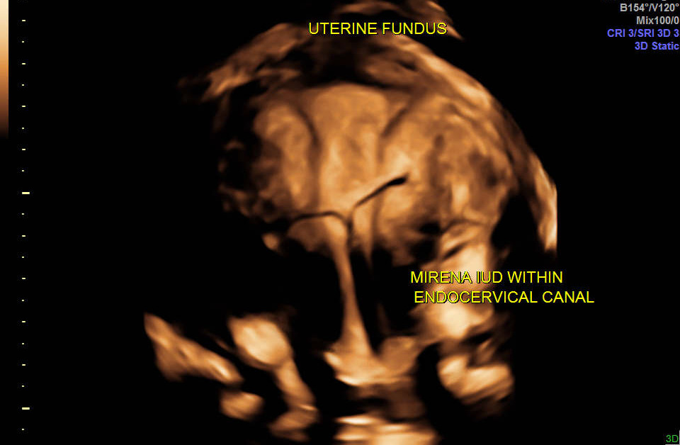
Figure 1. Partially expelled IUD.
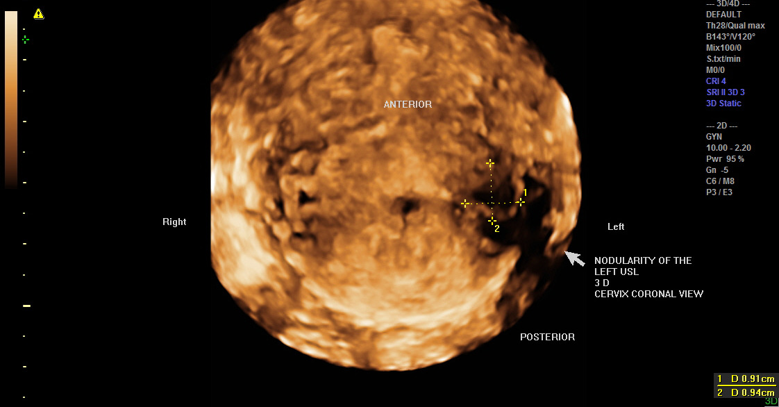
Figure 2. Pelvic pain and severe dysmenorrhea led to this sonographic exploration. Nodularity of the left uterosacral ligament is demonstrated in this rendering mode (pathologically confirmed).
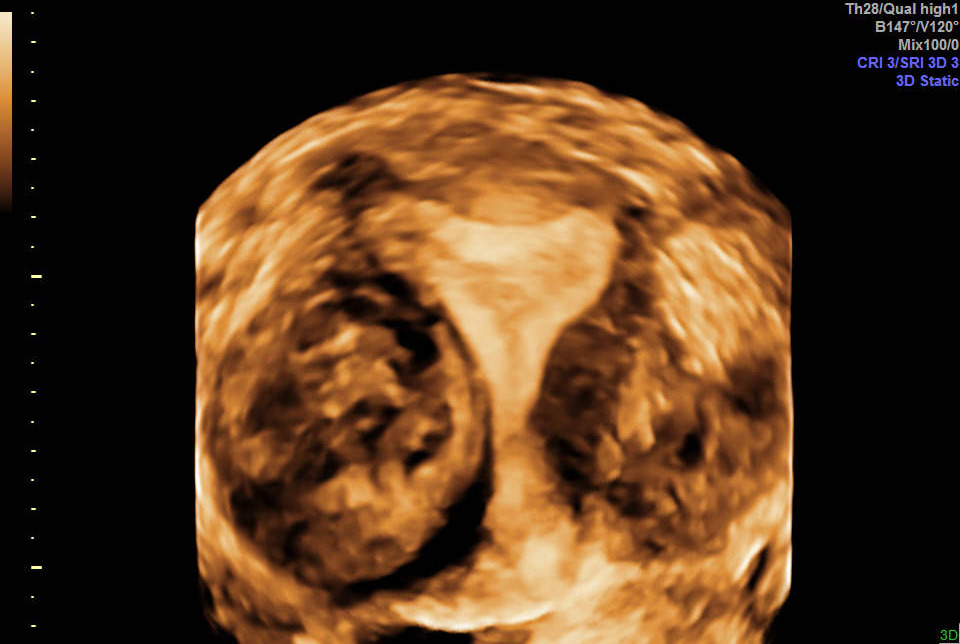
Figure 3. Intraluminal location of a fibroid can be demonstrated without saline infusion sonography or MRI.
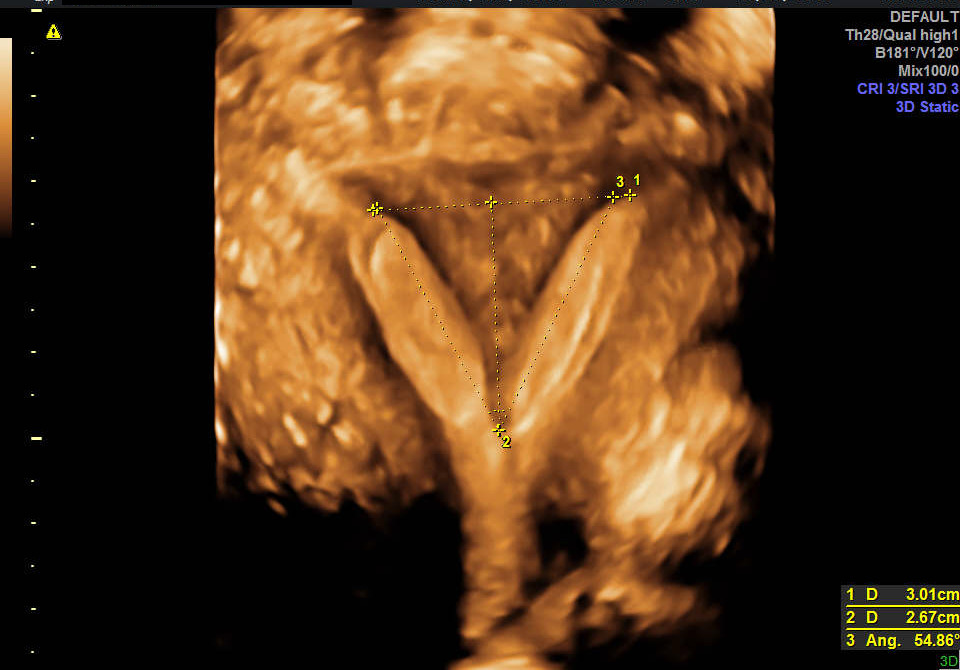
Figure 4. Inability to diagnose with Gray-Scale 2 D ultrasound. “Glass Body Rendering” convincingly demonstrated the septate uterus.
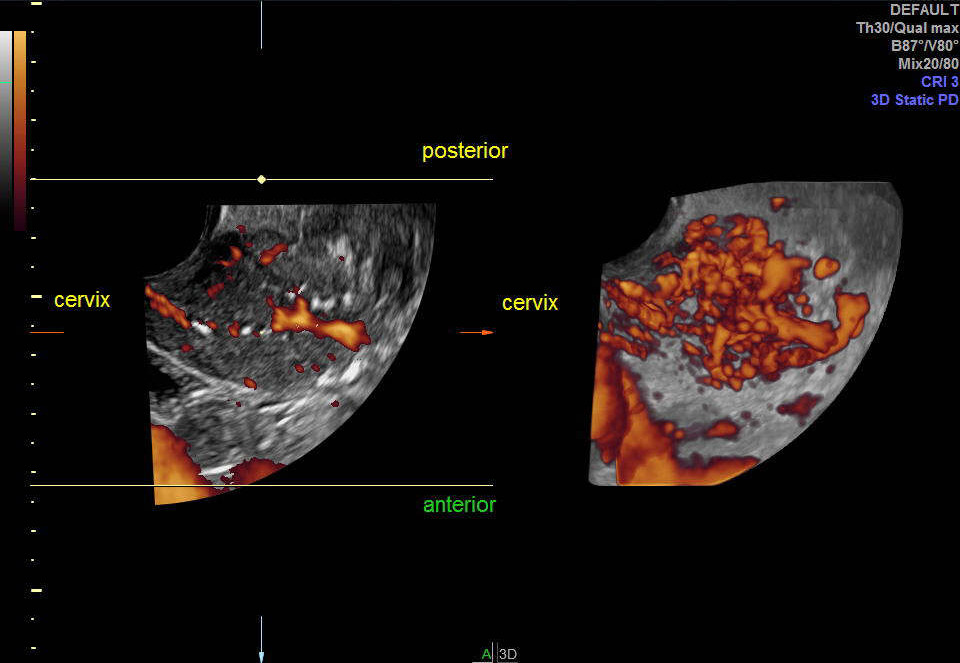
Figure 5. After 2 negative 2 D ultrasounds – and unable to perform endometrial biopsy due to cervical stenosis, the diagnosis of endometrial cancer, Stage IB, was suspected with 3DTVS. Suspected diagnosis was confirmed with pathology.
We are sure to see the future evolution of the use of ultrasound imaging in this specialty, with even greater expansion of technology, in currently unimaginable ways. A few rendering 3D images accompanying this message illustrates the sort of image resolution and clinical benefit that is possible.
References
2. Levine EM, Fernandez CM, Pham M, Shashoua A, Locher S. Deep Infiltrating Endometriosis: Making the Diagnosis. Journal of Diagnostic Medical Sonography. 2019 Nov;35(6):511-3.
3. Fernandez CM, Levine EM, Dini M, Bannon K, Butler S, Locher S. Predictive Value of Three-Dimensional Transvaginal Sonography for Staging of Endometrial Neoplasia. Journal of Diagnostic Medical Sonography. 2018 Nov;34(6):496-500.