

Abstract
POEMS syndrome is a rare paraneoplastic multisystemic disorder caused by an underlying monoclonal plasma-cell dyscrasia and characterized by Polyneuropathy, Organomegaly, Endocrinopathy, Monoclonal plasma cell disorder, and Skin changes. Because early manifestations often overlap with other conditions such as chronic inflammatory demyelinating polyneuropathy (CIDP) and multiple myeloma, diagnosis is frequently delayed.
Here, we describe the case of a 53-year-old women who initially presented with progressive upper-extremity weakness and sensorimotor neuropathy that was diagnosed and treated first as CIDP with IVIG. Her symptoms progressed to include lower extremities weakness requiring wheelchair use along with systemic abnormalities including menorrhagia with iron-deficiency anemia, thrombocytosis, venous thromboembolism and pulmonary embolism (PE). Further evaluation revealed elevated serum free light chains kappa and lambda levels with elevated kappa/lambda ratio, bone marrow biopsy demonstrated elevated plasma cells with flow cytometry consistent with monoclonal plasma cells population kappa-light chain restricted, and markedly elevated plasma vascular endothelial growth factor (VEGF) levels satisfying current diagnostic criteria for POEMS syndrome. Treatment with lenalidomide and dexamethasone resulted in rapid normalization of VEGF and clinical neurologic improvement. Despite sustained biochemical control, the patient developed significant long-term complications including bilateral foot drop, contractures, extremities lymphedema, and progressive restrictive pulmonary dysfunction.
Keywords
POEMS, Polyneuropathy, VEGF, Plasma cell dyscrasia, CIDP, Lenalidomide, Restrictive lung disease, Case report
Introduction
POEMS syndrome, also known as Crow-fukase syndrome, Takatsuki disease, osteosclerotic myeloma, or PEP syndrome, is a multisystem plasma-cell disorder defined by the constellation of peripheral polyneuropathy (P), organomegaly (O), endocrinopathy (E), monoclonal protein (M), and skin changes (S). Not all features are present at initial presentation, and the syndrome remains frequently under-recognized or diagnosed late. Reported median delays from symptoms onset to diagnosis have ranged from 13 to 18 months [1], with misclassification as chronic inflammatory demyelinating polyneuropathy (CIDP), being the most common pitfall, given that neuropathy is typically the earliest and most disabling manifestation [2].
Current diagnostic criteria require both mandatory major criteria of demyelinating polyneuropathy and a clonal plasma-cell disorder along with at least one additional major criterion (sclerotic bone lesion, markedly elevated VEGF, or Castleman disease) and at least one minor criterion [3]. Recognition of this tiered framework is critical because POEMS responds to plasma-cell–directed therapies rather than the immunomodulatory agents used for CIDP. Practical tools including plasma VEGF measurement, skeletal imaging, and clonality assessment can substantially reduce diagnostic delay and redirect patients toward effective treatment [4].
Herein, we present the case of a patient with an extended course of progressive, IVIG-refractory neuropathy before POEMS syndrome was finally diagnosed few months after seeking medical advice. The case illustrates the clinical clues that should raise suspicion for POEMS, including poor IVIG response, thrombocytosis, thromboembolic events, a sclerotic bone lesion, lambda-restricted plasma-cell disease, and markedly elevated VEGF. We also provide a focused review of the literature on POEMS neuropathy, biomarker-based diagnosis, multisystem involvement, and treatment.
Case Presentation
A 53-year-old morbidly obese woman, BMI 53.7 kg/m² (normal range 18.5-24.9 kg/ m²), with a past medical history of POEMS Syndrome, gastroesophageal reflux disease, hiatal hernia, irritable bowel syndrome, hypothyroidism, primary hyperparathyroidism status post parathyroidectomy, obstructive sleep apnea on auto-CPAP, deep-vein thrombosis, pulmonary embolism, Chronic hypoxic respiratory failure requiring 2 L of Oxygen, and stage I endometrial adenocarcinoma, status post hysterectomy, presented to clinic in a wheelchair for evaluation of progressive exertional dyspnea and longstanding bilateral extremity weakness. She denied any history of smoking, alcohol use, or illicit drug use. Her medications at the time of presentation included lenalidomide, amlodipine, aspirin 81 mg, carvedilol, furosemide, gabapentin, levothyroxine, potassium chloride, tramadol, warfarin, albuterol as needed, portable supplemental oxygen, and auto-CPAP nightly.
The patient reported that her illness began approximately 12 years prior when she noted mild weakness of the right upper extremity and an increased tendency to drop objects, attributed at the time to lateral epicondylitis and not initially investigated medically. Two years later she was hospitalized for menorrhagia of approximately one month’s duration, resulting in iron-deficiency anemia (iron 16 mg/dL [normal 60–170 mg/dL], ferritin 7 ng/mL [normal 12–50 ng/mL in women]) and marked thrombocytosis (platelet count 1.08 million/μL [normal 150,000–400,000/μL]) which was treated with iron infusion and maintained on oral iron supplement. She later developed progressive bilateral lower-extremity swelling, dyspnea, and increasing difficulty ambulating, requiring a walker. The patient was subsequently hospitalized again with deep-vein thrombosis, pulmonary embolism. Her lower extremities weakness has progressed and was unable to hold herself up on her feet.
On examination, the patient appeared in no acute distress and was wheelchair-bound. Cardiovascular, respiratory and abdominal examinations were unremarkable. Neurological examination revealed intact cranial nerves. Upper extremity strength was 5/5 proximally and in the forearms; grip was preserved. Lower extremity strength was 2/5 bilaterally with bilateral foot drop and absent ankle dorsiflexion. Sensation in lower extremities was diminished to light touch and vibration in a length-dependent distribution. Deep tendon reflexes were 1+ (out of 4+) in both upper extremities and absent in the lower extremities; plantar responses were downward bilaterally. Skin examination revealed residual hyperpigmentation without active rash. There were bilateral lower-extremity non-pitting edema and mild increased turgor of the bilateral hands with fixed flexion contractures of the fingers (claw-hand deformity) bilaterally (Figure 1).
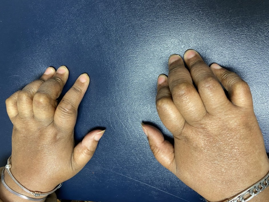
Figure 1. Bilateral claw-hand contractures.
At the time of her initial presentation 10 years ago, hematologic evaluation revealed elevated both serum free light chain kappa and lambda with elevated kappa/lambda ratio and a normal serum protein electrophoresis. Hemoglobin was 9.8 g/dL (normal 12.0–16.0 g/dL). Peripheral blood smear showed microcytic, hypochromic red cells with innumerable platelets and no abnormal myeloid or lymphoid cells. Total serum immunoglobulins: IgG 1,147 mg/dL (normal 700–1,600 mg/dL), IgA 253 mg/dL (normal 70–400 mg/dL), IgM 47 mg/dL (normal 40–230 mg/dL). Serum free light chains: kappa 219.4 mg/L (normal 3.3–19.4 mg/L), lambda 66.9 mg/L (normal 5.7–26.3 mg/L), kappa/lambda ratio 3.28 (normal 0.26–1.65). Thyroid function tests confirmed hypothyroidism. Serum calcium was elevated 15.1 mg/dL (normal 8.5–10.5 mg/dL). CT of the neck identified a 2.3 cm parathyroid adenoma. Urinalysis showed no significant proteinuria. She was diagnosed with primary hyperparathyroidism requiring bisphosphonate therapy; parathyroidectomy was subsequently performed.
Initial serum protein electrophoresis (SPEP) was normal with no detectable M-spike. However, serum immunoelectrophoresis revealed an elevated serum kappa immunoglobulin of 1,640 mg/dL (normal 574–1,267 mg/dL) and elevated serum lambda immunoglobulin of 763 mg/dL (normal 269-638), and urine immunoelectrophoresis demonstrating elevated kappa light chains at 11.8 mg/dL (normal <1.9 mg/dL), while lambda was less than 5 mg/dL (normal <6.7 mg/dL). These results are summarized in Table 1.
|
Test |
Patient Value |
Normal Reference Range |
Interpretation |
|
SPEP (serum protein electrophoresis) |
Normal; no M-spike detected |
No monoclonal band |
No monoclonal protein detected on SPEP |
|
Serum immunoelectrophoresis — IgG Kappa |
1,640 mg/dL |
574–1,267 mg/dL |
Elevated |
|
Serum immunoelectrophoresis — IgG Lambda |
763 mg/dL |
269–638 mg/dL |
Elevated |
|
Serum free light chains — Kappa |
219.4 mg/L |
3.3–19.4 mg/L |
Elevated |
|
Serum free light chains — Lambda |
66.9 mg/L |
5.7–26.3 mg/L |
Elevated |
|
Kappa/Lambda ratio |
3.28 |
0.26-1.65 |
Elevated |
|
Urine immunoelectrophoresis — Kappa |
11.8 mg/dL |
<1.9 mg/dL |
Elevated; kappa light chains in urine |
|
Urine immunoelectrophoresis — Lambda |
<5 mg/dL |
<5.0 mg/dL |
Normal |
|
SPEP: Serum Protein Electrophoresis; UPEP: Urine Protein Electrophoresis; IFE: Immunofixation Electrophoresis; IgG: Immunoglobulin G. Serum immunoelectrophoresis and urine immunoelectrophoresis values obtained at initial hospitalization prior to treatment. |
|||
CT of the thoracic spine, obtained following a fall, demonstrated a lucent lesion at the right posterior elements of T1, sclerotic lesions at the lower thoracic vertebral bodies and L1, and additional sclerotic foci in the pelvis and sacrum. MRI of the thoracic spine confirmed an enhancing lesion involving the T1 pedicle, facet, and lamina, which remained stable on serial imaging over approximately nine months. MRI of the lumbar spine showed inflammatory changes in the paraspinal musculature and nerve root enhancement. Bone scan was unremarkable. CT of the chest, abdomen and pelvis showed small left pleural effusion, moderate hepatosplenomegaly, a few mildly enlarged right axillary lymph nodes measuring 3´1.9 cm, and left inguinal lymphadenopathy. Diagnostic left thoracentesis with removal of 800 ml serous fluid demonstrated exudative pleural effusion and cytology was negative for malignancy.
Lumbar puncture (LP) demonstrated cytoalbuminologic dissociation consistent with a demyelinating or inflammatory process. Full CSF values with reference ranges are summarized in Table 2.
|
CSF Parameter |
Patient Value |
Normal Reference Range |
Interpretation |
|
Protein |
155 mg/dL |
15–45 mg/dL |
Markedly elevated — cytoalbuminologic dissociation |
|
Glucose |
49 mg/dL |
50–80 mg/dL (or ≥60% serum glucose) |
Low-normal |
|
WBC count |
2 cells/μL |
0–5 cells/μL |
Normal |
|
RBC count |
1 cell/μL |
0 cells/μL |
Trace, likely traumatic tap |
|
Lymphocytes |
52% |
60–80% |
Normal differential |
|
Monocytes |
48% |
15–45% |
Mildly elevated |
|
Albumin |
67.7 mg/dL |
6.6–35.0 mg/dL |
Elevated — blood–brain barrier disruption |
|
IgG |
20.5 mg/dL |
0.0–4.0 mg/dL |
Markedly elevated |
|
IgG Index |
0.68 |
<0.66 |
Elevated — intrathecal IgG production |
|
IgG Synthesis Rate |
32.3 mg/24 h |
<8.0 mg/24 h |
Elevated — intrathecal synthesis confirmed |
|
LDH |
74 U/L |
10–40 U/L |
Mildly elevated |
|
Serum albumin (paired) |
2.8 g/dL |
3.5–5.0 g/dL |
Low — reflects systemic hypoalbuminemia |
|
Serum IgG (paired) |
1,250 mg/dL |
700–1,600 mg/dL |
Normal |
|
CSF: Cerebrospinal Fluid; WBC: White Blood Cells; RBC: Red Blood Cells; IgG: Immunoglobulin G; LDH: Lactate Dehydrogenase. Cytoalbuminologic dissociation is defined as elevated protein with normal or near-normal cell count. |
|||
Nerve conduction studies (NCS) were technically limited by severe bilateral lower-extremity edema, precluding responses in the tibial, peroneal, sural, and superficial peroneal nerves on the left. Upper extremity NCS revealed markedly diminished conduction velocities of both ulnar and both median nerves with prolonged F-wave responses and diminished conduction of left radial nerve with prolonged F-wave responses. EMG, while was suboptimal study, demonstrated diffuse muscle membrane instability with sustained positive waves and fibrillation potentials, myopathic motor unit potentials in both upper and lower extremities, and markedly reduced recruitment of motor units on volitional activity along both upper extremities muscles while unable to do any volitional activity along both lower extremities. There were no fasciculations or joint potentials noted. The above findings are suggestive of severe peripheral neuropathy secondary to possible chronic inflammatory demyelinating polyneuropathy (CIDP) with associated myopathic changes and no evidence of motor neuron disease. IVIG was initiated at 2 g/kg over five days; the patient reported questionable and transient benefit. A second course of IVIG was administered without meaningful neurologic improvement.
Initial bone marrow biopsy was non-diagnostic (hemodiluted, hypocellular sample); flow cytometry was negative for lymphoproliferative disorder, though a reference laboratory report noted kappa light-chain restriction. Biopsy of the T1 lesion, performed after anticoagulation was held, demonstrated morphology consistent with a solitary plasmacytoma with lambda light-chain restriction on immunostaining. A left inguinal lymph node core biopsy showed few plasma cells present demonstrating kappa light chain predominance. Repeat bone marrow biopsy showed 7% plasma cells with a monoclonal population and flow cytometry immunophenotyping demonstrated kappa restriction. Plasma VEGF was subsequently measured and returned markedly elevated at greater than 12,000 pg/mL (reference range ≤200 pg/mL) (Table 3).
|
Timepoint |
Plasma VEGF (pg/mL) |
Clinical Context |
|
Pre-treatment (at diagnosis) |
>12,000 |
POEMS syndrome confirmed; lenalidomide/dexamethasone initiated |
|
Several months after treatment initiation |
81 |
Early biochemical response; lower-extremity weakness improving |
|
Approximately 7 months after initiation |
32 |
VEGF normalized; stem cell collection performed |
|
Annual monitoring (on maintenance lenalidomide) |
Normal (repeated) |
Sustained biochemical control; serial monitoring continued |
|
Most recent (~7–8 years post-diagnosis) |
39.3 |
Maintained suppression despite lenalidomide hold for endometrial surgery and restart |
|
VEGF: Vascular Endothelial Growth Factor. Reference range ≤96.2 pg/mL (Mayo Clinic Laboratories). All values represent plasma VEGF. |
||
A diagnosis of POEMS syndrome was established at this time, four months after her hospital presentation, based on fulfillment of all current diagnostic criteria (Table 5, in the Discussion section): both mandatory major criteria (demyelinating polyneuropathy and clonal plasma-cell disorder), two additional major criteria (sclerotic bone lesion at T1 and markedly elevated plasma VEGF), and five minor criteria (thrombocytosis, extravascular volume overload, endocrinopathy (hypothyroidism, hyperparathyroidism), skin changes (hyperpigmentation). She also has thromboembolic events. This fulfillment is summarized in Table 5.
Treatment with lenalidomide and dexamethasone was initiated. Within weeks, the patient reported meaningful improvement in bilateral lower-extremity weakness. Follow-up plasma VEGF normalized rapidly (Table 1). Dexamethasone was gradually tapered. Peripheral stem cells were harvested and cryopreserved; autologous stem-cell transplantation (ASCT) was deferred pending recovery of activities of daily living. Maintenance lenalidomide was continued. Warfarin anticoagulation was maintained given restricted ambulation and persistent thrombotic risk.
Most recent plasma VEGF remains suppressed at 39.3 pg/mL (reference ≤96.2 pg/mL, Mayo Clinic Laboratories).
Progressive restrictive pulmonary dysfunction developed over the course of her illness. Serial pulmonary function testing (PFT) documented a progressive restrictive defect and reduction in diffusion capacity (DLCO) over six years. Recent NIF measurement was −36 cmH2O (normal more negative than −60 to −70 cmH2O), indicating severe respiratory muscle weakness. Upright versus supine spirometry revealed a significant positional fall in forced vital capacity (FVC), consistent with diaphragmatic weakness (Tables 4a and 4b). Flow volume loop demonstrated restrictive defect (Figure 2). Recent arterial blood gas analysis on 2 L of Oxygen demonstrated PH 7.41 (normal 7.35–7.45), PCO2 64.1 mmHg (normal 35–45 mmHg), PO2 75.6 mmHg (normal 80–100 mmHg on room air), consistent with chronic hypercapnic hypoxic ventilatory failure from neuromuscular respiratory dysfunction.
|
Parameter |
Timepoint 1 (≈2 yr post-Dx) |
Timepoint 2 (≈4 yr post-Dx) |
Timepoint 3 (≈7 yr post-Dx) |
|
FVC (% predicted) |
58% |
56% |
47% |
|
FEV1 (% predicted) |
64% |
57% |
53% |
|
FEV1/FVC (%) |
90% |
83% |
90% |
|
DLCO (% predicted) |
79% |
43% |
67% |
|
FVC: Forced Vital Capacity; FEV1: Forced Expiratory Volume in 1 second; Dx: Diagnosis. Values shown as percent of predicted. |
|||
|
Parameter |
Upright (% predicted) |
Supine (% predicted) |
Positional Change |
|
FVC |
33% |
23% |
−30 % (fall ≥20% indicates diaphragm weakness) |
| FEV1 |
38% |
26% |
−32% |
|
FEV1/FVC (%) |
94% |
92% |
Preserved |
|
A positional fall in FVC ≥20% from upright to supine is the accepted threshold for clinically significant diaphragmatic weakness. FVC: Forced Vital Capacity; FEV1: Forced Expiratory Volume in 1 second. |
|||
Despite sustained biochemical control, the patient developed significant irreversible morbidity with persistent bilateral foot drop, bilateral claw-hand contractures (Figure 1), lower-extremity weakness and lymphedema managed with elevation and compression garments. She remains wheelchair-dependent for longer distances but ambulates short distances with a walker while continuing physical therapy.
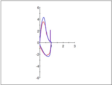
Figure 2. Flow volume loop.
Discussion
Diagnostic criteria and delayed recognition
POEMS syndrome diagnosis follows a tiered framework requiring both mandatory major criteria (demyelinating polyneuropathy and a monoclonal plasma-cell disorder, most commonly lambda-restricted), at least one additional major criterion (sclerotic bone lesion, markedly elevated VEGF, or Castleman disease), and at least one minor criterion (organomegaly, extravascular volume overload, endocrinopathy, skin changes, papilledema, thrombocytosis, or polycythemia). The diagnostic criteria and their operationalization in the present case are summarized in Table 5.
|
Criteria Category |
Criterion |
Our Patient Finding |
|
Mandatory Major (both required)
|
1. Polyneuropathy, typically demyelinating |
Severe symmetric sensorimotor neuropathy |
|
2. Monoclonal plasma-cell disorder |
λ-restricted clone on bone marrow biopsy; plasmacytoma at T1 |
|
|
Other Major (≥1 required)
|
3. Sclerotic bone lesions |
T1 vertebral body lesion; additional sclerotic foci at T10, L1, S1, pelvis |
|
4. Markedly elevated plasma VEGF |
>12,000 pg/mL (reference <200 pg/mL) |
|
|
5. Castleman disease: angiofollicular lymph node hyperplasia |
Not identified |
|
|
Minor (≥1 required)
|
6. Organomegaly: hepatomegaly, splenomegaly, lymphadenopathy |
Lymphadenopathy |
|
7. Thrombocytosis/polycythemia |
Marked thrombocytosis (platelets 1.08 million/μL) |
|
|
8. Extravascular volume overload |
Pleural effusion; lower-extremity peripheral edema |
|
|
9. Endocrinopathy |
Hypothyroidism; primary hyperparathyroidism |
|
|
10. Skin changes |
Hyperpigmentation |
|
|
11. Papilledema |
Not identified |
|
|
Other Manifestations |
Restrictive lung disease, pulmonary hypertension, thromboembolic disease arterial more common than venous. |
DVT, pulmonary embolism |
|
VEGF: Vascular Endothelial Growth Factor; DVT: Deep Vein Thrombosis; λ: Lambda. |
||
Prior series have established that diagnosis anchored solely on the POEMS acronym can fail to identify nonclassical variants, and that an algorithmic approach centering on demyelinating polyneuropathy and clonal plasma-cell disease is more reliable. Diagnostic precision remains imperfect despite established criteria. One review of 629 patients found that only half of those initially meeting criteria went on to develop classical POEMS; lambda-restricted clones and sclerotic bone lesions were more common among true POEMS patients, whereas kappa light chains and vasculitic nerve changes were more common in false-positive cases [5]. Conversely, some patients with clinical POEMS features lack a detectable M-protein, particularly after corticosteroid use [6]. In patients with suspected CIDP presenting with systemic features or poor response to immunotherapy, POEMS-specific testing should be pursued early rather than treating POEMS as a diagnosis of exclusion.
POEMS neuropathy and differentiation from CIDP
POEMS polyneuropathy commonly resembles CIDP in its early stages, as both present with progressive, symmetric, length-dependent sensorimotor weakness. However, POEMS neuropathy tends to be more motor-predominant, more painful and disabling, and more frequently accompanied by systemic features. Approximately 60% of patients with POEMS are initially misdiagnosed with CIDP in large comparative series, delaying plasma-cell-directed treatment [7].
Progressive neuropathy refractory to IVIG, particularly when accompanied by thrombocytosis, thromboembolic disease, a sclerotic bone lesion, and lambda-restricted plasma-cell disease, should shift the diagnostic pathway toward plasma VEGF measurement, skeletal imaging, immunofixation, and bone marrow evaluation.
Electrodiagnostic studies can help distinguish POEMS from CIDP. POEMS neuropathy typically shows disproportionate reduction in compound muscle action potential (CMAP) amplitude relative to only mild distal latency prolongation, particularly in the lower extremities. In a retrospective comparison of 51 POEMS and 46 CIDP patients, absent tibial CMAPs and F-waves were present in 56% of POEMS cases versus 7% of CIDP cases, reflecting severe lower-limb axonal loss. POEMS is also characterized by proximal or trunk-predominant demyelination and a higher terminal latency index, whereas CIDP more often demonstrates distal accentuated slowing with multifocal conduction abnormalities. Conduction block and severe temporal dispersion, hallmarks of CIDP, are uncommon in POEMS [8]. Needle EMG may reveal fibrillation potentials and positive sharp waves consistent with active axonal degeneration, with direct implications for irreversibility of neurologic injury if treatment is delayed. While both POEMS and CIDP slow conduction, POEMS often shows a more uniform slowing across intermediated nerve segments. POEMS syndrome rarely exhibits sural sparing, leading to uniform and severe sensory amplitude reduction, while in CIDP, sural sensory nerve potentials (SNAPs) are often spared or preserved relative to other nerves [9].
Histopathologic findings correlate with these electrodiagnostic patterns. Sural nerve biopsies in POEMS show diffuse axonal degeneration with increased epineurial neovascularization, fewer onion bulbs, and less endoneurial inflammation compared with CIDP. Immunoelectron microscopy has demonstrated monoclonal IgG-lambda or IgA-lambda deposits within the endoneurium and subperineurial area without amyloid or vasculitis, distinguishing POEMS from amyloid neuropathy and vasculitic neuropathy [10].
VEGF and biomarker-based diagnosis
Plasma VEGF is the most diagnostically useful and clinically validated biomarker in POEMS syndrome. VEGF levels are typically 15 to 30 times greater than those in CIDP or Guillain-Barré syndrome and decrease with effective treatment [11]. Plasma rather than serum VEGF measurement is preferred, as platelet activation during clotting can falsely elevate serum values, especially in patients with thrombocytosis. A plasma VEGF cutoff of approximately 200 pg/mL has demonstrated approximately 95% specificity for POEMS . VEGF normalization is a key treatment endpoint and should be monitored serially, with a response defined as normalization below 200 pg/mL or a reduction of at least 50%.
Emerging biomarkers may complement VEGF. Procollagen type I N-terminal propeptide (P1NP) demonstrated 80% sensitivity and 91.5% specificity for POEMS at a threshold greater than 70 ng/mL, and may help distinguish POEMS from mimickers and track treatment response [12]. Interleukins 6 and 12 have been associated with vascular leak, overall disease activity, and neuropathogenesis, decreasing after effective treatment [13,14]. Matrix metalloproteinases and TIMP-1 may serve as markers of vascular remodeling and endothelial injury [15]. Until these biomarkers achieve widespread availability, plasma VEGF will remain the mainstay of diagnosis and monitoring.
Imaging, bone marrow, and pathology
Osteosclerotic lesions may be small, multifocal, or subtle and can be missed on limited skeletal surveys. Computed tomography is particularly effective at detecting lesions preferentially involving the vertebrae and pelvis [16]. PET/CT may identify metabolically active lesions amenable to biopsy when conventional imaging is unrevealing and has a potential role in monitoring treatment response and detecting early relapse [17–19]. Atypical osteolytic or mixed lytic-sclerotic lesions have been reported, and any suspicious lesion in the context of unexplained demyelinating neuropathy should be considered for biopsy [20].
Bone marrow findings in POEMS often differ from those in multiple myeloma. Plasma-cell burden may be low, serum free light-chain ratios can be normal despite lambda-restricted monoclonality, and standard myeloma screening algorithms may miss the clone [21]. Classic histologic findings include lambda-restricted plasma cells rimming lymphoid aggregates, megakaryocytic hyperplasia and atypia, and a JAK2 V617F-negative pattern distinguishing POEMS-related thrombocytosis from a true myeloproliferative neoplasm [22]. Sensitive testing with immunofixation, immunohistochemistry, in situ hybridization, and flow cytometry is necessary to detect small clones and should be interpreted alongside clinical and imaging findings [23].
Multisystem involvement
Endocrinopathy is present in over 80% of patients at diagnosis in large series, involving gonadal, thyroid, adrenal, and glucose metabolism [24]. Baseline assessment should include sex hormones with gonadotropins, thyroid function, morning cortisol, fasting glucose or hemoglobin A1c, prolactin, calcium, and parathyroid hormone, with longitudinal reassessment thereafter [25].
Thrombosis occurs in approximately one quarter of patients, with arterial events slightly more common than venous in some series; approximately 8% of patients develop ischemic stroke [26]. Risk factors include thrombocytosis, polycythemia, splenomegaly, effusions, prior thrombosis, and VEGF-mediated endothelial dysfunction [27]. Antiplatelet therapy is recommended for most patients, with anticoagulation added for those with additional risk factors.
Pulmonary complications represent an underappreciated source of morbidity. Recognized manifestations include restrictive ventilatory defects, impaired diffusing capacity, pulmonary hypertension, respiratory muscle weakness from severe polyneuropathy, and pleural effusions [28]. The pathophysiology involves increased vascular permeability from elevated VEGF, extravascular volume overload, and progressive neuromuscular impairment. Pulmonary hypertension has been shown to improve with effective disease treatment, with the degree of reversal correlating with disease severity at presentation and duration of endothelial dysfunction [29]. Serial pulmonary function testing—including upright and supine spirometry to evaluate for diaphragmatic weakness—diffusing capacity measurement, exertional oxygen assessment, and echocardiography are recommended at baseline and serially for all POEMS patients, even after biochemical remission [30]. Neuromuscular respiratory failure, evidenced by a positional FVC drop of 20% or more and a reduced MIP, should prompt evaluation for non-invasive ventilatory support.
Renal involvement includes glomerular changes resembling membranoproliferative glomerulonephritis with mesangial expansion and double-contoured capillary walls, driven predominantly by VEGF-mediated injury rather than immune complex deposition [31,32]. Baseline and longitudinal monitoring with urinalysis and serum creatinine is recommended.
Characteristic skin findings, including hyperpigmentation, hypertrichosis, sclerodermoid changes, and hemangiomas, can provide early diagnostic clues and may predate neuropathy [33]. Multiple cherry-red to violaceous hemangiomas in the context of CIDP-like symptoms should prompt VEGF measurement [34]. The AESOP syndrome (adenopathy and extensive skin patch overlying a plasmacytoma) may serve as an early diagnostic entry point in rare cases of POMES [35].
Treatment, response, and long-term monitoring
Treatment selection is guided by disease extent. Patients with an isolated plasmacytoma or limited discrete lesions without diffuse marrow involvement may be candidates for involved-field radiation alone, which can be curative in select cases [36]. Those with diffuse skeletal involvement, marrow infiltration, or progression following radiation require systemic plasma-cell-directed therapy.
High-dose melphalan followed by autologous stem-cell transplantation is the preferred approach for eligible patients with disseminated disease, associated with durable hematologic responses, VEGF reduction, neurologic improvement, and favorable long-term survival [37–40]. Melphalan with dexamethasone is a validated non-transplant option [41]. Lenalidomide with dexamethasone has demonstrated activity in newly diagnosed, relapsed, and refractory settings with multiple reports of rapid VEGF decline and organ-function improvement [42–44].
Other active agents include bortezomib, which offers rapid plasma-cell cytoreduction but must be used cautiously given its neuropathy risk; subcutaneous administration and dose reduction are often warranted [45–47]. Daratumumab has shown activity in refractory and selected newly diagnosed cases with emerging reports of VEGF improvement and neurologic benefit, though larger studies are needed [48–50]. Thalidomide reduces VEGF and improves symptoms but carries a significant neuropathy burden, limiting its use compared with newer immunomodulatory agents [51].
Response assessment in POEMS requires evaluation across multiple domains, as no single parameter captures overall disease activity. Hematologic response is assessed using IMWG-style criteria, though depth of marrow response does not necessarily predict longitudinal outcome. VEGF normalization to below 200 pg/mL or a decrease of 50% or more defines biomarker response. Neurologic response should be tracked with standardized scoring systems and nerve-conduction studies, as clinical improvement commonly lags behind biomarker response or may not occur at all. Organ-specific surveillance encompassing volume status, pulmonary function, papilledema, renal function, and endocrine parameters should be performed systematically.
Comparison with multiple myeloma
POEMS shares its plasma-cell dyscrasia underpinning with multiple myeloma yet differs substantially in presentation and diagnostic approach. Classic multiple myeloma is defined by CRAB criteria (hypercalcemia, renal insufficiency, anemia, lytic bone lesions), none of which are primary features of POEMS. POEMS features neuropathy as the central manifestation alongside osteosclerotic or mixed lesions, lambda-restricted clonality, thrombocytosis, endocrinopathy, edema, and skin changes. Standard myeloma screening algorithms are inadequate for POEMS detection, particularly in low-burden disease where cytokine-mediated organ effects substantially outpace quantitative tumor burden [52].
Future directions
Future treatment approaches should target the cytokine-angiogenic environment and underlying molecular features rather than simply reducing plasma-cell burden. Anti-VEGF therapy with bevacizumab has produced mixed results and symptomatic worsening in some patients, suggesting direct VEGF inhibition may be too far downstream of the primary pathogenic process.
Upstream cytokine inhibition is under early investigation; IL-6 inhibition has led to more rapid resolution of effusions and edema, and IL-12/23 blockade has been associated with concomitant reductions in IL-12 and VEGF with motor improvement in small case series.
Restricted lambda light-chain gene usage (IGLV1-40 and IGLV1-44) may enable minimal residual disease monitoring via next-generation sequencing [53]. Composite response criteria incorporating VEGF, P1NP, cytokines, and clonal biomarkers may eventually allow earlier relapse detection and safer treatment de-escalation in deep responders. Anti-CD38 agents such as daratumumab warrant prospective study across treatment lines, and response-adaptive treatment pathways that escalate or de-escalate therapy based on hematologic, cytokine, and neurologic trajectories merit future investigation.
While VEGF remains the standard diagnostic and monitoring biomarker for POEMS syndrome, recent clinical developments have highlighted interleukin-6 (IL-6), N-Terminal propeptide of type I collagen (P1NP) and Interlukin-12 (IL-12) as a potential new diagnostic and prognostic biomarkers [54,55].
Conclusion
This case highlights the diagnostic challenges of POEMS syndrome and emphasizes the importance of considering this condition in patients presenting with CIDP-like neuropathy who exhibit systemic features such as thrombocytosis, extravascular volume overload, endocrine abnormalities, skin changes, thromboembolic events, sclerotic bone lesions, or poor response to immunomodulatory therapy. Early recognition, early measurement of plasma VEGF levels, and evaluation for plasma-cell disorders with bone marrow assessment, skeletal imaging and immunofixation are critical to establishing the diagnosis and initiating effective plasma-cell–directed therapy to reduce irreversible neurologic and organ damage.
References
2. Dispenzieri A. POEMS syndrome: 2017 update on diagnosis, risk stratification, and management. Am J Hematol. 2017;92(8):814–29.
3. Dispenzieri A. POEMS syndrome: 2021 update on diagnosis, risk stratification, and management. Am J Hematol. 2021;96(7):872–88.
4. D'Souza A, Hayman SR, Buadi F, Mauermann M, Lacy MQ, Gertz MA, et al. The utility of plasma vascular endothelial growth factor levels in the diagnosis and follow-up of patients with POEMS syndrome. Blood. 2011;118(17):4663–5.
5. Ofran Y, Elinav E. POEMS syndrome: failure of newly suggested diagnostic criteria to anticipate the development of the syndrome. Am J Hematol. 2005;79(4):316–8.
6. Charli-Joseph Y, Fernández-Sánchez M, Saeb-Lima M, Orozco-Topete R. POEMS syndrome: are current diagnostic criteria too exclusive? J Am Acad Dermatol. 2011;65(2):415–7.
7. Nasu S, Misawa S, Sekiguchi Y, Shibuya K, Kanai K, Fujimaki Y, et al. Different neurological and physiological profiles in POEMS syndrome and chronic inflammatory demyelinating polyneuropathy. J Neurol Neurosurg Psychiatry. 2012;83(5):476–9.
8. Piccione EA, Engelstad J, Dyck PJ, Mauermann ML, Dispenzieri A, Dyck PJ. Nerve pathologic features differentiate POEMS syndrome from CIDP. Acta Neuropathol Commun. 2016;4(1):116.
9. Guo X, Qin X, Zhang Y, Huang C, Yu G. Electrophysiological features of POEMS syndrome and chronic inflammatory demyelinating polyneuropathy. Journal of clinical neuroscience 21(2014)587–90.
10. Adams D, Said G. Ultrastructural characterisation of the M protein in nerve biopsy of patients with POEMS syndrome. J Neurol Neurosurg Psychiatry. 1998;64(6):809–12.
11. Watanabe O, Maruyama I, Arimura K, Kitajima I, Arimura H, Hanatani M, et al. Overproduction of vascular endothelial growth factor/vascular permeability factor is causative in Crow-Fukase syndrome. Muscle Nerve. 1998;21(11):1390–7.
12. Wang C, Zhou YL, Cai H, Cheng XQ, Zhang W, Kang WY, et al. Markedly elevated serum total N-terminal propeptide of type I collagen is a novel marker for the diagnosis and follow up of patients with POEMS syndrome. Haematologica. 2014;99(6):e78–e80.
13. Shikama N, Isono A, Otsuka Y, Terano T, Hirai A. A case of POEMS syndrome with high concentrations of interleukin-6 in pericardial fluid. J Intern Med. 2001;250(2):170–3.
14. Hitoshi S, Suzuki K, Sakuta M. Elevated serum interleukin-6 in POEMS syndrome reflects the activity of the disease. Intern Med. 1994;33(10):583–7.
15. Michizono K, Umehara F, Hashiguchi T, Arimura K, Matsuura E, Watanabe O, et al. Circulating levels of MMP-1, -2, -3, -9, and TIMP-1 are increased in POEMS syndrome. Neurology. 2001;56(6):807–10.
16. Shibuya K, Misawa S, Horikoshi T, Kanai K, Isose S, Nasu S, et al. Detection of bone lesions by CT in POEMS syndrome. Intern Med. 2011;50(13):1393–6.
17. Albertí MA, Martinez-Yélamos S, Fernandez A, Vidaller A, Narváez JA, Cano LM, et al. 18F-FDG PET/CT in the evaluation of POEMS syndrome. Eur J Radiol. 2010;76(2):180–2.
18. Montoriol PF, Cachin F, Michel JL, Soubrier M. Two more cases of evaluation of POEMS syndrome using 18-FDG PET/CT. Eur J Radiol. 2011;80(3):861–4.
19. Stefanelli A, Treglia G, Leccisotti L, Laurenti L, Luigetti M, Sabatelli M, et al. Usefulness of F-18 FDG PET/CT in the follow-up of POEMS syndrome after autologous peripheral blood stem cell transplantation. Clin Nucl Med. 2012;37(2):181–3.
20. Narváez JA, Majós C, Narváez J, Valls C, Fernandez-Cabrera L. POEMS syndrome: unusual radiographic, scintigraphic and CT features. Eur Radiol. 1998;8(1):134–6.
21. Stankowski-Drengler T, Gertz MA, Katzmann JA, Lacy MQ, Kumar S, Leung N, et al. Serum immunoglobulin free light chain measurements and heavy chain isotype usage provide insight into disease biology in patients with POEMS syndrome. Am J Hematol. 2010;85(6):431–4.
22. Dao LN, Hanson CA, Dispenzieri A, Morice WG, Kurtin PJ, Hoyer JD. Bone marrow histopathology in POEMS syndrome: a distinctive combination of plasma cell, lymphoid, and myeloid findings in 87 patients. Blood. 2011;117(24):6438–44.
23. Li J, Zhou DB, Huang Z, Jiao L, Duan MH, Zhang W, et al. Clinical characteristics and long-term outcome of patients with POEMS syndrome in China. Ann Hematol. 2011;90(7):819–26.
24. Gandhi GY, Basu R, Dispenzieri A, Basu A, Montori VM, Brennan MD. Endocrinopathy in POEMS syndrome: the Mayo Clinic experience. Mayo Clin Proc. 2007;82(7):836–42.
25. Caimari F, Keddie S, Lunn MP, D'Sa S, Baldeweg SE. Prevalence and course of endocrinopathy in POEMS syndrome. J Clin Endocrinol Metab. 2019;104(6):2140–6.
26. Mellors PW, Kourelis T, Go RS, Muchtar E, Gertz MA, Kumar SK, et al. Characteristics and risk factors for thrombosis in POEMS syndrome: a retrospective evaluation of 230 patients. Am J Hematol. 2022;97(2):209–15.
27. Soubrier M, Guillon R, Dubost JJ, Serre AF, Ristori JM, Boyer L, et al. Arterial obliteration in POEMS syndrome: possible role of vascular endothelial growth factor. J Rheumatol. 1998;25(4):813–15.
28. Allam JS, Kennedy CC, Aksamit TR, Dispenzieri A. Pulmonary manifestations in patients with POEMS syndrome: a retrospective review of 137 patients. Chest. 2008;133(4):969–74.
29. Niimi H, Arimura K, Jonosono M, Hashiguchi T, Kawabata M, Osame M, et al. VEGF is causative for pulmonary hypertension in a patient with Crow-Fukase syndrome. Intern Med. 2000;39(12):1101–04.
30. Chandrashekaran S, Dispenzieri A, Cha SS, Kennedy CC. Pulmonary morbidity improves after autologous stem cell transplantation in POEMS syndrome. Respir Med. 2015;109(1):122–30.
31. Nakamoto Y, Imai H, Yasuda T, Wakui H, Miura AB. A spectrum of clinicopathological features of nephropathy associated with POEMS syndrome. Nephrol Dial Transplant. 1999;14(10):2370–8.
32. Soubrier M, Sauron C, Souweine B, Larroche C, Wechsler B, Guillevin L, et al. Growth factors and proinflammatory cytokines in the renal involvement of POEMS syndrome. Am J Kidney Dis. 1999;34(4):633–8.
33. Miest RY, Comfere NI, Dispenzieri A, Lohse CM, el-Azhary RA. Cutaneous manifestations in patients with POEMS syndrome. Int J Dermatol. 2013;52(11):1349–56.
34. Lenormand C, Marzolf G, Lipsker D. AESOP syndrome: a potential life-saving and early clue to the diagnosis of POEMS syndrome. Clin Dermatol. 2021;39(2):215–19.
35. Lipsker D, Rondeau M, Massard G, Grosshans E. The AESOP syndrome: report of 4 cases of a new syndrome revealing POEMS syndrome at a curable stage. Medicine (Baltimore). 2003;82(1):51–9.
36. Humeniuk MS, Gertz MA, Lacy MQ, Kyle RA, Witzig TE, Kumar SK, et al. Outcomes of patients with POEMS syndrome treated initially with radiation. Blood. 2013;122(1):68–73.
37. Ohwada C, Sakaida E, Kawajiri-Manako C, Nagao Y, Oshima-Hasegawa N, Togasaki E, et al. Long-term evaluation of physical improvement and survival of autologous stem cell transplantation in POEMS syndrome. Blood. 2018;131(19):2173–6.
38. Cook G, Iacobelli S, van Biezen A, Ziagkos D, LeBlond V, Abraham J, et al. High-dose therapy and autologous stem cell transplantation in patients with POEMS syndrome: a retrospective study of the Plasma Cell Disorder sub-committee of the Chronic Malignancy Working Party of the European Society for Blood & Marrow Transplantation. Haematologica. 2017;102(1):160–7.
39. Karam C, Klein CJ, Dispenzieri A, Dyck PJ, Mandrekar J, D'Souza A, et al. Polyneuropathy improvement following autologous stem cell transplantation for POEMS syndrome. Neurology. 2015;84(19):1981–7.
40. Kuwabara S, Misawa S, Kanai K, Kikkawa Y, Nishimura M, Nakaseko C, et al. Autologous peripheral blood stem cell transplantation for POEMS syndrome. Neurology. 2006;66(1):105–7.
41. Li J, Zhang W, Jiao L, Duan MH, Guan HZ, Zhu WG, et al. Combination of melphalan and dexamethasone for patients with newly diagnosed POEMS syndrome. Blood. 2011;117(24):6445–9.
42. Li J, Huang XF, Cai QQ, Wang C, Cai H, Zhao H, et al. A prospective phase II study of low dose lenalidomide plus dexamethasone in patients with newly diagnosed POEMS syndrome. Am J Hematol. 2018;93(6):803–9.
43. Royer B, Merlusca L, Abraham J, Musset L, Haroche J, Choquet S, et al. Efficacy of lenalidomide in POEMS syndrome: a retrospective study of 20 patients. Am J Hematol. 2013;88(3):207–12.
44. Nozza A, Terenghi F, Gallia F, Adami F, Briani C, Merlini G, et al. Lenalidomide and dexamethasone in patients with POEMS syndrome: results of a prospective, open-label trial. Br J Haematol. 2017;179(5):748–55.
45. He H, Fu W, Du J, Jiang H, Hou J. Successful treatment of newly diagnosed POEMS syndrome with reduced-dose bortezomib based regimen. Br J Haematol. 2018;181(1):126–8.
46. Zeng K, Yang JR, Li J, Wei Q, Yang YM, Liu T, et al. Effective induction therapy with subcutaneous administration of bortezomib for newly diagnosed POEMS syndrome: a case report and a review of the literature. Acta Haematol. 2013;129(2):101–5.
47. Warsame R, Kohut IE, Dispenzieri A. Successful use of cyclophosphamide, bortezomib, and dexamethasone to treat a case of relapsed POEMS. Eur J Haematol. 2012;88(6):549–50.
48. Tiew HW, Sampath VS, Gallardo CA, Christopher D, Chan SSW, Wong SW, et al. Single-agent daratumumab for refractory POEMS syndrome. Am J Hematol. 2022;97(6):E189–91.
49. Khan M, Stone K, van Rhee F. Daratumumab for POEMS syndrome. Mayo Clin Proc. 2018;93(4):542–4.
50. Khwaja J, Keh R, Smyth D, Lunn MP, D'Sa S, Sive J. Daratumumab-bortezomib-dexamethasone use in relapsed POEMS syndrome. EJHaem. 2022;3(3):1021–4.
51. Misawa S, Sato Y, Katayama K, Nagashima K, Aoyagi R, Sekiguchi Y, et al. Safety and efficacy of thalidomide in patients with POEMS syndrome: a multicentre, randomised, double-blind, placebo-controlled trial. Lancet Neurol. 2016;15(11):1129–37.
52. Bender S, Javaugue V, Saintamand A, Ayala MV, Alizadeh M, Filloux M, et al. Immunoglobulin variable domain high-throughput sequencing reveals specific novel mutational patterns in POEMS syndrome. Blood. 2020;135(20):1750–8.
53. Abe D, Nakaseko C, Takeuchi M, Tanaka H, Ohwada C, Sakaida E, et al. Restrictive usage of monoclonal immunoglobulin lambda light chain germline in POEMS syndrome. Blood. 2008;112(3):836–39.
54. Cook J, Warsame R, Omar M, Buadi FK, Abdallah N, Dingli D, et al. Interleukin-6 is a highly prognostic biomarker for POEMS syndrome. Leukemia. 2025 Sep;39(9):2281–4.
55. Wang C, Zhou YL, Cai H, Cheng XQ, Zhang W, Kang WY, et al. Markedly elevated serum total N-terminal propeptide of type I collagen is a novel marker for the diagnosis and follow up of patients with POEMS syndrome. Haematologica. 2014 Jun;99(6):e78–80.