Abstract
Vulvovaginal hematoma is not very common in modern obstetric practice due to increasing care, management and careful observation of mothers during and following delivery but when it occurs it can rapidly evolve into a life-threatening condition if it is not noticed early or managed promptly.
Among others, and if not in shock patient commonly presents with significant perineal pain and, depending on the location, there is always visible swelling, the index patient presented in shock 8 hours following unsupervised delivery.
The primary goals of management include prompt commencement of resuscitation, prevention of further blood loss, minimize tissue damage, relieve pain, and reduce the risk of infection. Management generally however, could be conservative for small blood collections, but surgery is usually inevitable when it is acute and rapidly expand in size.
Keywords
Vulvovaginal, Haematoma, Episiotomy, Shock, Exploration, Figure-of eight
Introduction
Vulvovaginal haematoma following vagina delivery can cause a serious life-threatening complication including maternal mortality if not promptly diagnosed or properly managed yet, is no more commonly seen, especially in modern obstetric practice due to improvement in quality of care and management in labour with careful observation of mothers in labour ward following delivery [1,2]. On several occasions, the diagnoses are missed or are misdiagnosed [3], this is because, it is not very common and the symptoms may be initially vague and nonspecific, also bleeding is usually not revealed [2], leading to diagnostic difficulties, until there is swelling hence delayed treatment. Vulva haematoma among others is one of the four T's mnemonic widely accepted as causes of Postpartum Haemorrhage, which are uterine atony (Tone), laceration, hematoma, rupture (Trauma), retained tissue (Tissue); and coagulopathy (Thrombi) [4,5]. Clinically, an estimated case series report incidence of 1:500 to 1:12,500 vaginal births [1] have been reported, majority of these cases can be managed conservatively, surgical intervention is however required in 1 out of 1000 vaginal deliveries [2].
Documented risk factors for vulva haematoma are the following nulliparity, poorly supervised second stage of labour, prolonged second stage of labour, delayed or poorly repaired episiotomy, instrumental delivery and lacerations, others include; delivery of a macrosomic baby, maternal age of 30 years and above and varicosities of the genital tract [6,7]. Usually, patient presents with vague symptoms ranging from above to a classical presentation of shock even without an obvious bleeding, 4-6 hours following delivery.
We therefore present a 31-year-old Nulliparous, who was referred in shock on account of vulvovaginal haematoma following vaginal delivery.
Case Presentation
We present a 31-year-old unbooked now Para 1 who was referred to the teaching hospital from a maternity centre in shock, 6 hours after a spontaneous vaginal delivery of a live female neonate with a birth weight of 3900gm. The labour was spontaneous and lasted for about 8 hours, it was said to have been supervised and conducted by a birth attendant at home but was taken to the maternity centre, when the woman developed postpartum haemorrhage secondary to a genital tract laceration. The genital tract laceration was repaired at the said maternity centre but this was poorly done, even though bleeding subsided, she was suddenly found in shock on the bed, this necessitated her referrer to the teaching hospital for expert management. On admission, examination revealed a young woman who was conscious but drowsy, pale, dehydrated with cold calm extremities. She was afebrile; temperature was 35.3oc, she was tachypneic with a respiratory rate 34 cycles /min, tachycardic with a pulse rate of 118 beats/min, regular but of small volume and a blood pressure of 80/30 mmHg.
Abdominal examination and pelvic examination revealed a well contracted uterus that was about 20week pregnancy size and a blood smeared vulva with an obviously tender, swelling involving the pubic area, though more on the left, bilateral labia minus and majus measuring about 10 cm by 8 cm respectively. An assessment of postpartum vulvovaginal haematoma with a preceding postpartum haemorrhage in shock was made. Resuscitative measure was commenced and instituted immediately. Intravenous access established, blood sample was obtained for packed cell volume (PCV) (which was 14%), grouping and cross-matching of three units of whole blood. She commenced blood transfusion, had surgical exploration and in total had 4units of blood transfused.
The intraoperative findings included the following: an extensive left Vulvovaginal haematoma of about 14 cm by 12 cm, it completely occluded the vaginal cavity, right from the orifice, with stretched and tensed vaginal wall and the overlying perineal skin (Figures 1). There was no significant bleeding from the vagina. A careful incision was made with evacuation of the haematoma and exploration of the cavity was done, about 2500mls of clotted blood was evacuated from the cavity. Haemostasis was achieved with a ‘’figure of eight”. The space was completely obliterated with deep sutures, in layers and the overlying skin approximated carefully, avoiding injury to the urethra. The vagina was packed with gauze to further assist with haemostasis. She had broad spectrum antibiotics, analgesics and transfusion of four units of blood, she was subsequently discharged home on the fourth day following a satisfactory postoperative recovery (Figure 2).
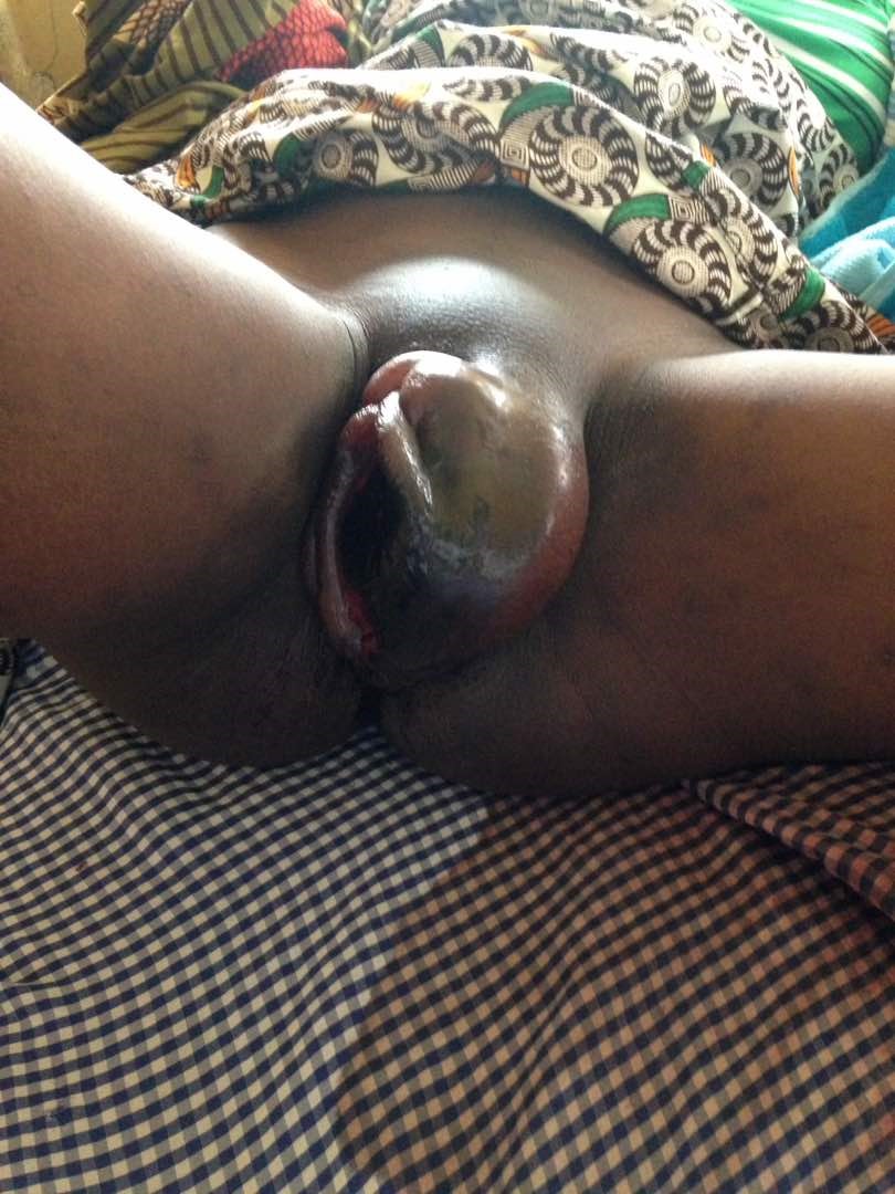
Figure 1: Vulvovaginal haematoma, with complete occlusion of the vaginal cavity, right from the orifice, with stretched and tensed vaginal wall and the overlying perineal skin.
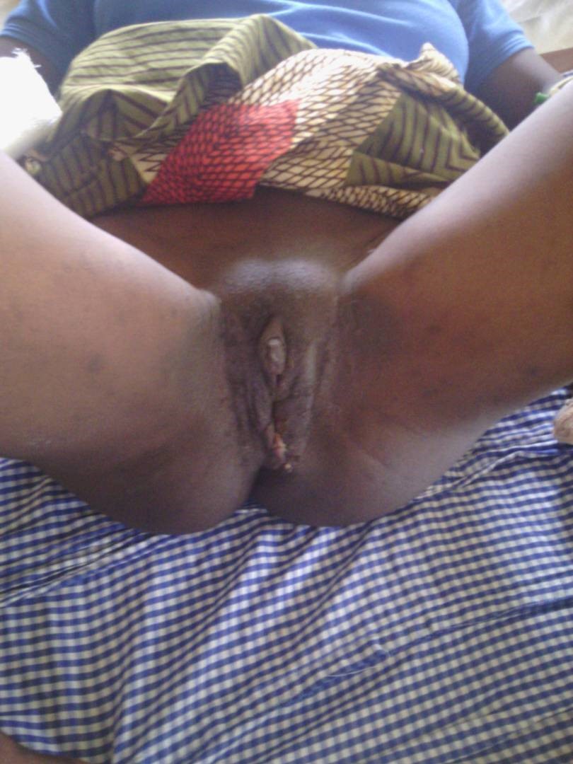
Figure 2: Resolved haematoma following evacuation of the haematoma, exploration of the cavity and subsequent repair.
Discussion
Episiotomy (especially when the repair is delayed or poorly repaired), vulva or perineal laceration or trauma from instrument-assisted vaginal delivery may result in haematoma collection in the vulva or retroperitoneum in addition to the rich vascular supplies in the perineum which could further make the bleeding worse(5, 6), and this aggravates the hematoma collection leading usually to this entity called vulvovaginal haematoma, this was seen in this case, in which an initial poor repair of genital laceration was done at the referring maternity centre. Other risk factors for severe perineal trauma and its possible sequelae, vulvovaginal hematomas include multiple gestation, prolonged second stage of labour, clotting derangements, macrosomia, vulvovaginal varicosities, cephalopelvic disproportion and poorly supervised labour [5,8]. Usually the development of vulvovaginal hematomas may take hours after delivery and most often initially misdiagnosed as vulva swelling or oedema [9] until later when delayed formation of the haematoma becomes obvious and late diagnosis or recognition and treatment may result in adverse severe consequences with increased risk of maternal morbidity and mortality [5], which is what happens in most cases, when it becomes suspected, only when attention is drawn to them that the woman collapses in shock [10], this is true for this patient who was referred in shock due to delay in diagnosis and initiation of treatment, in addition to sudden presentation in shock because the vagina tissue is soft and pliable, haematoma collection can become so extensive and progress to occlude the vaginal orifice leading to acute urinary retention [2].
Management of vulvovaginal haematoma depends on the patient’s presentation [5,11], which include; the size of the haematoma, hemodynamic stability of the woman and availability of resources including medical expertise, usually a conservative measure is put in place, if the haematoma is <5 cm, static i.e. not rapidly increasing in size [2] and if patient is hemodynamically stable; this includes provision of analgesia to reduce the pain, empirical antibiotics, ice pack, pressure dressing, bladder drainage to prevent acute retention and if necessary blood transfusion [5] however, as seen in this case if the patient present in shock a quick surgical exploration is needed and must be considered without hesitation while carrying out other resuscitative measures including blood transfusion if necessary, in addition evidence has shown that if swelling is greater than 5 cm, conservative management is not appropriate [2] because it is associated with prolonged hospital stay, increased need for antibiotics and blood transfusion, and greater recourse to surgical intervention [12].
At exploration, the incision is best placed as medially as possible to reduce obvious scarring [3], and this must be done carefully to prevent dyspareunia in future. During the repair, there is need to explore all obvious bleeding, a ‘’figure-of eight” suturing (which was used in this case) can be used as the case may be to secure heamostasis, drain may be used for 12-24 hours in some instances where some residual bleeding persists [13]. The repair should be carefully done to obliterate the dead space in order to prevent a repeat collection which could lead to abscess formation, a close monitoring of the surgical site, evaluation of the vital signs and a strict monitoring of input and output must be put in place. In rare cases, ligation of the internal iliac artery or even hysterectomy may be done [3] or use of angiographic embolization for cases of on-going bleeding not controlled by conventional surgical techniques [13]. After repair, broad-spectrum antibiotics should be given to prevent infection and judicious use of analgesia with close observation of the patient are imperative postoperatively.
Acknowledgments
We would like to acknowledge the whole team members involved in the management.
Funding
None.
Availability of Data and Materials
The datasets created during and/or analyzed during the current study available from the corresponding author on reasonable request and acknowledged.
Ethics Approval and Consent to Participate
The research and ethics committee of the department of obstetrics and Gynecology, Obafemi Awolowo University/Teaching Hospitals Complex, Ile-Ife. Nigeria, granted us the ethical clearance to publish this case report.
Consent for Publication
Written informed consent was obtained from the patient for the procedure, publication of this case report and accompanying images.
Competing Interests
We declare no competing interests.
References
2. Awoleke JO, Ipinnimo OM. Vulvovaginal Infralevator Haematoma Mimicking the Second Stage of Labour. Case Reports in Obstetrics and Gynecology. 2017 Jan 18;1-3.
3. Mawhinney S, Holman R. Puerperal genital haematoma: a commonly missed diagnosis. The Obstetrician & Gynaecologist. 2007 Jul;9(3):195-200.
4. Evensen A, Anderson JM, Fontaine P. Postpartum hemorrhage: prevention and treatment. American Family Physician. 2017 Apr 1;95(7):442-9.
5. Tseng JY, Lin IC, Lin JH, Chang CM, Chao WT, Wang PH. Optimal approach for management of postpartum vulva hematoma: Report of three cases. Taiwanese Journal of Obstetrics and Gynecology. 2020 Sep 1;59(5):780-3.
6. Thakar R, Sultan AH. Postpartum problems and the role of a perineal clinic. In: Perineal and anal sphincter trauma. Springer: London. 2009; pp. 65-79.
7. Saleem Z, Rydhström H. Vaginal hematoma during parturition: a population-based study. Acta Obstetricia et Gynecologica Scandinavica. 2004 Jan 1;83(6):560-2.
8. Gurtovaya Y, Hanna H, Wagley A. Spontaneous intrapartum vulvar haematoma. Midwives. 2013 Jul 1;16(5):48-9.
9. Nelson EL, Parker AN, Dudley DJ. Spontaneous vulvar hematoma during pregnancy: a case report. The Journal of Reproductive Medicine. 2012 Jan 1;57(1-2):74-6.
10. Lyons AW. Post-partum hematoma. New England Journal of Medicine. 1949 Mar 24;240(12):461-3.
11. Propst AM, Thorp Jr JM. Traumatic vulvar hematomas: conservative versus surgical management. Southern Medical Journal. 1998 Feb 1;91(2):144-6.
12. Benrubi G, Neuman C, Nuss RC, Thompson RJ. Vulvar and vaginal hematomas: a retrospective study of conservative versus operative management. Southern Medical Journal. 1987 Aug 1;80(8):991-4.
13. Zahn CM, Hankins GD, Yeomans ER. Vulvovaginal hematomas complicating delivery. Rationale for drainage of the hematoma cavity. The Journal of Reproductive Medicine. 1996 Aug 1;41(8):569-74.