

Abstract
TGF-β plays an important role in keeping HIV-1 in a hidden, inactive state by suppressing the immune system and creating conditions that prevent the virus from reactivating. However, exactly how TGF-β does this is not well understood. In this study, we used the U1 cell line to explore how TGF-β affects HIV-1 reactivation. We used real-time PCR and Western blotting to measure changes in viral activity and related cell pathways. We found that cells expressing TGF-β had lower levels of HIV-1 RNA compared to control cells. This effect appears to be due to TGF-β lowering the levels of certain host transcription factors like NFAT and Sp1, which are needed for activating HIV-1, while having no effect on AP-1 and NF-κBp65. TGF-β also blocks pathways like P-TEFb and Jak/Stat, which are important for turning on and copying HIV-1 genes. In addition, TGF-β reduces cell death by blocking both main types of apoptosis pathways and increasing Bcl-xL levels, which help cells survive. Together, these effects help HIV-1 stay inactive and hidden in the body. This makes it harder to cure HIV because the virus can remain in a dormant state. Our results suggest that targeting TGF-β could help develop new treatments to wake up and remove hidden HIV-1. Understanding how TGF-β controls HIV-1 latency and cell survival may help improve strategies to fully eliminate the virus.
Keywords
HIV-1, Latency, TGF-β, Transcription factor, Apoptosis, Bcl-xL
Introduction
HIV-1 is the main strain responsible for the global HIV/AIDS pandemic. Over time, the epidemic has changed, with different patterns of spread in different regions. As of 2023, about 39.9 million people were living with HIV worldwide. In the same year, there were around 1.3 million new HIV infections and 630,000 deaths from AIDS-related illnesses [1]. Antiretroviral therapy (ART) is still the most effective way to treat HIV-1. It works by stopping the virus from multiplying, lowering the amount of virus in the blood to undetectable levels, and preventing HIV from developing into AIDS [2]. When taken consistently, ART helps people with HIV live as long as people without the virus. It also strengthens the immune system, lowers the risk of other infections, and improves quality of life. However, ART is not a cure. If treatment is stopped, the virus can come back. If people do not take their medicine as prescribed, the virus can become resistant to the drugs, which makes treatment more complicated and expensive [3]. One major challenge is that ART cannot remove hidden HIV reservoirs—cells where the virus stays inactive or ‘asleep’. These hidden viruses, especially in long-lived memory CD4+ T cells, are the biggest barrier to a cure. The virus can stay inside the DNA of these cells without making new virus particles, so the immune system and ART cannot detect or remove it. If treatment stops, the hidden virus can ‘wake up’ and cause infection again. Other cells, like macrophages, dendritic cells, CD8+ T cells, and microglial cells, can also carry hidden HIV [4,5].
HIV-1 latency is controlled by complex processes involving several cellular pathways. These include the NF-κB, PKC, MAPK/ERK, TCR, JAK/STAT, and PI3K/Akt pathways, which help decide whether the virus stays hidden or becomes active again [6]. These pathways affect transcription factors and gene activity that are important for the virus to make new copies of itself [4,5]. Among the cells that act as reservoirs, macrophages are especially important. These long-lived immune cells can keep HIV-1 alive in the body for long periods. The U1 cell line, which is a model of HIV-infected monocytes and macrophages, is often used to study how HIV latency is created and maintained [7,8]. U1 cells help researchers understand how the virus hides in macrophages and are widely used to test strategies like the “shock and kill” method, which aims to wake up hidden virus and destroy it [9]. They are also used to test drugs that could either reactivate the virus or keep it dormant in macrophage reservoirs.
Transforming Growth Factor Beta (TGF-β) is a multifunctional cytokine that helps control immune responses, cell growth, cell development, and programmed cell death (apoptosis) [10]. While TGF-β is important for keeping the immune system balanced, it can also play a role in disease if its signals do not work properly. In HIV infection, TGF-β has been shown to help the virus stay hidden (latent) by affecting how T cells are activated, how genes are turned on or off, and how chromatin (the structure that packages DNA) is arranged [11]. Experiments have shown that TGF-β helps HIV-1 stay latent in human cells grown in the lab [12] and in animal models [13].
In our study, we used U1 cells to examine how TGF-β affects HIV-1 latency. We found that TGF-β helps keep the virus dormant by lowering the activity of two key host factors, NFAT and Sp1, which are needed to activate the virus. This happens through TCR signaling pathways. As a result, the virus can't produce its RNA and stays inactive. TGF-β also blocks the P-TEFb pathway, which is important for making full-length viral transcripts. Besides stopping the virus from replicating, TGF-β helps infected cells avoid dying. It does this by blocking caspase-3, an enzyme that starts cell death, and by boosting Bcl-xL, a protein that helps cells survive. Together, these effects let infected cells live longer and keep the virus hidden. Overall, TGF-β both suppresses HIV replication and supports the survival of latently infected cells. This makes it an important factor in HIV's long-term persistence and a possible target for future treatments aimed at curing the infection.
Materials and Methods
Chemicals and reagents
We obtained the following antibodies from Abcam (Waltham, MA): rabbit polyclonal/mouse monoclonal antibodies against Caspase-3, Caspase-8, Jak1, Jak2, NF-kB p65, PLCγ-1, PKC, and TCRα. Additionally, we acquired mouse monoclonal antibodies against Bax, Bcl2, Bcl-xL, gp130, Fas, FADD, FLIP, p53, XIAP from Santa Cruz Biotechnology (Santa Cruz, CA). Rabbit polyclonal antibodies against Akt, AP-1, Brd4, CD4, CDK9, Cyclin T1, Erk1/2, JNK, HIV-1 Tat, NFAT, p38, PCAF, PI3K, p-Rpb-CTD, Stat1, Stat3, Stat5, Sp-1, TGF-β, TF, GAPDH, and ZAP70 were bought from Cell Signaling Technology, Inc (Danvers, MA). Galunisertib (LY2157299) and WEHI-539 hydrochloride were obtained from Millipore Sigma (Rockville, MD). All other chemicals were sourced from Sigma (St. Louis, MO).
U1 Cell culture and treatments
U1 (promonocytic) cells, a subclone of U937 chronically infected with HIV-1, were obtained from the National Institutes of Health AIDS Research Reference and Reagent Program (Germantown, MD) and cultured at 37°C in 5% CO2 in RPMI 1640 medium containing 10% fetal calf serum, 2 mM glutamine, 50 μg/ml penicillin, and 50 μg/ml streptomycin.
For adenoviral infections, U1 cells were seeded at 2 × 10⁵ cells/mL for 24 hours and then cultured in medium containing 10% fetal calf serum and 105 CPU/mL of an adenoviral vector carrying either the TGF-β, Bcl-xL gene or LacZ gene. Infected cells were incubated for 3 and 7 days indicated.
For Bcl-xL inhibition, U1 cells were seeded at 2 × 10⁵ cells/mL for 24 hours and then cultured in medium containing 10% fetal calf serum and 100 nM of WEHI-539 hydrochloride, followed by incubation for 7 days.
RNA isolation and real-time PCR
Viral RNA was isolated from 140 µl of culture supernatant using the QIAamp Viral RNA Mini Kit (Valencia, CA 91355) according to the manufacturer’s protocol. Five out of the 50 µl of the extracted RNA was used as templates for real-time RT-PCR. Known concentrations of HIV-1 (MN) viral RNA (serially diluted: 108 to 100 copies) were used as templates. Quantitative RT-PCR was performed to generate a standard curve. Each value shown in the figures represents the average concentration of 6 reactions in triple isolated repeats based on the standard curve.
Primers and TaqMan probes were designed for the gag p24, which is the variable region among most of the HIV-1 subtype B isolate sequences according to the GenBank database. The forward primer was 5’-GACATCAAGCAGCCATGCAA-3’, corresponding to nucleotides 1367–1386, and the reverse primer was 5’-CTATCCCATTCTGCAGCTTCCT-3’, corresponding to nucleotides 1430–1409. The Taq-Man probes were composed of the oligonucleotide sequence 5’-ATTGATGGTCTCTTTTAACA-3’, corresponding to nucleotides 1488–1507, coupled with a reporter dye [6-carboxy fluorescein] (FAM) at the 5’ end and a non-fluorescent quencher and a minor groove binder (MGB), which is a Tm enhancer, at the 3’ end. The nucleic acids were amplified and detected in an automated TaqMan 7500 Analyzer using QuantiTectTM Probe RT-PCR kit (Qiagen Inc., Valencia, CA). The 25 µl PCR mixture consisted of 100 nM primers and 100 nM probe and underwent the following conditions: 95°C for 10 min, 45 cycles of two-step PCR at 95°C for 15 s and at 60°C for 1 min.
Western blot analysis
Proteins were isolated from U1 cells with RIPA buffer (1×PBS, 1% (v/v) NP-40, 0.5% (w/v) sodium deoxycholate, 0.1% (w/v) SDS, 0.1 mg/ml PMSF, 30 μl/ml aprotinin, 1 mM sodium orthovanadate). Equal amounts of protein were boiled in the loading buffer (100 mM Tris–HCl, 200 mM DTT, 4% SDS, 0.2% bromphenol blue, 20% glycerol), separated on SDS-PAGE and blotted onto polyvinylidene difluoride membranes. Data represented are from three independent experiments. The relative quantitation of protein expression was determined using ImageJ from NIH website (https://imagej.nih.gov/ij/).
Statistical analysis
The unpaired Student’s t test was used for data analyses as indicated, and p-value <0.05 (*) and p-value <0.01 (**) were considered significant and very significant, respectively.
Results
TGF-β plays a crucial role in promoting HIV-1 latency by actively suppressing transcriptional activity [12,13]. In contrast, inhibiting TGF-β has been shown to enhance HIV-1 replication and effectively reactivate latent HIV-1 [14]. As demonstrated in Supplementary Figure 1, U1 cells that were incubated with Galunisertib, a potent TGF-β inhibitor, exhibited a markedly significant increase in HIV-1 RNA production compared to the control group (Mock). This finding suggests that blocking TGF-β signaling can potentially disrupt HIV-1 latency, leading to increased viral transcription and replication.
TGF-β suppresses HIV-1 reactivation by downregulating Sp1 and NFAT transcription factors
A report suggests that TGF-β increases viral burden and promotes HIV-1 latency in primary differentiated human bronchial epithelial cells [12]. To determine whether TGF-β can reactivate HIV-1 replication from latency in U1 cells—a widely accepted in vitro model of HIV-1 latency—we infected U1 cells with an adenovirus carrying either the TGF-β gene or the LacZ gene (control) for 3 or 7 days. As shown in Figure 1, U1 cells infected with the adenovirus carrying the TGF-β gene exhibited increased TGF-β expression compared to those infected with the adenovirus carrying the LacZ gene (Figure 1A). Notably, this increased TGF-β expression inhibited HIV-1 RNA production at both day 3 and day 7, compared to the LacZ control (Figures 1B and 1C), suggesting that TGF-β suppresses HIV-1 replication, thereby promoting HIV-1 latency.
HIV-1 replication relies heavily on host cell transcription factors that regulate viral gene expression [6]. Key factors include NF-κB, which enhances viral transcription by binding to the HIV-1 long terminal repeat (LTR) promoter in activated T cells; Sp1, which binds to GC-rich regions in the LTR for basal transcription; NFAT, which promotes transcription in T cells following receptor activation; and AP-1, which is involved in regulating gene expression and is linked to T cell activation pathways. These factors are essential for efficient HIV-1 replication within host cells [6]. To examine the effects of TGF-β on the expression of AP-1, NFAT, NF-κBp65, and Sp1 in U1 cells, total proteins were extracted from U1 cells infected with an adenovirus carrying either the TGF-β1 gene or the LacZ gene (control) for 7 days (Figure 1C). These proteins were then used for Western blotting. As shown in Figure 1D, TGF-β expression downregulated NFAT and Sp1, upregulated AP-1, and had no significant effect on NF-κBp65. These results suggest that the downregulation of both NFAT and Sp1 has a stronger effect on the inactivation of HIV-1 replication than the upregulation of AP-1 on the activation of HIV-1 latency.
The data suggest that TGF-β promotes HIV-1 latency by selectively inhibiting the host transcription factors NFAT and Sp1 in U1 cells, while not affecting the activity of NF-κBp65. This indicates that the suppression of NFAT and Sp1 plays a critical role in the regulation of HIV-1 latency, whereas AP-1 and NF-κBp65 do not appear to contribute to this process in the same way.
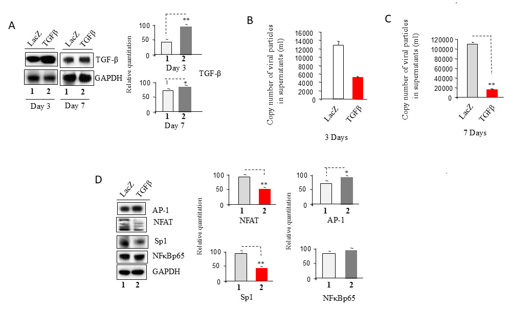
Figure 1. TGF-β suppresses HIV-1 reactivation by downregulating Sp1 and NFAT transcription factors. U1 cells were infected with an adenovirus carrying the LacZ or TGF-β gene and then incubated for 3 or 7 days as indicated. (A) Cell pellets were used to isolate total protein, which was then subjected to Western blot analysis to detect TGF-β. (B) & (C) A total of 140 µl of culture supernatants containing HIV-1 particles was used to isolate viral RNA for RT-PCR analysis to assess viral replication. (D) U1 cells were infected with an adenovirus carrying the LacZ or TGF-β gene and incubated for 7 days. Total cell lysates were subjected to Western blot analysis to detect AP-1, NFAT, NF-kB p65, and Sp-1.
TGF-β expression inhibits the activation of TCR-related signaling pathway
T-cell receptor (TCR) signaling plays a crucial role in regulating HIV-1 replication and latency in CD4+ T cells, macrophages, and other cell types by influencing viral reactivation and immune response through complex signaling cascades [6]. TCR activation—triggered by an antigen, superantigen, or artificial stimulation (e.g., anti-CD3/CD28 antibodies)—initiates intracellular signaling that can reverse latency and promote viral reactivation. This process dephosphorylates NFAT, allowing it to enter the nucleus and bind to the HIV-1 LTR, thereby initiating transcription [6]. As shown in Figures 2A and 2B, after a 7-day infection with adenovirus carrying either the LacZ or TGF-β gene, U1 cells exhibited downregulation of CD4 expression (Figure 2A) and inhibition of TCR, PLCγ1, PKC, and ZAP-70 expression in TCR-signaling pathways (Figure 2B), which regulate NFAT transcriptional activity.
The Mitogen-Activated Protein Kinase (MAPK) pathway, comprising ERK, JNK, and p38 MAPKs, plays a crucial role in regulating HIV-1 latency and reactivation within CD4+ T cells [6]. This pathway is triggered by TCR signaling and other external stimuli, leading to viral reactivation through transcription factor activation. ERK1/2 facilitates HIV-1 transcription by phosphorylating transcription factors that bind to the viral LTR [6]. Meanwhile, JNK activates c-Jun, which forms the AP-1 complex, further promoting transcription. While inhibiting p38 MAPK can restrict HIV-1 replication in latently infected cells, excessive activation of p38 contributes to T-cell dysfunction and inflammation in chronic HIV cases, ultimately enhancing transcription of the integrated HIV-1 genome and driving viral reactivation [6]. After a 7-day infection with adenovirus carrying either the LacZ or TGF-β gene, U1 cells exhibited downregulation of ERK1/2, p38, and JNK expression in the TGF-β group compared to the LacZ controls (Figure 2C).
TCR stimulation activates the PI3K-Akt signaling pathway, enhancing cellular metabolism and translation to support HIV replication [6]. As shown in Figure 2D, after a 7-day infection with adenovirus carrying either the LacZ or TGF-β gene, U1 cells exhibited downregulation of PI3K and Akt expression. This suggests that TGF-β expression may inhibit the PI3K-Akt pathway, potentially impacting HIV replication dynamics.
These findings suggest that TGF-β plays a significant role in promoting HIV-1 latency through multiple mechanisms. First, it downregulates CD4 expression, which may reduce the ability of HIV-1 to bind to host cells, thereby limiting viral entry. Additionally, TGF-β inhibits key TCR-related signaling pathways, including the TCR, MAPK, and PI3K-Akt pathways. This inhibition leads to a substantial reduction in the activity of host transcription factors, which are essential for viral transcription and replication. Collectively, these mechanisms contribute to the establishment and maintenance of HIV-1 latency.
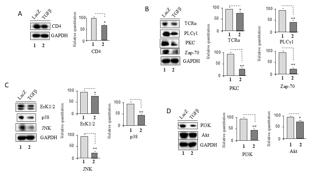
Figure 2. TGF-β expression inhibits the activation of TCR-related signaling pathway. U1 cells were infected with an adenovirus carrying the LacZ or TGF-β gene and incubated for 7 days. Total cell lysates were subjected to Western blot analysis to detect (A) CD4; (B) TCRα, PKC, PLCγ1 and ZAP 70; (C) ERK1/2, p38 and JNK; and (D) PI3K and Akt.
TGF-β expression suppresses P-TEFb signaling pathways
The Positive Transcription Elongation Factor b (P-TEFb), composed of Cyclin T1 and CDK9, is crucial for HIV-1 transcription and the shift from latency to active replication [15]. It phosphorylates RNA polymerase II (RNAP II), enabling transcription elongation. HIV-1 Tat (Trans-Activator of Transcription) recruits P-TEFb to enhance viral gene expression, while in latent infection, P-TEFb is sequestered in the inactive 7SK snRNP complex, blocking transcription. Reactivation occurs when P-TEFb is released, triggered by Tat, cytokines, or signaling pathways like PKC and MAPK/ERK. Tat boosts P-TEFb activity by enhancing RNAP II phosphorylation, whereas BRD4 competes for P-TEFb binding, suppressing transcription [15]. This pathway regulates HIV-1 latency and reactivation, determining whether the virus remains dormant or actively transcribes RNA [15].
To examine the effects of TGF-β on P-TEFb Signaling Pathway in U1 cells, total proteins were extracted from U1 cells infected with an adenovirus carrying either the TGF-β1 gene or the LacZ gene (control) for 7 days (Figure 1C). These proteins were then used for Western blotting. As shown in Figure 3, TGF-β expression downregulated Brd4, CDK9 and Cyclin T1 (Figure 3A), decreased phosphorylation of C-terminal domain of RNAP II (p-RPb-CTD, the phosphorylated C-terminal domain of RNAP II) (Figure 3B).
HIV-1 Tat can also interact with host factors like PCAF, a histone acetyltransferase (HAT), to regulate viral gene expression. Tat binds to TAR RNA and recruits PCAF to the HIV-1 LTR, where PCAF acetylates histone H3 [16]. This loosens chromatin, making it more accessible for transcription factors and RNAP II, enhancing HIV-1 transcription. In latent infection, low Tat levels keep PCAF activity minimal, maintaining repressive chromatin [16]. When Tat is present, it recruits PCAF, reverses repression, and reactivates HIV-1 transcription [16]. As shown in Figure 3C, TGF-β inhibited HIV-1 Tat and PCAF expression relative to LacZ control in U1 cells.
These results suggest that TGF-β plays a significant role in downregulating Tat, a key regulatory protein of HIV-1. This downregulation leads to the tightening of chromatin, rendering it inaccessible to crucial transcription factors and RNAP II. As a result, this chromatin compaction reduces the efficiency of HIV-1 transcription. Moreover, TGF-β also interferes with the P-TEFb signaling pathway by directly decreasing the expression levels of critical components such as Brd4 and CDK9. This disruption impairs the phosphorylation of RNAP II, a modification essential for the proper transcription and elongation of HIV-1 RNA. Thus, TGF-β contributes to multiple layers of regulation that collectively suppress HIV-1 transcription and replication.
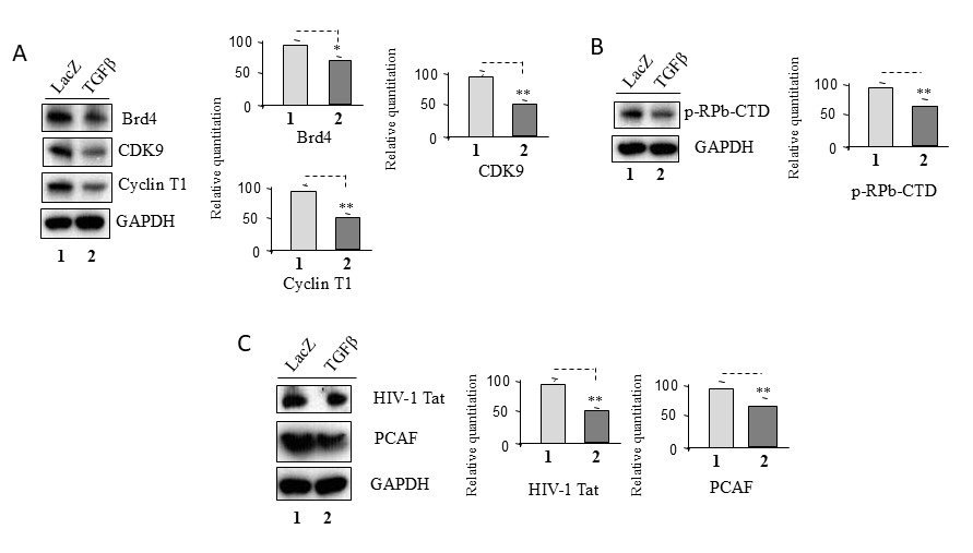
Figure 3. TGF-β expression suppresses P-TEFb signaling pathways. U1 cells were infected with an adenovirus carrying the LacZ or TGF-β gene and incubated for 7 days. Total cell lysates were subjected to Western blot analysis to detect (A) Brd4, CDK9 and Cyclin T1; (B) p-Rpb-CTD; (C) HIV-1 Tat and PCAF.
TGF-β expression inhibits gp130/Jak/STAT signaling pathways
The gp130/Jak signaling pathway is important for HIV-1 latency and reactivation in CD4+ T cells and other immune cells [17]. The pathway can be activated by certain cytokines, which trigger JAK phosphorylation, leading to the activation of STAT5, STAT3 and STAT1 [6,17]. These transcription factors promote immune activation and inflammation, which may influence the latent HIV-1 reservoir and key transcription factors (TFs) that regulate HIV-1 transcription [6,17]. In resting CD4+ T cells, HIV-1 stays dormant, but gp130/Jak signaling can activate pro-inflammatory cytokines that trigger HIV-1 expression [18]. Inflammatory signals from this pathway can also reactivate the virus in inactive cells, potentially increasing viral replication [6,18]. JAK/STAT signaling is involved in immune responses and can impact HIV-1 replication by modulating viral gene transcription, with STAT5, STAT3 and STAT1 helping to reactivate the virus from latency by binding to the HIV-1 LTR and influencing transcription [18]. As shown in Figure 4, after a 7-day infection with an adenovirus carrying either the LacZ or TGF-β gene, U1 cells exhibited a significant downregulation of gp130, Jak1, and Jak2 expression (Figure 4A). This was accompanied by a marked inhibition of Stat1, Stat3, and Stat5 signaling pathways (Figure 4B), ultimately leading to a substantial decrease in the expression of the host transcription factor, TF (Figure 4C).
These findings suggest that TGF-β blocks JAK/STAT signaling pathways, affecting HIV-1 latency by suppressing immune activation and reducing the homeostatic proliferation of latent reservoirs. TGF-β downregulates STAT5, STAT3, and STAT1, each of which has distinct yet interconnected effects on viral persistence and immune regulation. It influences the establishment, maintenance, and potential reactivation of latency in different ways. Furthermore, TGF-β impacts TF that plays a critical role in controlling the balance between maintaining viral silence and initiating the reactivation of HIV transcription, thereby contributing to the overall dynamics of the viral lifecycle.
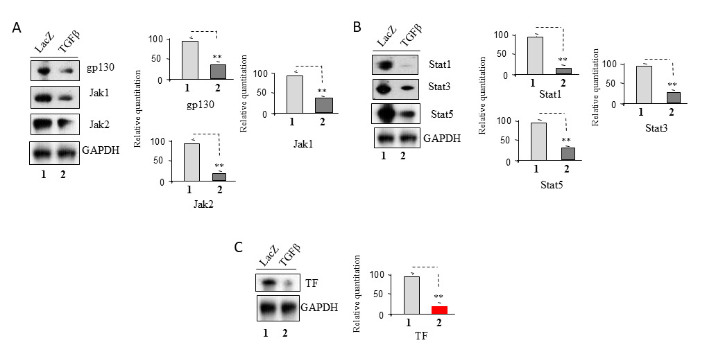
Figure 4. TGF-β expression blocks gp130/Jak/STAT signaling pathways. U1 cells were infected with an adenovirus carrying the LacZ or TGF-β gene and incubated for 7 days. Total cell lysates were subjected to Western blot analysis to detect (A) gp130, Jak1 and Jak2; (B) Stat1, Stat3 and Stat5; (C) TF.
TGF-β expression downregulates apoptotic signaling pathways
Apoptosis has two main pathways: intrinsic (mitochondrial) and extrinsic (death receptor). The intrinsic pathway responds to stress like DNA damage or infection, balancing pro-apoptotic (Bax) and anti-apoptotic (Bcl-2, Bcl-xL) proteins [19]. When activated, mitochondria release cytochrome c, forming the apoptosome, which activates caspase 3, leading to cell death [19]. HIV-1 manipulates this by promoting CD4+ T-cell death or preventing it in infected cells (by increasing Bcl-2) [20]. The extrinsic pathway starts with external signals, where Fas binds FasL, activating caspase-8 and triggering cell death. HIV-1 increases FasL to kill uninfected T cells while protecting infected ones, aiding its survival [20]. Previously, we reported that pro-apoptotic proteins involved in apoptotic pathways can reactivate HIV-1 replication from latency, whereas the downregulation or inhibition of these proteins can enhance HIV-1 latency in lymphoma [21,22].
Caspase-3, a classic marker of activation of apoptotic signaling pathways, plays a central role in both the intrinsic and extrinsic apoptotic pathways [19]. After a 7-day infection with an adenovirus carrying either the LacZ or TGF-β gene, U1 cells showed a significant downregulation of caspase-3 (Figure 5A). This downregulation occurred through the inhibition of the intrinsic pathway, as evidenced by the reduced expression of p53 and Bax (Figure 5B), and the extrinsic pathway, as demonstrated by the decreased expression of Fas, FADD, and caspase-8 (Figure 5C).
Anti-apoptotic proteins play a critical role in promoting HIV-1 latency and enabling the virus to persist in a dormant state within host cells by preventing apoptosis, which protects infected cells from programmed cell death [21,22]. Key proteins involved include Bcl-2 and Bcl-xL from the Bcl-2 family, which block mitochondrial apoptosis; FLIP, which inhibits caspase-8 in the extrinsic pathway; IAPs (c-IAP1, c-IAP2, and XIAP), which prevent caspase activation [19]. These proteins help HIV-1 evade immune clearance by maintaining infected cell viability and supporting the virus’s persistence in latent reservoirs, especially in CD4+ T cells [23,24]. By manipulating these proteins, HIV-1 can promote latency, making it harder for the immune system to eliminate the virus. Targeting these proteins may offer therapeutic strategies to reactivate latent HIV-1 or reduce viral reservoirs by inducing apoptosis in infected cells [23–25]. As shown in Figure 5D, TGF-β expression downregulated Bcl-2, FLIP, and XIAP, while significantly upregulating Bcl-xL.
The data strongly suggest that TGF-β plays a pivotal role in inhibiting both the intrinsic and extrinsic apoptotic pathways that are triggered by HIV-1 infection, potentially influencing the regulation of cell death responses in this context. However, the downregulation of key apoptotic regulators such as Bcl-2, FLIP, and XIAP, which have been shown to promote apoptosis, was insufficient to overcome the protective effects of the higher expression of Bcl-xL. Bcl-xL may significantly inhibit the activation of pro-apoptotic proteins, including caspase-3, thus counteracting the apoptotic signals and further contributing to the survival of infected cells.
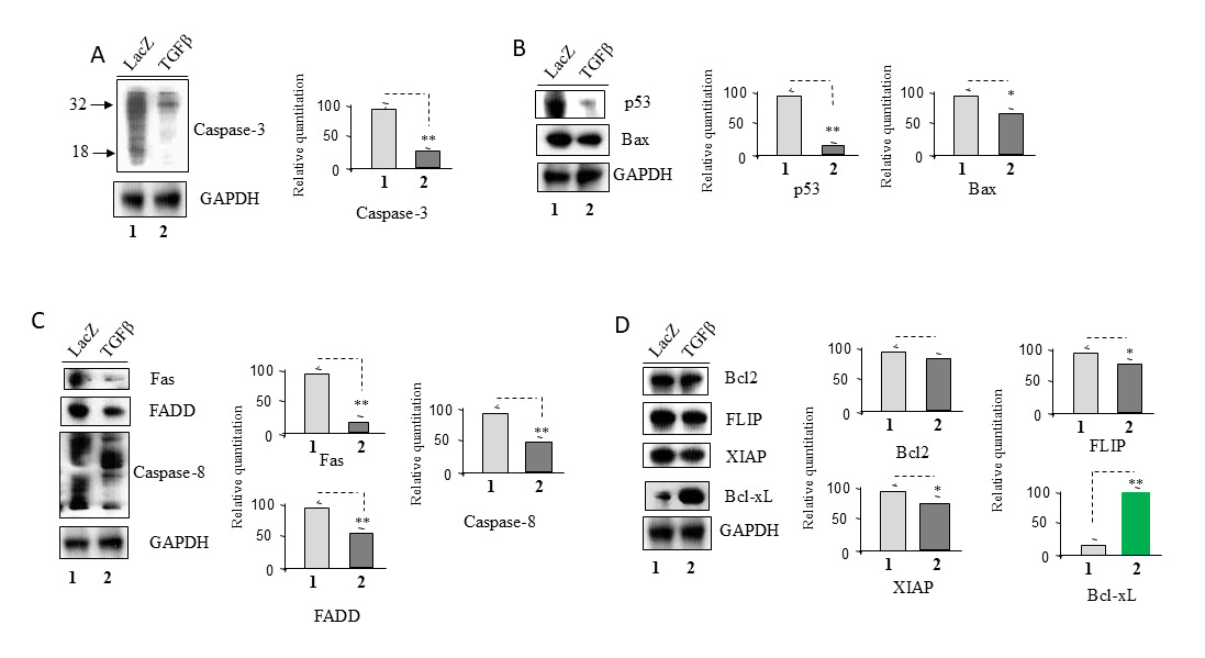
Figure 5. TGF-β expression downregulates apoptotic signaling pathways. U1 cells were infected with an adenovirus carrying the LacZ or TGF-β gene and incubated for 7 days. Total cell lysates were subjected to Western blot analysis to detect (A) Caspase-3; (B) p53 and Bax; (C) Fas, FADD and Caspase-8; (D) Bcl2, FLIP, XIAP and Bcl-xL.
Bcl-xL reduces HIV-1 replication from latency
Bcl-xL and Bcl-2 are anti-apoptotic proteins in the Bcl-2 family that regulate cell death, sharing structural similarities but differing in function and expression. Both contain four Bcl-2 homology domains (BH1–BH4) essential for apoptosis inhibition [26]. Bcl-xL is a splice variant of the Bcl-x gene with multiple isoforms, while Bcl-2 comes from the Bcl-2 gene without alternative splicing [26]. Bcl-2 is broadly expressed, particularly in immune cells, where it supports survival, while Bcl-xL is more tissue-specific, mainly found in the brain, heart, and muscles, and crucial for neuron and blood cell survival [27]. Bcl-xL is often upregulated in certain infected cells, especially macrophages, helping them resist apoptosis and persist as viral reservoirs [27].
Previously, we reported that Bcl-xL can inhibit HIV-1 replication in lymphoma [21]. To examine the effects of Bcl-xL on U1 cells, total proteins were extracted from U1 cells infected with an adenovirus carrying either the Bcl-xL gene or the LacZ gene (control) for seven days. These proteins were then used for Western blotting. As shown in Figure 6A, Bcl-xL expression significantly decreased HIV-1 RNA production, suggesting that the anti-apoptotic molecule Bcl-xL can inhibit HIV-1 replication and promote HIV-1 latency. Additionally, U1 cells were treated with the Bcl-xL inhibitor, WEHI-539 hydrochloride, and incubated for seven days. As shown in Figure 6B, inhibition of Bcl-xL reactivated HIV-1 replication from latency.
These data suggest that Bcl-xL has the potential to inhibit HIV-1 replication, thereby reducing viral proliferation, while also promoting HIV-1 latency, which may play a crucial role in the persistence of the virus within the host.
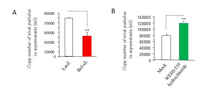
Figure 6. Bcl-xL reduces HIV-1 replication from latency. (A) U1 cells were infected with an adenovirus carrying the LacZ or Bcl-xL gene and incubated for 7 days. A total of 140 µl of culture supernatants containing HIV-1 particles was used to isolate viral RNA for RT-PCR analysis to assess viral replication. (B) U1 cells were incubated in medium containing 100 nM of WEHI-539 hydrochloride or DMSO as controls (Mock) for 7 days. A total of 140 µl of culture supernatants containing HIV-1 particles was used to isolate viral RNA for RT-PCR analysis to assess viral replication.
Discussion
TGF-β exerts its effects binding to TGF-β receptors on the surface of target cells [10,28], which can activate Smad-dependent pathways (canonical TGF-β signaling pathways) [29] or Smad-independent pathways (Non-Canonical TGF-β signaling pathways) [30]. The Smad-dependent pathway is the classical and most well-known signaling route for TGF-β. Upon receptor activation, TGF-β type I receptor phosphorylates Smad(s), triggering their activation [29]. TGF-β signaling can also activate Smad-independent pathways, which play significant roles in cellular processes [30]. In our experiment condition, TGF-β acts through Non-Canonical TGF-β signaling pathways with downregulation of Smad2, Smad3 and Smad4 in U1 cells (Supplementary Figure 2), and their mechanisms remain unknown.
Studying the effects of TGF-β on HIV-1 latency is essential for understanding HIV persistence and developing potential cures. TGF-β, a key immunosuppressive cytokine, promotes HIV latency by inducing a quiescent state in CD4+ T cells and suppressing viral reactivation, making it harder to eliminate latent reservoirs [11,12]. It also plays a role in maintaining latency in specific immune cells and tissue reservoirs, such as the gut and brain [14]. Insights into TGF-β’s influence can help refine HIV cure strategies, whether by enhancing latency reversal or reinforcing latency maintenance, ultimately contributing to more effective therapeutic approaches [11]. Accumulating evidence indicates that TGF-β promotes HIV-1 latency both in vivo and in vitro [11,12,30,31], which is consistent with the results presented here in U1 cells. Our findings involve various cell signaling pathways, including TCR-related signaling pathways, P-TEFb pathways, JAK/STAT pathways, and apoptotic pathways. We also found that TGF-β plays a similar role in inhibiting HIV-1 replication, where its inhibition leads to the reactivation of HIV-1 from latency in lymphoma, ACH2 cells, another well-accepted in vitro latency model (Supplementary Figure 3).
PKC is a key regulator of HIV-1 transcription, replication, and latency reactivation, playing a crucial role in signal transduction, immune responses, and inflammation [32]. PKC is activated by diacylglycerol (DAG) and phorbol esters, which trigger signaling pathways that enhance viral transcription [32]. It activates the NF-κB pathway by phosphorylating IκB kinase, leading to NF-κB's entry into the nucleus and binding to the HIV-1 LTR to promote viral transcription [33]. Additionally, PKC activates the MAPK/ERK pathway [34], increasing AP-1 activity to further boost transcription [35]. PKC also collaborates with the HIV-1 Tat protein to amplify viral transcription, making it a potential therapeutic target for HIV cure research [36]. However, TGF-β downregulated PKC (Figure 2B) and ERK1/2 (Figure 2C) in U1 cells, while the expression levels of AP-1 (Figure 1D) were higher in the TGF-β group compared to the LacZ group. Additionally, there was no significant difference in NF-κBp65 protein levels (Figure 1D) between the TGF-β and LacZ control groups. Further studies are needed to investigate these discrepancies.
Recent findings indicate that anti-apoptotic molecules enhance viral DNA seeding while reducing viral RNA production [22]. This supports the notion that HIV dynamically adapts to its cellular environment to optimize proliferation and persistence [21,22]. When the virus detects a pro-apoptotic state signaling the imminent demise of its host cell, it accelerates replication and virion production [21]. Conversely, in the presence of active anti-apoptotic factors that promote cell survival, HIV favors the establishment of latent reservoirs [21,22]. Notably, studies have shown that CD4+ T cells from aviremic HIV-infected individuals on ART exhibit significantly higher levels of Bcl-2 compared to those from viremic patients and even uninfected controls [23,37]. TGF-β inhibited apoptotic signaling pathways in U1 cells with downregulation of pro-apoptotic proteins (Figures 5B and 5C). However, TGF-β also reduces the expression of Bcl-2, FLIP, and XIAP (Figure 5D)—molecules known to inhibit the activity of pro-apoptotic proteins. Further studies are needed to investigate this paradox.
Bcl-xL, an anti-apoptotic protein in the Bcl-2 family [26,27], plays a crucial role in HIV-1 latency by inhibiting apoptosis, especially in memory CD4+ T cells, which serve as major reservoirs for the virus [23]. By preventing cell death, Bcl-xL allows latently infected cells to persist, stabilizing the viral reservoir and complicating eradication efforts [38,39]. It also hinders the effectiveness of latency-reversing agents (LRAs), which rely on apoptosis to eliminate reactivated cells, increasing the risk of reseeding the reservoir rather than clearing it [38,39]. TGF-β upregulates Bcl-xL in U1 cells, further supporting its role in latency. However, it is not widely known that Bcl-xL may also regulate HIV-1 transcription through interactions with factors such as NF-κB and STAT5, and it plays a role in maintaining chromatin silencing [40]. Additionally, Bcl-xL engages with pathways like TGF-β and NF-κB, influencing HIV reactivation [41], and is involved in autophagy, which helps maintain latency [42]. Furthermore, targeting Bcl-xL with inhibitors could enhance "shock-and-kill" strategies by promoting the death of reactivated cells, while modulating Bcl-xL may improve "block-and-lock" strategies to keep the virus dormant [38]. Measuring Bcl-xL levels in HIV-1 reservoirs and comparing expression across patient groups could provide valuable insights into its role in persistence.
In conclusion, TGF-β plays a crucial role in HIV-1 latency by promoting immune suppression, enhancing latency establishment, and inhibiting viral reactivation through non-SMAD signaling and epigenetic modifications. TGF-β expression exhibits a more pronounced inhibition of HIV-1 reactivation compared to controls. This reduced reactivation is associated with decreased HIV-1 RNA transcription from the host cell genome, involving the downregulation of various pathways, including TCR/PI3K-Akt-related and MAPK-mediated pathways (Erk1/2, p38, and JNK), as well as Jak-Stat pathways (Stat1, Stat3, and Stat5, and TF). Furthermore, TGF-β expression is linked to a reduction in full-length HIV-1 mRNA synthesis due to decreased p-TEFb signaling, marked by the downregulation of Brd4, CDK9, Cyclin T1, and p-RPb-CTD. Concurrently, TGF-β expression downregulates PCAF, which functions as a histone acetyltransferase (HAT) for chromatin acetylation, ultimately suppressing the reactivation of HIV-1 from latency. TGF-β expression also plays a role in apoptosis regulation, contributing to reduced apoptotic signaling. These findings highlight that TGF-β can inactivate HIV-1 from latency while preventing cell death. Importantly, TGF-β expression increases Bcl-xL levels, which can contribute to HIV-1 latency by promoting the survival of infected cells, inhibiting apoptosis, and supporting the maintenance of latent reservoirs, thereby hindering viral clearance. Clinically, TGF-β reinforces HIV-1 latency by sustaining viral persistence in reservoirs through immune suppression and epigenetic silencing, posing a significant challenge for eradication strategies and cure efforts.
Funding
This work was not supported by any external grants.
Acknowledgments
The authors wish to acknowledge Dr. Yideng Liang and Dr. Santanu Biswas for their critical review of this manuscript. The findings and conclusions in this article have not been formally disseminated by the Food and Drug Administration and should not be construed to represent any Agency determination or policy.
Author Contributions
XW and IH conceived the project. XW performed experiments. XW formatted datasets and analyzed data. XW and IH participated in manuscript writing, engaged in data interpretation, and contributed to the intellectual content of this work.
Conflict of Interests
The authors declare no competing interests.
Ethical Approval
Not available.
Data Availability
No datasets were generated or analyzed during the current study.
Abbreviations
AIDS: Acquired Immunodeficiency Syndrome; Akt: Protein Kinase B; AP-1: Activator Protein 1; ART: Antiretroviral Therapy; Bax: Bcl-2-associated X protein; Bcl2: B-cell lymphoma protein; Bcl-xL: B-cell lymphoma-extra large; Brd4: Bromodomain containing protein 4; CD: Cluster of Differentiation; CDK9: Cyclin-Dependent Kinase 9; CTD: Carboxyl-Terminal Domain; ERK: Extracellular Signal-Regulated Kinase; FADD: Fas-Associated Death Domain; FLIP: Fas-Associated Death Domain-Like Interleukin-1beta Converting Enzyme-like Inhibitory Protein; gp130: Glycoprotein 130; HAT: Histone Acetyl Transferase; HIV-1: Human Immunodeficiency Virus type-1; IAP: Inhibitor of Apoptosis Protein; Jak: Janus Kinase; JNK: c-Jun N-terminal Kinase; LRA: latency Reversing Agent; LTR: Long Terminal Repeat; MAPK: Mitogen-Activated Protein Kinase; NFAT: Nuclear Factor of Activated T-cells; NF-kB: Nuclear Factor Κb; P53: Tumor Protein p53; PBMCs: Peripheral Blood Mononuclear Cells; PCAF: P300/CBP-Associated Factor; PCR: Polymerase Chain Reaction; PI3K: Phosphoinositide 3-kinase; PKC: Protein Kinase C; PLCγ-1: Phospholipase C gamma1; pol II: RNA Polymerase II; p-Rpb-CTD: Phosphorylated RNA Polymerase II C-terminal Domain; P-TEFb: positive Transcription Elongation Factor b; RNAP: RNA Polymerase II; Sp1: Specificity Protein 1; STAT: Signal Transducer and Activator of Transcription; TAT: Trans-Activator of Transcription; TCR: T-cell Receptor; TGF-β: Transforming Growth Factor beta; TF: Transcription Factor; U1: Promonocytic Cell; XIAP: X-linked Inhibitor of Apoptosis Protein; Zap-70: Zeta-chain-associated protein kinase 70
References
2. HIVinfo. HIV treatment: the basics [Internet]. Bethesda (MD): National Institutes of Health; 2025 Jan 14. Available from: https://hivinfo.nih.gov/understanding-hiv/fact-sheets/hiv-treatment-basics
3. Gunst JD, Gohil J, Li JZ, Bosch RJ, White Catherine Seamon A, Chun TW, et al. Time to HIV viral rebound and frequency of post-treatment control after analytical interruption of antiretroviral therapy: an individual data-based meta-analysis of 24 prospective studies. Nat Commun. 2025 Jan 21;16(1):906.
4. Ruelas DS, Greene WC. An integrated overview of HIV-1 latency. Cell. 2013 Oct 24;155(3):519–29.
5. Ranga U, Panchapakesan A, Saini C. HIV-1 subtypes and latent reservoirs. Curr Opin HIV AIDS. 2024 Mar 1;19(2):87–92.
6. Khanal S, Schank M, El Gazzar M, Moorman JP, Yao ZQ. HIV-1 Latency and Viral Reservoirs: Existing Reversal Approaches and Potential Technologies, Targets, and Pathways Involved in HIV Latency Studies. Cells. 2021 Feb 23;10(2):475.
7. Chandra PK, Gerlach SL, Wu C, Khurana N, Swientoniewski LT, Abdel-Mageed AB, et al. Mesenchymal stem cells are attracted to latent HIV-1-infected cells and enable virus reactivation via a non-canonical PI3K-NFκB signaling pathway. Sci Rep. 2018 Oct 2;8(1):14702.
8. Wang X, Zhao J, Mbondji C, Hewlett I. p53 expression activation of HIV-1 latency in U1 cells. Int J Virol AIDS. 2017;4:1510036.
9. Fujinaga K, Cary DC. Experimental Systems for Measuring HIV Latency and Reactivation. Viruses. 2020 Nov 9;12(11):1279.
10. Massagué J, Sheppard D. TGF-β signaling in health and disease. Cell. 2023 Sep 14;186(19):4007–37.
11. Yim LY, Lam KS, Luk TY, Mo Y, Lu X, Wang J, et al. Transforming Growth Factor β Signaling Promotes HIV-1 Infection in Activated and Resting Memory CD4+ T Cells. J Virol. 2023 May 31;97(5):e0027023.
12. Chinnapaiyan S, Dutta RK, Nair M, Chand HS, Rahman I, Unwalla HJ. TGF-β1 increases viral burden and promotes HIV-1 latency in primary differentiated human bronchial epithelial cells. Sci Rep. 2019 Aug 29;9(1):12552.
13. Kim J, Bose D, Araínga M, Haque MR, Fennessey CM, Caddell RA, et al. Publisher Correction: TGF-β blockade drives a transitional effector phenotype in T cells reversing SIV latency and decreasing SIV reservoirs in vivo. Nat Commun. 2024 Oct 10;15(1):8787.
14. Samer S, Thomas Y, Araínga M, Carter C, Shirreff LM, Arif MS, et al. Blockade of TGF-β signaling reactivates HIV-1/SIV reservoirs and immune responses in vivo. JCI Insight. 2023 Nov 22;8(22):e176882.
15. Flores O, Lee G, Kessler J, Miller M, Schlief W, Tomassini J, et al. Host-cell positive transcription elongation factor b kinase activity is essential and limiting for HIV type 1 replication. Proc Natl Acad Sci U S A. 1999 Jun 22;96(13):7208–13.
16. Mujtaba S, He Y, Zeng L, Farooq A, Carlson JE, Ott M, et al. Structural basis of lysine-acetylated HIV-1 Tat recognition by PCAF bromodomain. Mol Cell. 2002 Mar;9(3):575–86.
17. Wang X, Zhao J, Hewlett I. Phorbol-12-Myristate-13-Acetate (PMA) Reactivates Replication from HIV-1 Latency and Induces Jurkat Cell Death. J Cell Signal. 2024;5(1):30–40.
18. Gargan S, Ahmed S, Mahony R, Bannan C, Napoletano S, O'Farrelly C, et al. HIV-1 Promotes the Degradation of Components of the Type 1 IFN JAK/STAT Pathway and Blocks Anti-viral ISG Induction. EBioMedicine. 2018 Apr;30:203–16.
19. Elmore S. Apoptosis: a review of programmed cell death. Toxicol Pathol. 2007 Jun;35(4):495–516.
20. Bell DJ, Dockrell DH. Apoptosis in HIV-1 infection. J Eur Acad Dermatol Venereol. 2003 Mar;17(2):178–83.
21. Wang X, Ragupathy V, Zhao J, Hewlett I. Molecules from apoptotic pathways modulate HIV-1 replication in Jurkat cells. Biochem Biophys Res Commun. 2011 Oct 14;414(1):20–4.
22. Wang X, Zhao J, Biswas S, Devadas K, Hewlett I. Components of apoptotic pathways modulate HIV-1 latency in Jurkat cells. Microbes Infect. 2022 Apr-May;24(3):104912.
23. Chandrasekar AP, Cummins NW, Badley AD. The Role of the BCL-2 Family of Proteins in HIV-1 Pathogenesis and Persistence. Clin Microbiol Rev. 2019 Oct 30;33(1):e00107–19.
24. Reece MD, Song C, Hancock SC, Pereira Ribeiro S, Kulpa DA, Gavegnano C. Repurposing BCL-2 and Jak 1/2 inhibitors: Cure and treatment of HIV-1 and other viral infections. Front Immunol. 2022 Dec 9;13:1033672.
25. Cummins NW, Badley AD. Anti-apoptotic mechanisms of HIV: lessons and novel approaches to curing HIV. Cell Mol Life Sci. 2013 Sep;70(18):3355–63.
26. Warren CFA, Wong-Brown MW, Bowden NA. BCL-2 family isoforms in apoptosis and cancer. Cell Death Dis. 2019 Feb 21;10(3):177.
27. Fiebig AA, Zhu W, Hollerbach C, Leber B, Andrews DW. Bcl-XL is qualitatively different from and ten times more effective than Bcl-2 when expressed in a breast cancer cell line. BMC Cancer. 2006 Aug 23;6:213.
28. Tzavlaki K, Moustakas A. TGF-β Signaling. Biomolecules. 2020 Mar 23;10(3):487.
29. Derynck R, Budi EH. Specificity, versatility, and control of TGF-β family signaling. Sci Signal. 2019 Feb 26;12(570):eaav5183.
30. Zhang YE. Non-Smad Signaling Pathways of the TGF-β Family. Cold Spring Harb Perspect Biol. 2017 Feb 1;9(2):a022129.
31. Gokavi J, Sadawarte S, Shelke A, Kulkarni-Kale U, Thakar M, Saxena V. Inhibition of miR-155 Promotes TGF-β Mediated Suppression of HIV Release in the Cervical Epithelial Cells. Viruses. 2021 Nov 12;13(11):2266.
32. López-Huertas MR, Mateos E, Díaz-Gil G, Gómez-Esquer F, Sánchez del Cojo M, Alcamí J, et al. Protein kinase Ctheta is a specific target for inhibition of the HIV type 1 replication in CD4+ T lymphocytes. J Biol Chem. 2011 Aug 5;286(31):27363–77.
33. Jiang G, Dandekar S. Targeting NF-κB signaling with protein kinase C agonists as an emerging strategy for combating HIV latency. AIDS Res Hum Retroviruses. 2015 Jan;31(1):4–12.
34. Tsirimonaki E, Fedonidis C, Pneumaticos SG, Tragas AA, Michalopoulos I, Mangoura D. PKCε signalling activates ERK1/2, and regulates aggrecan, ADAMTS5, and miR377 gene expression in human nucleus pulposus cells. PLoS One. 2013 Nov 28;8(11):e82045.
35. Whitmarsh AJ, Davis RJ. Transcription factor AP-1 regulation by mitogen-activated protein kinase signal transduction pathways. J Mol Med (Berl). 1996 Oct;74(10):589–607.
36. Borgatti P, Zauli G, Cantley LC, Capitani S. Extracellular HIV-1 Tat protein induces a rapid and selective activation of protein kinase C (PKC)-alpha, and -epsilon and -zeta isoforms in PC12 cells. Biochem Biophys Res Commun. 1998 Jan 14;242(2):332–7.
37. Ren Y, Huang SH, Patel S, Alberto WDC, Magat D, Ahimovic D, et al. BCL-2 antagonism sensitizes cytotoxic T cell-resistant HIV reservoirs to elimination ex vivo. J Clin Invest. 2020 May 1;130(5):2542–59.
38. Ren Y, Huang SH, Macedo AB, Ward AR, Alberto WDC, Klevorn T, et al. Selective BCL-XL Antagonists Eliminate Infected Cells from a Primary-Cell Model of HIV Latency but Not from Ex Vivo Reservoirs. J Virol. 2021 Jul 12;95(15):e0242520.
39. Wyżewski Z, Świtlik W, Mielcarska MB, Gregorczyk-Zboroch KP. The Role of Bcl-xL Protein in Viral Infections. Int J Mol Sci. 2021 Feb 16;22(4):1956.
40. Socolovsky M, Fallon AE, Wang S, Brugnara C, Lodish HF. Fetal anemia and apoptosis of red cell progenitors in Stat5a-/-5b-/- mice: a direct role for Stat5 in Bcl-X(L) induction. Cell. 1999 Jul 23;98(2):181–91.
41. Spender LC, O'Brien DI, Simpson D, Dutt D, Gregory CD, Allday MJ, et al. TGF-beta induces apoptosis in human B cells by transcriptional regulation of BIK and BCL-XL. Cell Death Differ. 2009 Apr;16(4):593–602.
42. Chen M, Li M, Budai MM, Rice AP, Kimata JT, Mohan M, et al. Clearance of HIV-1 or SIV reservoirs by promotion of apoptosis and inhibition of autophagy: Targeting intracellular molecules in cure-directed strategies. J Leukoc Biol. 2022 Nov;112(5):1245–59.