

Abstract
Paracentesis is a routinely performed and generally safe procedure in patients with cirrhotic ascites. Hemorrhagic complications are uncommon but may occasionally result in significant morbidity and mortality. We report a case of a 51-year-old man with alcohol-related cirrhosis and moderate ascites who developed a rapidly enlarging anterior abdominal wall hematoma following diagnostic paracentesis. The patient presented with progressive swelling and ecchymosis over the paracentesis site associated with tachycardia and a significant decline in hemoglobin. Non-contrast computed tomography revealed a large left lateral anterior abdominal wall hematoma measuring 18.6 × 11.5 × 8.1 cm. CT angiography excluded active bleeding and pseudo aneurysm formation. The patient was managed conservatively with transfusion support including packed red blood cells, platelets, and fresh frozen plasma, resulting in clinical stabilization and gradual regression of the hematoma. This case highlights the importance of early recognition of hemorrhagic complications after paracentesis and emphasizes that conservative management may be successful in hemodynamically stable patients without evidence of active arterial bleeding.
Keywords
Paracentesis, Abdominal wall hematoma, Cirrhosis, Ascites, Hemorrhagic complications, Conservative management
Introduction
Paracentesis is one of the most commonly performed bedside procedures in patients with cirrhosis and ascites and is considered relatively safe, even in the presence of coagulopathy and thrombocytopenia [1,2]. The procedure is routinely used for diagnostic evaluation of ascitic fluid as well as therapeutic decompression in tense ascites. Reported major complication rates are low, generally below 1%, with hemorrhagic events being particularly uncommon [1,3].
Hemorrhagic complications associated with paracentesis include abdominal wall hematoma, hem peritoneum, and pseudo aneurysm formation involving the inferior epigastric artery or its branches, with abdominal wall hematoma being the most frequently encountered manifestation [1]. Predisposing factors include portal hypertension, abdominal wall collateralization, thrombocytopenia, renal dysfunction, and coagulation abnormalities frequently present in patients with advanced liver disease [4].
We report a case of a large anterior abdominal wall hematoma developing shortly after diagnostic paracentesis in a cirrhotic patient, which was successfully managed conservatively.
Case Report
A 51-year-old male with alcohol-related cirrhosis and moderate ascites underwent diagnostic paracentesis at a peripheral healthcare center. The procedure was performed in the left lower quadrant of the abdomen.
Within a few hours after the procedure, the patient noticed localized swelling at the puncture site associated with progressive skin discoloration. Over the next several hours, the swelling enlarged considerably, prompting referral to our tertiary care center.
At presentation, the patient was conscious and oriented. His pulse rate was 106 beats/minute and blood pressure was 106/80 mmHg without significant postural hypotension. Physical examination revealed a large tender swelling over the left lower anterior abdominal wall with overlying ecchymosis suggestive of expanding hematoma (Figure 1A).
Laboratory investigations demonstrated severe anemia with hemoglobin of 5.9 g/dL, thrombocytopenia with platelet count of 88,000/mm³, blood urea nitrogen of 62 mg/dL, serum creatinine of 1.4 mg/dL, and an international normalized ratio (INR) of 1.5.
Non-contrast computed tomography (CT) of the abdomen demonstrated a large left lateral anterior abdominal wall hematoma measuring 18.6 × 11.5 × 8.1 cm involving the abdominal wall musculature (Figures 1B and 1C). Subsequent computed tomography (CT) angiography did not reveal active contrast extravasation, pseudo aneurysm, or any identifiable arterial source of ongoing bleeding.
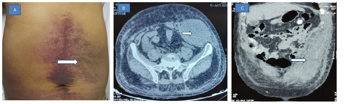
Figure 1. A. Arrow showing ecchymosis with swelling in left anterior abdominal wall. B. Noncontrast Axial computed tomography image showing hyperdense left anterior abdominal wall hematoma. C. Coronal computed tomography image showing large hematoma in relation to left anterior abdominal wall.
Given the enlarging hematoma, tachycardia, and significant decline in hemoglobin, the patient received supportive management including transfusion of packed red blood cells, platelets, and fresh frozen plasma. Close hemodynamic monitoring was undertaken. Since no active arterial bleeding was demonstrated and the patient remained hemodynamically stable, invasive radiological or surgical intervention was deferred.
Over the following days, the patient showed gradual clinical improvement with stabilization of vital parameters, improvement in hemoglobin levels, and reduction in hematoma size. He was subsequently discharged in satisfactory clinical condition with outpatient follow-up advice.
Discussion
Paracentesis is widely regarded as a low-risk bedside procedure in patients with cirrhosis. Despite frequent underlying abnormalities in coagulation parameters among cirrhotic patients, clinically significant bleeding remains uncommon [1,3]. Existing evidence suggests that conventional coagulation tests such as INR and platelet count correlate poorly with actual bleeding risk in cirrhosis because of the concept of “rebalanced hemostasis” [2].
Sharzehi et al. categorized hemorrhagic complications following paracentesis into abdominal wall hematomas, hemoperitoneum, and pseudo aneurysms, with abdominal wall hematomas being the most common manifestation [1]. Injury to the inferior epigastric artery or its branches during needle insertion is considered a principal mechanism [5].
Our patient possessed several recognized risk factors for bleeding, including cirrhosis-related thrombocytopenia, mild coagulopathy, and renal dysfunction. Although the procedure was intended as a diagnostic paracentesis, even small-caliber needle insertion can result in vascular injury, particularly when performed without ultrasound guidance or in the presence of abdominal wall collateral vessels [4].
Clinical presentation of abdominal wall hematoma may vary from localized pain and ecchymosis to hemodynamic instability and hemorrhagic shock. Progressive swelling at the puncture site associated with a drop in hemoglobin should immediately raise suspicion for post-procedural bleeding. Computed tomography (CT) imaging remains the diagnostic modality of choice as it accurately delineates the extent of hematoma and can identify active bleeding or pseudo aneurysm formation. Computed Tomography (CT) angiography is especially valuable in determining the need for transcatheter embolization [5].
Management depends largely on hemodynamic status and imaging findings. Hemodynamically stable patients without active arterial bleeding can often be managed conservatively with blood product transfusion, correction of coagulopathy, analgesia, and close monitoring [1,4]. Conversely, unstable patients or those with ongoing arterial extravasation may require interventional radiological embolization or surgical exploration.
The present case emphasizes several clinically important points. First, clinicians should maintain a high index of suspicion for hemorrhagic complications even after seemingly uncomplicated diagnostic paracentesis. Second, progressive localized swelling and ecchymosis should not be dismissed as minor post-procedural changes. Finally, conservative management can be highly effective in carefully selected stable patients.
Use of ultrasound guidance before paracentesis has been shown to reduce complication rates by identifying ascitic pockets and avoiding abdominal wall vessels [3]. Current guidelines increasingly advocate ultrasound-guided paracentesis as standard practice whenever feasible [2].
Conclusion
Anterior abdominal wall hematoma is a rare but potentially serious complication of paracentesis in patients with cirrhosis. Early recognition through careful clinical assessment and prompt imaging is essential to prevent adverse outcomes. Conservative management may be successful in hemodynamically stable patients without evidence of active bleeding. Routine use of ultrasound guidance and meticulous procedural technique may further minimize the risk of hemorrhagic complications.
Authors’ Contribution
Dr Vishal came up with the idea. Material preparation, data collection and analysis were performed by Dr Anshul. The first draft of the manuscript was written by Dr Rajesh and revised by Dr Brij. All authors read and approved the final manuscript.
Informed Consent
Patient provided informed consent to publish the included information.
Conflicts of Interest
None.
Declaration of Funding Source
None.
References
2. Runyon BA. Introduction to the revised American Association for the Study of Liver Diseases Practice Guideline management of adult patients with ascites due to cirrhosis 2012 update. Hepatology. 2013;57(4):1651–3.
3. Grabau CM, Crago SF, Hoff LK, Simon JA, Melton CA, Frankel RM, et al. Performance standards for therapeutic abdominal paracentesis. Hepatology. 2004;40(2):484–8.
4. Pache I, Bilodeau M. Severe hemorrhage following abdominal paracentesis for ascites in patients with liver disease. Aliment Pharmacol Ther. 2005;21(5):525–9.
5. Webster ST, Brown KL, Lucey MR, Nostrant TT. Hemorrhagic complications of large-volume paracentesis. Am J Gastroenterol. 1996;91(2):366–8