
Abstract
Chemosensory systems exhibit substantial plasticity that supports adaptive perception, sensory learning, and behavioral responses to changing environmental and physiological conditions. Although synaptic mechanisms have traditionally been viewed as the principal basis of chemosensory plasticity, accumulating evidence indicates that adaptive sensory function depends on coordinated interactions among circuit modulation, glial regulation, metabolic and inflammatory state, and structural remodeling. This review synthesizes experimental findings showing how cortical feedback reshapes olfactory and broader chemosensory representations, how astrocytes and microglia regulate synaptic, metabolic, inflammatory, and remodeling environments, and how neurogenic and other structural mechanisms contribute to longer-term adaptation in experimental olfactory systems. On this basis, the review proposes an integrated multiscale framework in which cortical, glial, metabolic, inflammatory, genetic, and structural processes act as interconnected regulators of chemosensory plasticity rather than isolated contributors. Pharmacologically relevant entry points include neuromodulatory control of cortical feedback, astrocytic glutamate handling, purinergic signaling, metabolic support, microglial inflammatory responses, complement-associated synaptic remodeling, and neurotransmitter-dependent regulation of neurogenic niche dynamics. The framework also considers sirtuin 1 (SIRT1) as a candidate genetic and metabolic regulator linking cellular energy state, inflammation, oxidative stress, autophagy, synaptic plasticity, aging, and neurodegeneration, while emphasizing that its direct role in human chemosensory recovery remains unproven. Because smell and taste disturbances are prominent in aging, neurodegenerative disease, metabolic disorders, and post-viral conditions, this framework provides a basis for identifying context-dependent therapeutic strategies that may support sensory adaptation and recovery while avoiding disruption of beneficial inflammatory or remodeling responses.
Keywords
Chemosensory plasticity, Neuropharmacology, Olfactory dysfunction, Astrocytes, Microglia, Neuroinflammation, Pharmacological modulation, SIRT1
Introduction — Chemosensory Plasticity Beyond Synapses: Toward Pharmacological Modulation
Chemosensory systems, encompassing olfaction and gustation, exhibit substantial plasticity that allows sensory perception and behavior to adapt to environmental stimuli, prior experience, and physiological state. This plasticity supports odor and taste discrimination, sensory learning, food-related decision making, hazard detection, and context-dependent behavior. Traditionally, chemosensory plasticity has been explained primarily through synaptic mechanisms, including long-term potentiation, long-term depression, and activity-dependent changes in synaptic strength within the olfactory bulb, piriform cortex, and gustatory cortex [1,2].
However, synaptic plasticity alone does not fully explain the persistence, flexibility, and disease vulnerability of chemosensory adaptation. Chemosensory circuits are continuously influenced by cortical feedback, neuromodulatory signaling, glial regulation, inflammatory state, metabolic support, and structural remodeling. Experimental studies increasingly show that these mechanisms interact across biological scales rather than acting independently. Cortical feedback modifies early sensory processing and sensory representations [3–8], astrocytes regulate neurotransmitter handling and metabolic support, microglia influence inflammatory signaling and synaptic remodeling [7,9], and neurogenic or structural remodeling contributes to longer-term circuit adaptation in experimental olfactory systems [5,10].
The central problem is that these mechanisms are often considered separately, making it difficult to explain how chemosensory circuits maintain adaptive plasticity or why that plasticity fails during aging, inflammation, metabolic stress, viral injury, and neurodegenerative disease. Existing reviews commonly emphasize individual mechanisms such as synaptic plasticity, neuroinflammation, glial function, or neurogenesis, but less attention has been given to how these processes interact as a coordinated regulatory system.
This review addresses that gap by proposing an integrated framework in which cortical feedback, glial regulation, and structural remodeling operate as interconnected regulators of chemosensory plasticity. Within this framework, cortical feedback shapes circuit activity and sensory gain, astrocytes and microglia regulate the synaptic, metabolic, and inflammatory environment, and neurogenic or other structural mechanisms support longer-term remodeling in experimental systems where these processes remain active. The emphasis is therefore not simply on identifying individual mechanisms, but on understanding how their interactions determine whether chemosensory plasticity remains adaptive, becomes impaired, or supports recovery.
This integrated view has direct pharmacological relevance. Chemosensory dysfunction occurs in aging, neurodegenerative disease, metabolic disorders, inflammatory states, and post-viral conditions, yet therapeutic options remain limited. Potential pharmacological entry points include neuromodulatory control of cortical feedback, astrocytic glutamate handling, purinergic signaling, metabolic support, microglial inflammatory responses, complement-associated synaptic remodeling, and regulation of neurogenic niche dynamics in experimental systems. The therapeutic challenge is therefore not simply to increase plasticity or suppress inflammation, but to restore conditions that support normal sensory adaptation while preserving beneficial circuit remodeling.
Accordingly, this review has three aims. First, it synthesizes experimental evidence on cortical feedback, astrocytes, microglia, and neurogenic or structural remodeling in chemosensory plasticity. Second, it examines how these mechanisms interact across molecular, cellular, circuit, and structural levels. Third, it identifies pharmacologically relevant pathways that may influence chemosensory adaptation, recovery, and disease-associated dysfunction, while considering the translational limitations of experimental models.
Review methodology
This article was developed as a narrative and integrative review rather than a systematic review or meta-analysis. Relevant literature was identified through targeted searches of PubMed and Google Scholar and through examination of reference lists from pertinent primary studies and reviews, with the literature updated through July 2026. Searches focused on combinations of terms related to chemosensory and olfactory plasticity, cortical feedback, sensory learning, astrocytes, microglia, neuroinflammation, metabolic regulation, neural stem cells, adult neurogenesis, olfactory bulb circuitry, pharmacological modulation, aging, neurodegeneration, and post-viral chemosensory dysfunction.
Priority was given to peer-reviewed experimental studies that directly addressed mechanisms of chemosensory circuit plasticity, glial regulation, activity-dependent remodeling, neurogenic or structural plasticity, or pharmacologically relevant pathways. Recent studies were emphasized where available, while established earlier studies were retained when necessary to define foundational mechanisms. Reviews were used primarily to provide broader context and to identify relevant primary literature. Evidence from experimental animal models was distinguished from evidence applicable to humans, particularly for adult SVZ-derived olfactory neurogenesis, where substantial species differences limit direct translation.
The selected literature was organized according to the major mechanistic levels examined in this review, including circuit regulation, glial regulation, structural remodeling, and their interactions. It was then evaluated in relation to pharmacological targets, disease-associated chemosensory dysfunction, and translational relevance.
Chemosensory Circuit Architecture as a Plastic Substrate
Organization of chemosensory circuits
Chemosensory processing is mediated by distributed neural circuits that transform chemical stimuli into perceptual and behavioral outputs. In the olfactory system, sensory information originates in olfactory receptor neurons, which project to the olfactory bulb, where inputs are organized into discrete glomerular units based on receptor identity [6,11]. Within the olfactory bulb, principal neurons (mitral and tufted cells) relay processed signals to higher-order regions, most notably the piriform cortex, which serves as a primary olfactory cortical area [6,11]. Unlike other sensory systems, olfactory pathways bypass thalamic relay nuclei and project directly to cortical and limbic structures, enabling rapid integration with memory, emotion, and behavioral circuits [6,12].
A distinguishing feature of chemosensory systems, particularly within the piriform cortex, is the absence of strict topographic organization. Unlike visual or somatosensory systems that rely on spatially ordered maps, olfactory representations are encoded through distributed and associative activity patterns across neuronal populations [8,12]. This organizational principle supports flexible and context-dependent encoding of odor information. Although gustatory pathways involve brainstem and thalamic relays prior to reaching the gustatory cortex, similar principles of distributed representation and adaptive coding apply across chemosensory modalities [2,13].
The major organizational features of chemosensory circuit architecture discussed in this section are summarized in Figure 1.
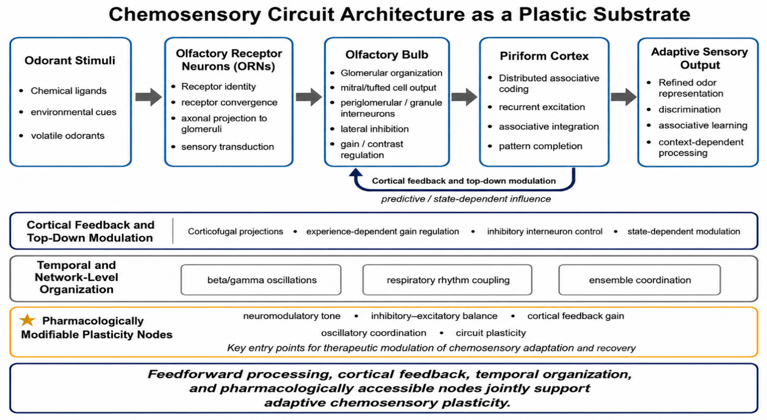
Figure 1. Chemosensory circuit architecture as a plastic substrate. Schematic overview of major circuit features that support chemosensory plasticity, with emphasis on the olfactory system. Odorant information is transmitted from olfactory receptor neurons to the olfactory bulb and then to the piriform cortex, where distributed associative coding supports adaptive odor representation. Cortical feedback provides top-down modulation of olfactory bulb activity, while temporal network mechanisms, including beta/gamma oscillations, respiratory rhythm coupling, and ensemble coordination, further shape sensory processing. The figure also highlights pharmacologically accessible nodes, including neuromodulatory tone, inhibitory–excitatory balance, cortical feedback gain, oscillatory coordination, and circuit plasticity, which may influence chemosensory adaptation and recovery. This figure was created by the author for the present manuscript and was not reproduced or adapted from another source.
Recurrent connectivity and feedback pathways
As illustrated in Figure 1, chemosensory circuits are organized as distributed and highly interconnected systems in which feedforward, recurrent, and top-down pathways jointly shape sensory processing. A defining feature of chemosensory circuits is the presence of extensive recurrent and feedback connectivity. In the olfactory system, the piriform cortex sends dense corticofugal projections back to the olfactory bulb, forming feedback pathways that modulate early sensory processing [6,8]. These projections target both excitatory and inhibitory neuronal populations, enabling dynamic regulation of signal gain, contrast enhancement, and temporal coordination within the bulb [6,8,14].
Local recurrent circuits further contribute to network dynamics. In the olfactory bulb, inhibitory interneurons such as granule and periglomerular cells mediate lateral inhibition and synchronize mitral and tufted cell activity, shaping odor representations [11,14]. In the piriform cortex, recurrent excitatory connections support associative processing, enabling pattern completion and the retrieval of learned odor representations from partial or degraded inputs [12,15]. Together, these recurrent and feedback interactions establish chemosensory circuits as highly interconnected systems in which information flow is continuously shaped by internal network states and prior experience.
Activity-dependent circuit reconfiguration
Chemosensory circuits exhibit substantial activity-dependent plasticity at multiple levels, including synaptic strength, connectivity patterns, and network organization. Experimental studies have demonstrated that sensory experience, learning, and environmental exposure can modify synaptic efficacy within both the olfactory bulb and piriform cortex, leading to changes in odor tuning, discrimination, and perceptual sensitivity [8,12,16,17]. These modifications include long-term potentiation and depression, as well as shifts in inhibitory–excitatory balance that influence circuit output and stability [6,8,16].
In addition to synaptic changes, activity-dependent processes can alter connectivity patterns and influence the recruitment of neuronal ensembles during sensory processing. Repeated odor exposure or associative learning refines neuronal population responses, enhancing selectivity and improving signal-to-noise ratios in odor representations [8,12,17]. Chemosensory processing is also strongly shaped by temporal dynamics, including oscillatory activity in the beta and gamma frequency ranges and coupling to respiratory rhythms, which organize neuronal firing and influence odor representation and discrimination [18–20]. These temporal features provide an additional dimension through which circuit activity is structured and modulated by experience.
Together, feedforward processing, recurrent connectivity, cortical feedback, and activity-dependent reconfiguration allow chemosensory circuits to update sensory representations according to experience, internal state, and behavioral context [6,8,12,16–18]. These circuit properties are further shaped by glial regulation and longer-term structural remodeling.
Experience-Dependent Plasticity in Chemosensory Systems
Behavioral and circuit-level adaptation to sensory experience
Chemosensory experience modifies detection, discrimination, preference, and learned associations. Repeated exposure can produce habituation, whereas perceptual or associative learning can improve discrimination and assign behavioral significance to odors or tastes [4,12,21]. These behavioral changes are accompanied by altered neuronal responsiveness, tuning, ensemble recruitment, and temporal coordination within the olfactory bulb, piriform cortex, gustatory cortex, and related chemosensory circuits [4,12,22].
Attention, behavioral relevance, reward, and internal state all influence experience-dependent plasticity. Chemosensory learning therefore changes circuit activity according to context rather than producing a fixed response to repeated sensory input [23].
Molecular and cellular mechanisms underlying experience-dependent plasticity
At the molecular and cellular levels, sensory activity engages calcium-dependent signaling, transcriptional regulation, neuromodulatory pathways, and changes in synaptic efficacy and neuronal excitability [5,24]. Cholinergic and other neuromodulatory systems help link plasticity to attention, reward, and behavioral state.
Glial regulation and, in experimental olfactory systems, neurogenic remodeling extend these effects beyond neurons. Astrocytes and microglia shape neurotransmitter availability, metabolic support, inflammatory signaling, and synaptic remodeling, whereas sensory experience can influence the survival and integration of newly generated neurons [5,7,10]. These mechanisms are examined in greater detail in the following sections.
Cortical Feedback as a Driver of Circuit Reconfiguration
Anatomical and functional organization of cortical feedback pathways
Cortical feedback is a major feature of chemosensory circuit organization, particularly in the olfactory system. The piriform cortex sends dense corticofugal projections to the olfactory bulb, targeting principal neurons and local interneurons, including granule and periglomerular cells [6,8,25]. Through these targets, cortical inputs regulate lateral inhibition, neuronal synchronization, and the temporal structure and specificity of mitral and tufted cell output [26–28]. These feedback pathways are modified by learning and repeated sensory exposure, allowing cortical control of early sensory processing to be tuned by prior experience and behavioral relevance [6,8,29].
Mechanisms of cortical modulation of sensory processing
Cortical feedback modifies chemosensory processing partly through regulation of inhibitory interneurons. Cortical inputs to olfactory bulb granule cells alter lateral inhibition and thereby adjust the contrast, selectivity, and gain of odor representations produced by mitral and tufted cells [8,29,30].
Corticofugal projections also influence the temporal organization of sensory activity, including beta- and gamma-frequency oscillations associated with odor processing and learning [8,31,32]. By modifying the timing and coordination of neuronal firing, cortical feedback helps organize how complex sensory information is encoded.
These effects are further shaped by cholinergic, noradrenergic, and other neuromodulatory inputs, which regulate the gain and plasticity of corticofugal signaling [19,24,33]. Through these neuromodulatory influences, cortical feedback can enhance behaviorally relevant signals, reduce responses to less relevant inputs, and adjust sensory processing according to attention, arousal, reward, and behavioral context [8,12,30,32,33].
Cortical feedback in learning and predictive processing
Cortical feedback contributes to learning-dependent plasticity by transmitting experience- and context-related signals from higher-order regions to early chemosensory circuits. During associative learning, these signals can modify olfactory bulb activity and refine sensory representations according to prior experience and behavioral relevance [8,12,34]. Predictive-processing models further suggest that cortical pathways convey expectations about incoming sensory input, although the precise mechanisms remain under investigation [35–37]. Experimental findings nevertheless support a role for cortical feedback in enhancing relevant representations, suppressing less informative signals, and improving odor discrimination and learning [8,12,34].
Cortical feedback therefore links experience, behavioral state, and sensory context to early chemosensory processing. Because these pathways are regulated by cholinergic, noradrenergic, and other neuromodulatory systems, they provide pharmacologically accessible mechanisms for modifying sensory gain, discrimination, learning, and adaptive circuit responses [19,24,33].
Glial Regulation of Chemosensory Circuit States as Pharmacological Entry Points
Astrocytes in chemosensory circuit modulation
Astrocytes are integral components of chemosensory circuits, contributing to the regulation of synaptic transmission, metabolic support, and extracellular homeostasis. Within the olfactory bulb and piriform cortex, astrocytes are positioned in close proximity to synapses, forming part of the “tripartite synapse,” in which pre- and postsynaptic neuronal elements are functionally integrated with astrocytic processes that regulate synaptic transmission and plasticity [38–40]. Through uptake and recycling of neurotransmitters such as glutamate and GABA, astrocytes regulate synaptic signaling and prevent excitotoxicity, thereby maintaining circuit stability [41–43].
These astrocytic functions are pharmacologically relevant because glutamate clearance, GABA–glutamate balance, transporter activity, and extracellular homeostasis directly shape neuronal excitability and plasticity. In chemosensory circuits, altered astrocytic regulation could influence sensory gain, discrimination, and vulnerability to inflammatory or metabolic disruption [18,44,45]. Thus, astrocyte-mediated neurotransmitter handling represents a potential entry point for modulating chemosensory circuit stability and maladaptive sensory processing.
In addition to neurotransmitter clearance, astrocytes modulate synaptic transmission through the release of gliotransmitters, including ATP, D-serine, and glutamate, which can influence synaptic strength and plasticity [40,46,47]. Astrocytes also play a key role in providing metabolic support to neurons by regulating glucose uptake, lactate production, and energy distribution within neural circuits [48–50]. Astrocyte–neuron metabolic coupling, including lactate shuttling, provides an additional mechanism through which glial cells support activity-dependent plasticity [48,49,51]. These functions are particularly relevant in chemosensory systems, where continuous sensory input requires efficient metabolic coordination between neuronal activity and energy supply.
From a therapeutic perspective, astrocyte–neuron metabolic coupling may be especially important in aging, metabolic disorders, and neurodegenerative disease, where impaired energy support can reduce circuit resilience [52,53]. Pharmacological strategies that influence astrocytic metabolism, lactate signaling, or activity-dependent energy supply may therefore affect the capacity of chemosensory circuits to maintain adaptive plasticity under physiological stress.
Experimental studies have demonstrated that astrocytic activity is dynamically regulated by sensory experience and can influence neuronal responsiveness and circuit function [54–56]. Changes in astrocytic calcium signaling and gliotransmission have been associated with modulation of synaptic efficacy and network activity in response to sensory stimulation, indicating that astrocytes contribute both to maintaining baseline circuit function and to experience-dependent modulation of chemosensory processing.
These observations position astrocytes not simply as supportive cells but as pharmacologically accessible regulators of sensory circuit state. Astrocytic calcium signaling, purinergic signaling, gliotransmission, glutamate handling, and metabolic support may each provide mechanisms through which drug action could influence chemosensory adaptation, recovery, or dysfunction.
Microglial roles in synaptic remodeling and immune signaling
Microglia serve as the primary immune cells of the central nervous system and play an active role in shaping neural circuits through synaptic remodeling and immune-related processes. In chemosensory systems, microglia continuously survey the local environment and respond to changes in neuronal activity, injury, or inflammation [57–59]. One of their key functions is the selective elimination of synaptic elements through activity-dependent pruning, which contributes to the refinement of neural circuits [57,60,61].
Experimental evidence indicates that microglia can modulate synaptic connectivity in response to sensory experience, participating in the removal of less active synapses while preserving or strengthening functionally relevant connections [7,58,62]. This process is mediated through molecular pathways involving complement proteins and other signaling mechanisms that tag synapses for elimination [60,63,64]. Through these actions, microglia contribute to the optimization of circuit architecture and the maintenance of efficient sensory processing.
This remodeling function has direct immunopharmacological relevance. Complement-associated synaptic tagging, microglial pruning, and activity-dependent remodeling are mechanisms through which inflammatory state can alter sensory circuit architecture [63]. In disease contexts, excessive or misdirected microglial remodeling may contribute to impaired chemosensory discrimination, persistent sensory dysfunction, or reduced recovery after injury.
In addition to synaptic remodeling, microglia release cytokines and other signaling molecules that influence neuronal excitability and plasticity [18,59,64,65]. While these immune-related functions are essential for maintaining tissue integrity, excessive or dysregulated microglial activation can disrupt circuit function and contribute to pathological conditions. Thus, microglial activity must be tightly regulated to balance adaptive plasticity with circuit stability. The principal astrocytic and microglial mechanisms that regulate chemosensory circuit states are summarized schematically in Figure 2. Because microglial responses are shaped by cytokine signaling, purinergic pathways, complement activity, and inflammatory priming, they represent an important class of potential immunopharmacological targets. However, therapeutic modulation of microglia requires caution, because suppressing inflammatory signaling indiscriminately may impair adaptive remodeling, while excessive activation may destabilize circuits and promote maladaptive plasticity.
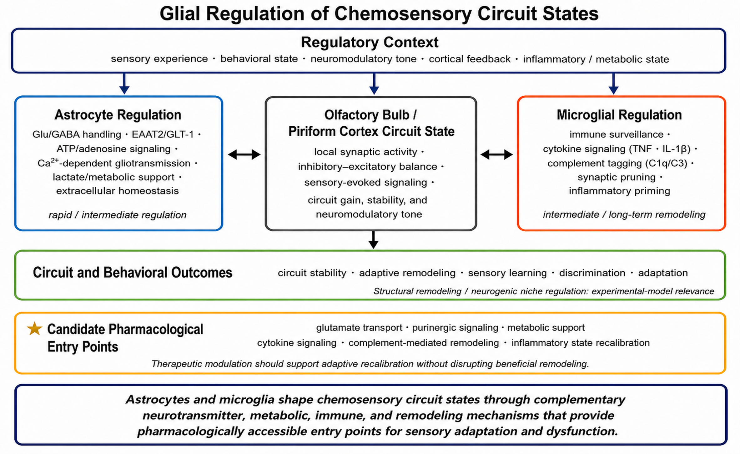
Figure 2. Glial regulation of chemosensory circuit states. Schematic overview of astrocyte and microglial mechanisms that regulate chemosensory circuit function and provide pharmacologically accessible entry points. Astrocytes influence circuit state through glutamate/GABA handling, EAAT2/GLT-1–associated transport, ATP/adenosine signaling, Ca2+-dependent gliotransmission, metabolic support, and extracellular homeostasis. Microglia contribute through immune surveillance, cytokine signaling, complement-associated tagging, synaptic pruning, and inflammatory priming. These mechanisms interact with local synaptic activity, inhibitory–excitatory balance, sensory-evoked signaling, circuit gain, and neuromodulatory tone to shape circuit stability, adaptive remodeling, sensory learning, discrimination, and adaptation. This figure was created by the author for the present manuscript and was not reproduced or adapted from another source.
Integrated glial regulation and pharmacological relevance
Astrocytes and microglia regulate complementary features of the local circuit environment. Astrocytes primarily support neurotransmitter handling, extracellular homeostasis, gliotransmission, and metabolic coupling, whereas microglia contribute to immune surveillance, cytokine signaling, complement-associated synaptic tagging, and activity-dependent remodeling [56,57,66]. Together, these cells help determine whether chemosensory circuits remain stable, undergo adaptive remodeling, or develop maladaptive changes.
Glial regulation is activity-dependent and context-sensitive. Sensory activity, cortical feedback, neuromodulatory tone, inflammatory state, and metabolic conditions can alter astrocytic and microglial responses [56,57,67–71]. Glial signaling can in turn modify neuronal excitability, synaptic efficacy, circuit gain, and structural remodeling [56,57,66–71]. Glia therefore shape the conditions under which chemosensory plasticity occurs rather than controlling it independently.
These functions identify several pharmacologically relevant pathways, including astrocytic glutamate transport, ATP and adenosine signaling, calcium-dependent gliotransmission, lactate-mediated metabolic support, microglial cytokine signaling, complement-associated remodeling, and inflammatory-state regulation. Treatment should aim to restore conditions that support healthy circuit function rather than broadly suppress glial or inflammatory activity, because these responses can either promote repair or contribute to dysfunction depending on their timing and context.
Neural Stem Cells and Structural Plasticity in Experimental Olfactory Systems
SVZ-derived neurogenesis and species limitations
Adult neurogenesis provides a well-characterized form of structural plasticity in rodent and other experimental mammalian olfactory systems. Neural stem and progenitor cells within the subventricular zone generate neuroblasts that migrate through the rostral migratory stream to the olfactory bulb [72–74]. After reaching the bulb, these cells differentiate primarily into granule and periglomerular interneurons and integrate into local circuits involved in sensory processing [10,75,76]. This pathway allows experimental olfactory circuits to modify their cellular composition in response to developmental, environmental, and activity-dependent signals.
The relevance of this pathway differs substantially across species and developmental stages. SVZ-to-olfactory-bulb neurogenesis is robust in adult rodents, whereas in humans it is prominent early in life but becomes sharply attenuated thereafter and appears extremely limited as a major source of ongoing olfactory bulb interneuron replacement in adulthood. Adult olfactory neurogenesis should therefore be interpreted here primarily as an experimental model of structural plasticity rather than as an established mechanism of adult human olfactory circuit renewal.
Activity-dependent and neurotransmitter regulation
Neurogenic output is regulated by interactions among intrinsic programs, local niche signals, and neural activity. The SVZ niche contains ependymal, vascular, extracellular-matrix, and glial components that influence stem-cell quiescence, activation, proliferation, and differentiation [77–79]. Sensory enrichment and learning can promote the survival and integration of newly generated neurons, whereas sensory deprivation can reduce neurogenic output and alter olfactory circuit composition [80–82]. Activity-dependent selection therefore helps retain neurons that become functionally engaged within active circuits [5,76,80].
Neurotransmitter systems provide major links between circuit state and stem-cell behavior. GABAergic signaling helps maintain quiescence and regulate the size and activity of the stem-cell pool [83–85]. Cholinergic inputs can promote activation of quiescent stem cells and increase proliferation through receptor-dependent mechanisms [86,87]. Dopaminergic and serotonergic pathways also contribute to niche regulation, although their effects depend on cell state, receptor expression, and experimental context [88–90]. Together, these signals allow short-term circuit activity and sustained sensory experience to influence proliferation, survival, differentiation, and neuronal integration.
Integration into olfactory circuits and sensory function
After migration to the olfactory bulb, adult-born neurons undergo synaptic maturation and integrate primarily into inhibitory granule and periglomerular cell networks. During a critical period of maturation, these neurons display heightened synaptic responsiveness and are preferentially recruited into active circuits [76,91,92]. Their survival and stabilization depend partly on sensory experience and local network activity, linking neuronal integration to functional demand [75,93].
Adult-born interneurons can influence inhibitory control, excitation–inhibition balance, synchronization, and the temporal organization of olfactory bulb activity [75,81,94]. Experimental disruption of adult neurogenesis can impair discrimination between similar odorants, whereas increased survival or recruitment of adult-born neurons has been associated with improved pattern separation, odor learning, and novelty encoding [81,94,95]. These findings support a role for newly generated neurons in refining sensory representations and updating circuit function in experimental models.
The relationship between circuit activity and neurogenesis is bidirectional. Sensory experience, cortical and subcortical activity, neuromodulatory signaling, and local inhibitory networks influence the neurogenic niche [86,87,96,97]. Once integrated, adult-born interneurons modify the same circuit activity that helped regulate their production and survival [75,81,94,98–100]. Neurogenesis therefore functions as an activity-coupled form of structural remodeling in experimental systems where this pathway remains active.
The relationship among circuit activity, neurogenic-niche regulation, neuronal integration, and sensory function is summarized in Figure 3.
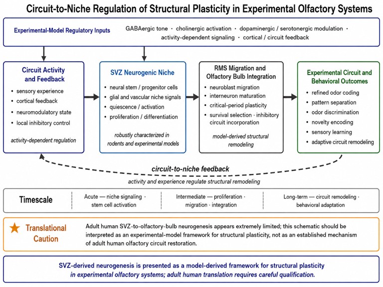
Figure 3. Circuit-to-niche regulation of structural plasticity in experimental olfactory systems. Circuit activity, cortical feedback, neuromodulatory signaling, and local niche conditions regulate SVZ stem-cell activation, proliferation, differentiation, migration, and olfactory bulb integration. Adult-born interneurons can support pattern separation, odor discrimination, novelty encoding, sensory learning, and circuit remodeling in experimental models. Because adult human SVZ-to-olfactory-bulb neurogenesis appears extremely limited, the pathway is presented as an experimental framework for structural plasticity rather than as an established mechanism of adult human olfactory restoration. This figure was created by the author for the present manuscript and was not reproduced or adapted from another source.
Translational interpretation
SVZ-derived olfactory neurogenesis is a robust mechanism of structural plasticity in rodents and other experimental mammalian models, but it should not be presented as an established restorative mechanism in adult humans. Its principal translational value lies in demonstrating how neural activity, neurotransmitter signaling, glial state, inflammation, metabolism, and niche biology can regulate structural remodeling and repair capacity across species and disease contexts. Pharmacological investigation of this pathway should therefore focus on general principles of niche regulation and structural plasticity rather than assume that robust adult human olfactory bulb neurogenesis can be therapeutically restored.
Integrated Framework for Chemosensory Plasticity
The evidence reviewed above supports an integrated model in which chemosensory plasticity emerges from interactions among circuit regulation, glial state, and structural remodeling. Cortical feedback rapidly modifies sensory gain, inhibition, temporal coordination, and context-dependent processing. Astrocytes and microglia regulate the synaptic, metabolic, and inflammatory conditions under which these circuit changes occur. In experimental systems where neurogenic remodeling remains active, neural stem cell-derived neurons provide a slower form of structural adaptation.
These mechanisms operate over different but overlapping timescales. Circuit modulation can occur rapidly, glial regulation can alter intermediate cellular, metabolic, and inflammatory conditions, and structural remodeling can support longer-term changes in circuit composition and function. Activity-dependent signaling, neuromodulatory pathways, metabolic coupling, inflammatory mediators, and niche-regulatory mechanisms connect these levels rather than allowing them to operate independently [6,8,9,45,55].
The effect of a drug will depend on the condition of the sensory circuit and its surrounding cellular environment. The same treatment may support adaptation in one setting but interfere with compensation or repair in another. Treatment strategies should therefore consider circuit integrity, glial and inflammatory conditions, metabolic support, disease stage, and whether the relevant form of structural remodeling is active in the experimental model or human tissue being studied.
The principal components and timescales of this framework are summarized in Table 1.
|
Component |
Principal function |
Predominant timescale |
Role in the integrated framework |
|
Cortical feedback |
Regulates sensory gain, inhibition, temporal coordination, and context-dependent processing [6,8,9,16,19,45,100] |
Rapid to intermediate |
Adjusts circuit activity according to experience, behavioral relevance, and internal state |
|
Astrocytes |
Regulate neurotransmitter handling, extracellular homeostasis, gliotransmission, and metabolic support [9,45,55,100] |
Rapid to intermediate |
Shape the synaptic and metabolic conditions under which circuit plasticity occurs |
|
Microglia |
Mediate immune surveillance, cytokine signaling, complement-associated tagging, and synaptic remodeling [58,101,102] |
Intermediate to long-term |
Influence circuit refinement, inflammatory state, and the balance between adaptive and maladaptive remodeling |
|
Neurogenic and structural remodeling in experimental systems |
Supports activity-dependent neuronal integration and longer-term changes in circuit composition [45,58,76,81,94,100–103] |
Long-term |
Provides a model of structural adaptation regulated by circuit activity, glial state, inflammation, metabolism, and niche biology; adult human extrapolation remains limited |
Neuropharmacological and Therapeutic Implications
The mechanisms reviewed above provide several possible targets for pharmacological intervention. At the circuit level, cholinergic, noradrenergic, dopaminergic, serotonergic, and GABAergic systems can alter cortical feedback, sensory gain, inhibition, attention, and learning. At the glial level, candidate targets include astrocytic glutamate transport, purinergic signaling, metabolic coupling, microglial cytokine responses, and complement-associated remodeling. In experimental neurogenic systems, neurotransmitter- and niche-regulatory pathways may also influence longer-term structural adaptation.
Circuit and neuromodulatory targets
Neuromodulatory systems provide pharmacological access to circuit-level plasticity. Cholinergic signaling can influence attention-dependent sensory processing and cortical feedback, whereas dopaminergic and noradrenergic pathways can modify salience, reward association, learning, and state-dependent responsiveness [66,104–108]. Serotonergic and GABAergic pathways may further regulate excitability and inhibitory–excitatory balance. Modulation of these systems could therefore affect odor and taste discrimination, sensory learning, predictive processing, and the recovery of sensory circuit function [40,109,110].
The effects of circuit-directed treatments are unlikely to be uniform. Their outcomes may depend on behavioral state, prior sensory experience, disease stage, and the integrity of inhibitory, glial, and metabolic support systems. In experimental olfactory models, neurotransmitter signaling can also regulate neural stem-cell activation, survival, and integration, providing a possible mechanism for longer-term structural adaptation [81,94,111]. However, this experimental mechanism should not be presented as an established method for restoring adult human olfactory bulb neurogenesis.
Glial, inflammatory, and metabolic targets
Astrocytic glutamate transport, GABA–glutamate balance, ATP and adenosine signaling, calcium-dependent gliotransmission, and lactate-mediated metabolic support represent candidate targets for modifying synaptic stability, circuit gain, and resilience [40,109,110]. Microglial cytokine signaling, inflammatory priming, complement-associated synaptic tagging, and activity-dependent pruning may influence whether circuits undergo adaptive refinement or maladaptive remodeling [49,64,112–114].
These pathways require context-sensitive modulation. Inflammation and glial activation are not uniformly harmful; they may support debris clearance, tissue repair, synaptic remodeling, and recovery under some conditions. Indiscriminate suppression could therefore interfere with beneficial responses, whereas excessive or persistent activation could destabilize circuit function. Similarly, interventions targeting metabolism may support circuit resilience but can also produce broad systemic effects. The therapeutic goal should be to restore glial, inflammatory, and metabolic conditions that support healthy sensory function rather than simply suppress these processes.
SIRT1 as a genetic and metabolic regulatory target
Genetic and epigenetic regulation may influence chemosensory plasticity by controlling the inflammatory, metabolic, and synaptic conditions under which sensory circuits adapt or recover. One candidate regulator is sirtuin 1 (SIRT1), an NAD+-dependent deacetylase encoded by the SIRT1 gene. SIRT1 links cellular energy status to transcriptional regulation and has been implicated in mitochondrial function, oxidative-stress responses, inflammatory signaling, autophagy, neuronal survival, and synaptic plasticity. These functions place SIRT1 at the intersection of several mechanisms already discussed in this review, including glial regulation, metabolic support, circuit resilience, aging, and neurodegeneration [115,116].
Direct evidence connecting SIRT1 to chemosensory recovery remains limited but is biologically relevant. In an experimental model of excitotoxic olfactory-bulb injury, recovery of olfactory function was associated with increased expression of SIRT1 and SIRT4 in the olfactory bulb. This association does not establish that increased sirtuin expression caused recovery, but it suggests that sirtuin-dependent metabolic and stress-response pathways may participate in tissue remodeling or functional restoration after olfactory injury [117]. SIRT1 may therefore provide a mechanistic link among metabolic state, inflammation, synaptic plasticity, and recovery-related circuit remodeling.
Pharmacological activation of SIRT1 has shown anti-inflammatory, antioxidant, metabolic, autophagic, and neuroprotective effects in several preclinical settings, whereas SIRT1 inhibition or loss of function can disrupt these protective pathways. However, the consequences of SIRT1 modulation are likely to depend on cell type, dose, timing, disease stage, and metabolic context. Current evidence does not establish that SIRT1 activators improve human olfactory or gustatory function, nor that SIRT1 inhibitors directly cause chemosensory impairment. SIRT1 should therefore be considered a candidate regulatory target for mechanistic investigation rather than a validated treatment for chemosensory dysfunction.
The principal candidate targets, potential outcomes, and translational cautions are summarized in Table 2.
|
Target domain |
Candidate pharmacological entry points |
Primary biological role |
Potential relevance to chemosensory plasticity or dysfunction |
Translational caution |
|
Neuromodulatory control of cortical feedback |
Cholinergic, dopaminergic, noradrenergic, serotonergic, and GABAergic signaling |
Regulates sensory gain, inhibitory–excitatory balance, attention, reward association, cortical feedback, and state-dependent plasticity |
May influence odor and taste discrimination, salience assignment, sensory learning, predictive processing, and recovery-related circuit recalibration |
Effects are likely context-dependent and may vary by disease stage, behavioral state, and circuit integrity |
|
Astrocytic neurotransmitter handling |
Glutamate uptake, GABA–glutamate balance, transporter activity, extracellular ion regulation |
Maintains synaptic stability, prevents excitotoxicity, and regulates neuronal excitability |
May affect sensory gain, discrimination, synaptic plasticity, and vulnerability to maladaptive circuit activity |
Excessive modulation could disrupt normal synaptic signaling or compensatory circuit adaptation |
|
Purinergic and gliotransmission pathways |
ATP/adenosine signaling, astrocytic calcium signaling, D-serine, glutamate release |
Couples glial activity to synaptic function, plasticity, and neuron–glia communication |
May regulate experience-dependent plasticity, adaptation, and local circuit responsiveness |
Purinergic signaling can support both adaptive repair and pathological inflammation depending on context |
|
Astrocyte–neuron metabolic support |
Glucose utilization, lactate signaling, mitochondrial support, activity-dependent energy supply |
Coordinates neuronal activity with metabolic demand and circuit resilience |
May influence plasticity under aging, metabolic stress, neurodegeneration, or post-injury recovery |
Metabolic interventions may have broad systemic effects and require disease-specific interpretation |
|
SIRT1-dependent genetic and metabolic regulation |
SIRT1 expression and activity; NAD+-dependent deacetylation; experimental SIRT1 activators or inhibitors |
Links cellular energy state with transcription, mitochondrial function, oxidative-stress responses, inflammation, autophagy, neuronal survival, and synaptic plasticity |
May influence metabolic resilience, inflammatory state, injury responses, synaptic remodeling, and recovery-related mechanisms in experimental chemosensory circuits |
Direct evidence in human olfactory or gustatory dysfunction is lacking; effects may depend on cell type, dose, timing, and disease stage |
|
Microglial inflammatory state |
Cytokine signaling, inflammatory priming, immune surveillance, microglial activation state |
Regulates immune tone, neuronal excitability, synaptic remodeling, and tissue response to injury |
May contribute to persistent chemosensory dysfunction, post-viral impairment, aging-related decline, and neurodegenerative sensory deficits |
Inflammation is not uniformly harmful; indiscriminate suppression may impair repair or adaptive remodeling |
|
Complement-associated synaptic remodeling |
Complement signaling, synaptic tagging, microglial pruning |
Refines circuit architecture by removing selected synaptic elements |
May influence circuit refinement, maladaptive pruning, sensory discrimination, and long-term network remodeling |
Excessive inhibition or activation may disrupt normal circuit maintenance and plasticity balance |
|
Species-dependent neurogenic and structural remodeling |
Neural stem cell niche regulation, neurotransmitter-dependent progenitor control, glial–niche interactions, inflammatory and metabolic regulation of structural plasticity |
Supports structural remodeling in experimental olfactory systems where SVZ-derived neurogenesis remains active |
Provides a model for long-term adaptation, circuit updating, and structural plasticity constraints across disease contexts |
Adult human SVZ-to-olfactory-bulb neurogenesis appears extremely limited; this should not be framed as an established human therapeutic restorative mechanism |
|
Multiscale combination approaches |
Pharmacological modulation combined with sensory training, behavioral intervention, metabolic support, or disease-specific treatment |
Coordinates circuit, glial, metabolic, inflammatory, and structural mechanisms |
May better support adaptive recalibration than targeting one mechanism alone |
Combination strategies require careful timing, patient selection, and mechanistic validation |
Context, timing, and combination strategies
The effect of a pharmacological intervention will depend on the origin, location, duration, and stage of chemosensory dysfunction. Olfactory or gustatory impairment may result from peripheral receptor or epithelial injury, altered central processing, inflammatory or metabolic disruption, or combinations of these mechanisms. The same target may therefore produce different outcomes depending on age, disease stage, inflammatory state, circuit integrity, and whether the underlying response is damaging or compensatory.
Combination approaches may be more effective than isolated pathway modulation. Pharmacological treatment could be combined with sensory training, behavioral intervention, metabolic support, or disease-specific therapy to promote recovery at both circuit and cellular levels. Such strategies will require careful timing, patient selection, and mechanistic validation to avoid increasing maladaptive plasticity or disrupting beneficial repair.
Implications for Chemosensory Dysfunction and Disease
Chemosensory dysfunction can arise from disruption at peripheral, circuit, glial, inflammatory, metabolic, and structural levels. The relative importance of these mechanisms differs across conditions, and the presence of the same sensory deficit does not necessarily indicate the same underlying biological process.
Post-viral and inflammatory dysfunction
Anosmia and hyposmia may involve altered olfactory bulb activity, reduced cortical feedback, disrupted synaptic organization, and changes in the local glial and inflammatory environment [6,8,30,118,119]. The resulting impairment can affect odor detection, discrimination, and adaptation, although the relative contributions of peripheral injury and central circuit dysfunction vary among patients and conditions.
Post-viral olfactory dysfunction, including that associated with SARS-CoV-2, illustrates this mechanistic complexity. Viral injury can disrupt peripheral sensory and supporting cells while also producing inflammatory changes that may affect central olfactory pathways [120–122]. Persistent anosmia, hyposmia, or distorted odor perception may therefore reflect incomplete peripheral repair, sustained inflammation, altered circuit activity, or interactions among these processes.
Aging and neurodegenerative disease
Aging is associated with declining odor sensitivity, discrimination, and adaptation. Age-related changes in synaptic regulation, glial activity, inflammatory tone, and metabolic support may reduce circuit responsiveness and resilience [123–125]. Reductions in SVZ-derived olfactory neurogenesis are well documented in experimental aging models, although their relevance to adult human olfactory decline remains uncertain [126–128].
Olfactory impairment frequently appears early in Parkinson’s disease and Alzheimer’s disease. In Parkinson’s disease, altered olfactory bulb circuitry and cortical processing occur alongside astrocytic and microglial activation and other inflammatory changes [129–134]. These alterations may impair odor identification, discrimination, and adaptive sensory processing.
In Alzheimer’s disease, disrupted olfactory pathway connectivity, altered network activity, and glial inflammatory responses may contribute to declining odor recognition [58,127,131–133,135–138]. Structural and neurogenic changes have also been reported in experimental models, but their functional importance and relevance to adult human chemosensory impairment remain incompletely resolved [58,123,127,130,131,139]. Olfactory dysfunction in neurodegenerative disease should therefore be interpreted as a feature of broader disease-associated circuit and cellular disruption rather than as evidence of one isolated mechanism.
SIRT1 may also connect aging and Alzheimer’s-related pathology with chemosensory circuit vulnerability. Experimental and broader neurodegenerative evidence indicates that SIRT1 can regulate inflammatory signaling, mitochondrial and metabolic function, oxidative stress, autophagy, synaptic plasticity, and pathways related to amyloid-β and tau biology [115,140]. These processes are relevant to olfactory dysfunction because aging and Alzheimer’s disease affect many of the same circuit, glial, inflammatory, and metabolic mechanisms discussed in this review. However, direct evidence showing that SIRT1 modulation prevents or reverses chemosensory impairment in patients with Alzheimer’s disease remains insufficient.
The major disease-related alterations are summarized in Table 3.
|
Condition |
Circuit-level alterations |
Glial / inflammatory features |
Species-dependent structural / neurogenic remodeling |
Functional chemosensory outcome |
|
Anosmia / hyposmia |
Altered olfactory bulb activity, reduced cortical feedback, disrupted synaptic organization [6,8] |
Local inflammatory responses and glial activation may impair circuit stability [7,141] |
Structural remodeling and repair-related processes may be secondarily affected depending on severity, duration, and site of dysfunction [10,122] |
Reduced odor detection, impaired discrimination, diminished sensory adaptation [120,121] |
|
Post-viral dysfunction / SARS-CoV-2 |
Peripheral sensory disruption with possible secondary central alterations in olfactory pathways [121,122] |
Prominent inflammatory and cellular disruption involving peripheral and central mechanisms [118,120] |
Potential disruption of reparative, inflammatory, and structural plasticity processes remains under investigation [118,122] |
Persistent anosmia, hyposmia, distorted odor perception, delayed sensory recovery [121,142] |
|
Aging |
Progressive decline in circuit plasticity, altered synaptic regulation, reduced adaptive responsiveness [123,125] |
Age-related changes in glial activity and inflammatory tone [58,125] |
Reduced structural plasticity and diminished SVZ-derived olfactory neurogenesis are documented in experimental models; adult human relevance remains uncertain [10,126] |
Decline in odor sensitivity, discrimination, and adaptation to novel sensory input [123,125,143] |
|
Parkinson’s disease |
Early alterations in olfactory bulb circuitry and cortical processing; impaired circuit-level plasticity [131,135,137] |
Increased microglial and astrocytic activation, neuroinflammatory disruption of local microenvironment [58,132] |
Impaired structural remodeling and altered neurogenic capacity have been reported mainly in experimental models, with human relevance requiring careful interpretation [127,137] |
Early olfactory dysfunction, impaired odor identification and discrimination [137,143] |
|
Alzheimer’s disease |
Disrupted olfactory pathway connectivity and altered network activity [129,144] |
Neuroinflammatory processes involving astrocytic and microglial responses [133,145,146] |
Structural plasticity and neurogenic processes may be altered, though their functional significance and relevance to adult human chemosensory dysfunction remain incompletely resolved [145,147] |
Early olfactory impairment, reduced odor recognition, progressive sensory decline [123,144] |
|
Metabolic / systemic disorders |
Altered neuronal excitability and impaired integration of sensory signals [50,148] |
Hormonal, metabolic, and inflammatory dysregulation affecting local circuit environment [50,58,148] |
Potential disruption of adaptive remodeling, metabolic support, and neurogenic niche regulation in experimental systems [10,149] |
Altered smell and taste perception, reduced sensory flexibility, impaired adaptive responses [143,148] |
Metabolic and systemic conditions
Metabolic and systemic disorders can affect olfactory and gustatory function through altered hormonal signaling, energy regulation, inflammation, and neuronal excitability [150–153]. These changes may impair sensory-signal integration, astrocyte–neuron metabolic support, and adaptive circuit responses. Effects on neurogenic-niche regulation have also been reported in experimental models, but their direct relevance to adult human chemosensory dysfunction requires careful interpretation [154–156].
Across these conditions, changes in circuit activity, glial state, inflammation, metabolism, and structural remodeling may function as causes, consequences, or compensatory responses. Their relative contribution is likely to vary with disease stage, age, and the location and duration of injury. Chemosensory dysfunction may therefore signal disruption across several biological levels rather than identify one specific cause.
Conclusion
Chemosensory plasticity cannot be explained by synaptic modification alone. The evidence reviewed here supports an integrated model in which cortical feedback regulates circuit activity, astrocytes and microglia shape the synaptic, metabolic, and inflammatory environment, and structural remodeling contributes to longer-term adaptation in experimental systems where these mechanisms remain active.
This framework identifies pharmacologically relevant targets at circuit, glial, inflammatory, metabolic, genetic, and niche-regulatory levels. SIRT1 illustrates how an NAD+-dependent regulatory pathway may connect metabolic state, inflammation, stress responses, synaptic plasticity, aging, and neurodegeneration, although its specific role in human chemosensory recovery remains unproven. The effects of all candidate interventions are likely to depend on disease stage, age, circuit integrity, and the surrounding cellular environment. Therapeutic strategies should therefore aim to restore adaptive sensory regulation rather than simply increase plasticity or suppress inflammation.
Adult SVZ-to-olfactory-bulb neurogenesis remains a valuable experimental model of structural plasticity but appears extremely limited as a restorative mechanism in adult humans. Future studies should link circuit manipulation, glial and inflammatory profiling, metabolic assessment, and behavioral measures to determine which mechanisms support recovery and which sustain chemosensory dysfunction.
Author Contributions
Moawiah M. Naffaa: Conceptualization, literature analysis, framework development, writing—original draft, and writing—review and editing.
Declarations
Funding
This research received no external funding.
Conflict of Interest
The author declares no conflict of interest.
Ethics statement
Ethical approval was not required for this review article because it did not involve new studies with human participants or animals performed by the author.
Data availability statement
No new data were generated or analyzed in this study.
Artificial intelligence generated content declaration
During preparation of this manuscript, the author used Google Gemini solely for language editing and improvement of phrasing. All scientific content, interpretation, organization, and final wording were reviewed and approved by the author, who takes full responsibility for the manuscript. No AI tool was used as an author or to replace the author’s intellectual contribution.
References
2. Kogan JF, Fontanini A. Learning enhances representations of taste-guided decisions in the mouse gustatory insular cortex. Curr Biol. 2024;34(9):1880–92 e5.
3. Dossat AM, Kokoska MM, Whitaker-Fornek JR, Sniffen SE, Kulkarni AS, Levitt ES, et al. Glucagon-Like Peptide-1 Receptors in the Gustatory Cortex Influence Food Intake. J Neurosci. 2023;43(23):4251–61.
4. Subramanian N, Leong LM, Salemi Mokri Boukani P, Storace DA. Recent odor experience selectively modulates olfactory sensitivity across the glomerular output in the mouse olfactory bulb. Chem Senses. 2025;50.
5. Bao S, Romero JM, Belfort BDW, Arenkiel BR. Signaling mechanisms underlying activity-dependent integration of adult-born neurons in the mouse olfactory bulb. Genesis. 2024;62(2):e23595.
6. Zak JD, Reddy G, Konanur V, Murthy VN. Distinct information conveyed to the olfactory bulb by feedforward input from the nose and feedback from the cortex. Nat Commun. 2024;15(1):3268.
7. Zhao D, Hu M, Liu S. Glial cells in the mammalian olfactory bulb. Front Cell Neurosci. 2024;18:1426094.
8. Hernandez DE, Ciuparu A, Garcia da Silva P, Velasquez CM, Rebouillat B, Gross MD, et al. Fast updating feedback from piriform cortex to the olfactory bulb relays multimodal identity and reward contingency signals during rule-reversal. Nat Commun. 2025;16(1):937.
9. Gomez-Sotres P, Skupio U, Dalla Tor T, Julio-Kalajzic F, Cannich A, Gisquet D, et al. Olfactory bulb astrocytes link social transmission of stress to cognitive adaptation in male mice. Nat Commun. 2024;15(1):7103.
10. Dejou J, Mandairon N, Didier A. Olfactory neurogenesis plays different parts at successive stages of life, implications for mental health. Front Neural Circuits. 2024;18:1467203.
11. Wang P, Li S, Li A. Odor representation and coding by the mitral/tufted cells in the olfactory bulb. J Zhejiang Univ Sci B. 2024;25(10):824–40.
12. Federman N, Romano SA, Amigo-Duran M, Salomon L, Marin-Burgin A. Acquisition of non-olfactory encoding improves odour discrimination in olfactory cortex. Nat Commun. 2024;15(1):5572.
13. Chen K, Kogan JF, Fontanini A. Spatially Distributed Representation of Taste Quality in the Gustatory Insular Cortex of Behaving Mice. Curr Biol. 2021;31(2):450.
14. Puche AC, Hook C, Zhou FW. Cell type-specific and frequency-dependent centrifugal modulation in olfactory bulb output neurons in vivo. J Neurophysiol. 2024;131(6):1226–39.
15. Bolding KA, Nagappan S, Han BX, Wang F, Franks KM. Recurrent circuitry is required to stabilize piriform cortex odor representations across brain states. Elife. 2020;9.
16. Terral G, Harrell E, Lepousez G, Wards Y, Huang D, Dolique T, et al. Endogenous cannabinoids in the piriform cortex tune olfactory perception. Nat Commun. 2024;15(1):1230.
17. Terrier C, Greco-Vuilloud J, Cavelius M, Thevenet M, Mandairon N, Didier A, et al. Long-term olfactory enrichment promotes non-olfactory cognition, noradrenergic plasticity and remodeling of brain functional connectivity in older mice. Neurobiol Aging. 2024;136:133–56.
18. A AD, Michelon F, Patella P, Petrucco L, Piasini E, Iurilli G. A mechanosensory feedback that uncouples external and self-generated sensory responses in the olfactory cortex. Cell Rep. 2024;43(4):114013.
19. Geng C, Li R, Li S, Liu P, Peng Y, Liu C, et al. Noradrenergic inputs from the locus coeruleus to anterior piriform cortex and the olfactory bulb modulate olfactory outputs. Nat Commun. 2025;16(1):260.
20. Gonzalez J, Torterolo P, Bolding KA, Tort ABL. Communication subspace dynamics of the canonical olfactory pathway. iScience. 2024;27(12):111275.
21. Schiff HC, Kogan JF, Isaac M, Czarnecki LA, Fontanini A, Maffei A. Experience-dependent plasticity of gustatory insular cortex circuits and taste preferences. Sci Adv. 2023;9(2):eade6561.
22. Gonzalez J, Torterolo P, Tort ABL. Mechanisms and functions of respiration-driven gamma oscillations in the primary olfactory cortex. Elife. 2023;12.
23. Okumura T, Kida I, Yokoi A, Nakai T, Nishimoto S, Touhara K, et al. Semantic context-dependent neural representations of odors in the human piriform cortex revealed by 7T MRI. Hum Brain Mapp. 2024;45(6):e26681.
24. Yu B, Yue Y, Ren C, Yun R, Lim B, Komiyama T. Cholinergic feedback for modality- and context-specific modulation of sensory representations. Science. 2025;388(6753):1324–9.
25. Boyd AM, Sturgill JF, Poo C, Isaacson JS. Cortical feedback control of olfactory bulb circuits. Neuron. 2012;76(6):1161–74.
26. Burton SD, Malyshko CM, Urban NN. Fast-spiking interneuron detonation drives high-fidelity inhibition in the olfactory bulb. PLoS Biol. 2024;22(8):e3002660.
27. Chae H, Banerjee A, Dussauze M, Albeanu DF. Long-range functional loops in the mouse olfactory system and their roles in computing odor identity. Neuron. 2022;110(23):3970–85 e7.
28. Chen Z, Padmanabhan K. Top-down feedback enables flexible coding strategies in the olfactory cortex. Cell Rep. 2022;38(12):110545.
29. Otazu GH, Chae H, Davis MB, Albeanu DF. Cortical Feedback Decorrelates Olfactory Bulb Output in Awake Mice. Neuron. 2015;86(6):1461–77.
30. Lindeman S, Fu X, Reinert JK, Fukunaga I. Value-related learning in the olfactory bulb occurs through pathway-dependent perisomatic inhibition of mitral cells. PLoS Biol. 2024;22(3):e3002536.
31. Gnaedinger A, Gurden H, Gourevitch B, Martin C. Multisensory learning between odor and sound enhances beta oscillations. Sci Rep. 2019;9(1):11236.
32. Norden F, Iravani B, Schaefer M, Winter AL, Lundqvist M, Arshamian A, et al. The human olfactory bulb communicates perceived odor valence to the piriform cortex in the gamma band and receives a refined representation back in the beta band. PLoS Biol. 2024;22(10):e3002849.
33. Garg R, Qiu Q, Yu CR. Basal forebrain cholinergic input mediates adaptive attention allocation to enhance olfactory discrimination. PLoS Biol. 2025;23(9):e3003374.
34. Jacobson GA, Rupprecht P, Friedrich RW. Experience-Dependent Plasticity of Odor Representations in the Telencephalon of Zebrafish. Curr Biol. 2018;28(1):1–14 e3.
35. Lyons SH, Gottfried JA. Predictive coding in the human olfactory system. Trends Cogn Sci. 2025;29(9):814–26.
36. Rajabi N, Zanettin I, Ribeiro AH, Vasco M, Bjorkman M, Lundstrom JN, et al. Exploring the feasibility of olfactory brain-computer interfaces. Sci Rep. 2025;15(1):18404.
37. Zelano C, Mohanty A, Gottfried JA. Olfactory predictive codes and stimulus templates in piriform cortex. Neuron. 2011;72(1):178–87.
38. Akinlaja YO, Nishiyama A. Glial modulation of synapse development and plasticity: oligodendrocyte precursor cells as a new player in the synaptic quintet. Front Cell Dev Biol. 2024;12:1418100.
39. Noriega-Prieto JA, Araque A. Sensing and Regulating Synaptic Activity by Astrocytes at Tripartite Synapse. Neurochem Res. 2021;46(10):2580–5.
40. Sanz-Galvez R, Falardeau D, Kolta A, Inglebert Y. The role of astrocytes from synaptic to non-synaptic plasticity. Front Cell Neurosci. 2024;18:1477985.
41. Andersen JV. The Glutamate/GABA-Glutamine Cycle: Insights, Updates, and Advances. J Neurochem. 2025;169(3):e70029.
42. Andersen JV, Schousboe A, Wellendorph P. Astrocytes regulate inhibitory neurotransmission through GABA uptake, metabolism, and recycling. Essays Biochem. 2023;67(1):77–91.
43. Limon ID, Angulo-Cruz I, Sanchez-Abdon L, Patricio-Martinez A. Disturbance of the Glutamate-Glutamine Cycle, Secondary to Hepatic Damage, Compromises Memory Function. Front Neurosci. 2021;15:578922.
44. Sardar D, Cheng YT, Woo J, Choi DJ, Lee ZF, Kwon W, et al. Induction of astrocytic Slc22a3 regulates sensory processing through histone serotonylation. Science. 2023;380(6650):eade0027.
45. Ung K, Huang TW, Lozzi B, Woo J, Hanson E, Pekarek B, et al. Olfactory bulb astrocytes mediate sensory circuit processing through Sox9 in the mouse brain. Nat Commun. 2021;12(1):5230.
46. Lei L, Wang YF, Chen CY, Wang YT, Zhang Y. Novel insight into astrocyte-mediated gliotransmission modulates the synaptic plasticity in major depressive disorder. Life Sci. 2024;355:122988.
47. Won W, Bhalla M, Lee JH, Lee CJ. Astrocytes as Key Regulators of Neural Signaling in Health and Disease. Annu Rev Neurosci. 2025;48(1):251–76.
48. Kim Y, Dube SE, Park CB. Brain energy homeostasis: the evolution of the astrocyte-neuron lactate shuttle hypothesis. Korean J Physiol Pharmacol. 2025;29(1):1–8.
49. Marty-Lombardi S, Lu S, Ambroziak W, Schrenk-Siemens K, Wang J, DePaoli-Roach AA, et al. Neuron-astrocyte metabolic coupling facilitates spinal plasticity and maintenance of inflammatory pain. Nat Metab. 2024;6(3):494–513.
50. Pang Y, Yang J, Liu J, Xie Z, Wang J. Metabolic interactions in the brain: the crucial roles of neurons, astrocytes, and microglia in health and disease. Front Neurosci. 2026;20:1731771.
51. Chamaa F, Magistretti PJ, Fiumelli H. Astrocyte-derived lactate in stress disorders. Neurobiol Dis. 2024;192:106417.
52. Acevedo A, Torres F, Kiwi M, Baeza-Lehnert F, Barros LF, Lee-Liu D, et al. Metabolic switch in the aging astrocyte supported via integrative approach comprising network and transcriptome analyses. Aging (Albany NY). 2023;15(19):9896–912.
53. Sun Y, Wang Y, Chen ST, Chen YJ, Shen J, Yao WB, et al. Modulation of the Astrocyte-Neuron Lactate Shuttle System contributes to Neuroprotective action of Fibroblast Growth Factor 21. Theranostics. 2020;10(18):8430–45.
54. Miguel-Quesada C, Zaforas M, Herrera-Perez S, Lines J, Fernandez-Lopez E, Alonso-Calvino E, et al. Astrocytes adjust the dynamic range of cortical network activity to control modality-specific sensory information processing. Cell Rep. 2023;42(8):112950.
55. Ogino T, Agetsuma M, Sawada M, Inada H, Nabekura J, Sawamoto K. Astrocytic activation increases blood flow in the adult olfactory bulb. Mol Brain. 2024;17(1):52.
56. Rosa JM, Aguilar J. Rethinking Sensory Information Processing: The Essential Role of Astrocytes. J Neurochem. 2025;169(6):e70113.
57. Duran Laforet V, Schafer DP. Microglia: Activity-dependent regulators of neural circuits. Ann N Y Acad Sci. 2024;1533(1):38–50.
58. Meller SJ, Greer CA. Olfactory Development and Dysfunction: Involvement of Microglia. Physiology (Bethesda). 2025;40(2):0.
59. Zhao S, Umpierre AD, Wu LJ. Tuning neural circuits and behaviors by microglia in the adult brain. Trends Neurosci. 2024;47(3):181–94.
60. Nandi S, de Deus JL, Faborode OS, Nandi S. Synaptic Pruning by Microglia: Lessons from Genetic Studies in Mice. Dev Neurosci. 2025;47(5):362–82.
61. Rogerson-Wood L, Sawatari A, Leamey CA. Microglia: Mediators of experience-driven corrective neuroplasticity. IBRO Neurosci Rep. 2025;19:91–100.
62. Grier BD, Belluscio L, Cheetham CE. Olfactory Sensory Activity Modulates Microglial-Neuronal Interactions during Dopaminergic Cell Loss in the Olfactory Bulb. Front Cell Neurosci. 2016;10:178.
63. Tillmon H, Soteros BM, Shen L, Cong Q, Wollet M, General J, et al. Complement and microglia activation mediate stress-induced synapse loss in layer 2/3 of the medial prefrontal cortex in male mice. Nat Commun. 2024;15(1):9803.
64. Yang G, Xu X, Gao W, Wang X, Zhao Y, Xu Y. Microglia-orchestrated neuroinflammation and synaptic remodeling: roles of pro-inflammatory cytokines and receptors in neurodegeneration. Front Cell Neurosci. 2025;19:1700692.
65. Pena-Ortega F. Microglial modulation of neuronal network function and plasticity. J Neurophysiol. 2025;133(2):661–80.
66. Faust TE, Lee YH, O'Connor CD, Boyle MA, Gunner G, Duran-Laforet V, et al. Microglia-astrocyte crosstalk regulates synapse remodeling via Wnt signaling. Cell. 2025;188(19):5212–30e21.
67. Khakh BS. On astrocyte-neuron interactions: Broad insights from the striatum. Neuron. 2025;113(19):3079–107.
68. Lines J, Corkrum M, Aguilar J, Araque A. The Duality of Astrocyte Neuromodulation: Astrocytes Sense Neuromodulators and Are Neuromodulators. J Neurochem. 2025;169(4):e70054.
69. Rasmussen RN, Asiminas A, Carlsen EMM, Kjaerby C, Smith NA. Astrocytes: integrators of arousal state and sensory context. Trends Neurosci. 2023;46(6):418–25.
70. Taylor CR, Tse V, Willoughby DD, Levesque M, Vaidyanathan TV, Paz JT, et al. Cortical astrocyte histamine-1-receptors regulate intracellular calcium and extracellular adenosine dynamics across sleep and wake. PLoS Biol. 2025;23(10):e3003376.
71. Naffaa MM. Glial-Dopamine crosstalk: Astrocytic and microglial gatekeepers of neuroinflammation, plasticity, and motivation. AIMS Neuroscience. 2026;13:64–118.
72. Gregory JD, Kunkhyen T, Sweat SC, Huang JS, Brechbill TR, Cheetham CEJ. New Neurons in the Postnatal Olfactory System: Functions in the Healthy and Regenerating Brain. Brain Sci. 2025;15(6):597.
73. Li K, Zheng Y, Cai S, Fan Z, Yang J, Liu Y, et al. The subventricular zone structure, function and implications for neurological disease. Genes Dis. 2025;12(3):101398.
74. Purvis EM, Garcia-Epelboim AD, Krizman EN, O'Donnell JC, Cullen DK. A three-dimensional tissue-engineered rostral migratory stream as an in vitro platform for subventricular zone-derived cell migration. Front Bioeng Biotechnol. 2024;12:1410717.
75. Shani-Narkiss H, Vinograd A, Landau ID, Tasaka G, Yayon N, Terletsky S, et al. Young adult-born neurons improve odor coding by mitral cells. Nat Commun. 2020;11(1):5867.
76. Tufo C, Cheah M, Lipovsek M, Byrne DJ, Kothandapani K, Browne LP, et al. Functional Maturation and Experience-Dependent Plasticity in Adult-Born Olfactory Bulb Dopaminergic Neurons. Eur J Neurosci. 2025;62(1):e70188.
77. Baig S, Nadaf J, Allache R, Le PU, Luo M, Djedid A, et al. Identity and nature of neural stem cells in the adult human subventricular zone. iScience. 2024;27(4):109342.
78. Nath S, Martinez Santamaria JC, Chu YH, Choi JS, Conforti P, Lin JD, et al. Interaction between subventricular zone microglia and neural stem cells impacts the neurogenic response in a mouse model of cortical ischemic stroke. Nat Commun. 2024;15(1):9095.
79. Quaresima S, Istiaq A, Jono H, Cacci E, Ohta K, Lupo G. Assessing the Role of Ependymal and Vascular Cells as Sources of Extracellular Cues Regulating the Mouse Ventricular-Subventricular Zone Neurogenic Niche. Front Cell Dev Biol. 2022;10:845567.
80. Alonso M, Viollet C, Gabellec MM, Meas-Yedid V, Olivo-Marin JC, Lledo PM. Olfactory discrimination learning increases the survival of adult-born neurons in the olfactory bulb. J Neurosci. 2006;26(41):10508–13.
81. Ferreira A, Constantinescu VS, Malvaut S, Saghatelyan A, Hardy SV. Distinct forms of structural plasticity of adult-born interneuron spines in the mouse olfactory bulb induced by different odor learning paradigms. Commun Biol. 2024;7(1):420.
82. Nakamura Y, Miwa T, Shiga H, Sakata H, Shigeta D, Hatta T. Histological changes in the olfactory bulb and rostral migratory stream due to interruption of olfactory input. Auris Nasus Larynx. 2024;51(3):517–24.
83. Alfonso J, Le Magueresse C, Zuccotti A, Khodosevich K, Monyer H. Diazepam binding inhibitor promotes progenitor proliferation in the postnatal SVZ by reducing GABA signaling. Cell Stem Cell. 2012;10(1):76–87.
84. Liu X, Wang Q, Haydar TF, Bordey A. Nonsynaptic GABA signaling in postnatal subventricular zone controls proliferation of GFAP-expressing progenitors. Nat Neurosci. 2005;8(9):1179–87.
85. Young SZ, Platel JC, Nielsen JV, Jensen NA, Bordey A. GABA(A) Increases Calcium in Subventricular Zone Astrocyte-Like Cells Through L- and T-Type Voltage-Gated Calcium Channels. Front Cell Neurosci. 2010;4:8.
86. Naffaa MM, Khan RR, Kuo CT, Yin HH. Cortical regulation of neurogenesis and cell proliferation in the ventral subventricular zone. Cell Rep. 2023;42(7):112783.
87. Naffaa MM, Yin HH. A cholinergic signaling pathway underlying cortical circuit activation of quiescent neural stem cells in the lateral ventricle. Sci Signal. 2024;17(855):eadk8810.
88. Hitoshi S, Maruta N, Higashi M, Kumar A, Kato N, Ikenaka K. Antidepressant drugs reverse the loss of adult neural stem cells following chronic stress. J Neurosci Res. 2007;85(16):3574–85.
89. Lao CL, Lu CS, Chen JC. Dopamine D3 receptor activation promotes neural stem/progenitor cell proliferation through AKT and ERK1/2 pathways and expands type-B and -C cells in adult subventricular zone. Glia. 2013;61(4):475–89.
90. O'Keeffe GC, Barker RA. Dopamine stimulates epidermal growth factor release from adult neural precursor cells derived from the subventricular zone by a disintegrin and metalloprotease. Neuroreport. 2011;22(18):956–8.
91. Hanson E, Swanson J, Arenkiel BR. Sensory experience shapes the integration of adult-born neurons into the olfactory bulb. J Nat Sci. 2017;3(8):e422.
92. Kelsch W, Lin CW, Mosley CP, Lois C. A critical period for activity-dependent synaptic development during olfactory bulb adult neurogenesis. J Neurosci. 2009;29(38):11852–8.
93. Li K, Figarella K, Su X, Kovalchuk Y, Gorzolka J, Neher JJ, et al. Endogenous but not sensory-driven activity controls migration, morphogenesis and survival of adult-born juxtaglomerular neurons in the mouse olfactory bulb. Cell Mol Life Sci. 2023;80(4):98.
94. Li WL, Chu MW, Wu A, Suzuki Y, Imayoshi I, Komiyama T. Adult-born neurons facilitate olfactory bulb pattern separation during task engagement. Elife. 2018;7:e33006.
95. Forest J, Moreno M, Cavelius M, Chalencon L, Ziessel A, Sacquet J, et al. Short-term availability of adult-born neurons for memory encoding. Nat Commun. 2019;10(1):5609.
96. Delgado AC, Maldonado-Soto AR, Silva-Vargas V, Mizrak D, von Kanel T, Tan KR, et al. Release of stem cells from quiescence reveals gliogenic domains in the adult mouse brain. Science. 2021;372(6547):1205–9.
97. Naffaa MM. Significance of the anterior cingulate cortex in neurogenesis plasticity: Connections, functions, and disorders across postnatal and adult stages. Bioessays. 2024;46(3):e2300160.
98. Breton-Provencher V, Lemasson M, Peralta MR 3rd, Saghatelyan A. Interneurons produced in adulthood are required for the normal functioning of the olfactory bulb network and for the execution of selected olfactory behaviors. J Neurosci. 2009;29(48):15245–57.
99. Bugeon S, Haubold C, Ryzynski A, Cremer H, Platel JC. Intrinsic Neuronal Activity during Migration Controls the Recruitment of Specific Interneuron Subtypes in the Postnatal Mouse Olfactory Bulb. J Neurosci. 2021;41(12):2630–44.
100. Wu A, Yu B, Chen Q, Matthews GA, Lu C, Campbell E, et al. Context-dependent plasticity of adult-born neurons regulated by cortical feedback. Sci Adv. 2020;6(42):eabc8319.
101. Ware K, Peter J, Yazell J, Thapa C, Taranov A, Bedolla A, et al. Inhibition of TGF-beta signaling in microglia stimulates hippocampal adult neurogenesis and reduces anxiety-like behavior in adult mice. Nat Commun. 2026;17(1):1440.
102. Zhou H, Wang L, Yang Y, Ye F, Zhao X, Zhu K, et al. Cathepsin B overexpression and lysosomal leakage in inflammatory microglia promote neuroinflammation in olfactory dysfunction by triggering mitochondrial dysfunction and pyroptosis. Brain Behav Immun. 2026;132:106188.
103. Pardillo-Diaz R, Perez-Garcia P, Ortego-Dominguez M, Gomez-Oliva R, Martinez-Gomez N, Dominguez-Garcia S, et al. The subventricular zone neurogenic niche provides adult born functional neurons to repair cortical brain injuries in response to diterpenoid therapy. Stem Cell Res Ther. 2025;16(1):1.
104. Bischoff S, Moyaert M, Clijsters M, Vanderbroek A, Van Gerven L. Treatment of COVID-19 Associated Olfactory Dysfunction: A Systematic Review. Curr Allergy Asthma Rep. 2024;25(1):2.
105. Kim S, Finlay JB, Ko T, Goldstein BJ. Long-term olfactory loss post-COVID-19: Pathobiology and potential therapeutic strategies. World J Otorhinolaryngol Head Neck Surg. 2024;10(2):148–55.
106. Brunert D, Rothermel M. Extrinsic neuromodulation in the rodent olfactory bulb. Cell Tissue Res. 2021;383(1):507–24.
107. Collins L, Francis J, Emanuel B, McCormick DA. Cholinergic and noradrenergic axonal activity contains a behavioral-state signal that is coordinated across the dorsal cortex. Elife. 2023;12.
108. Slater C, Liu Y, Weiss E, Yu K, Wang Q. The Neuromodulatory Role of the Noradrenergic and Cholinergic Systems and Their Interplay in Cognitive Functions: A Focused Review. Brain Sci. 2022;12(7):890.
109. Guedes JR, Ferreira PA, Costa JM, Cardoso AL, Peca J. Microglia-dependent remodeling of neuronal circuits. J Neurochem. 2022;163(2):74–93.
110. Rangel-Gomez M, Alberini CM, Deneen B, Drummond GT, Manninen T, Sur M, et al. Neuron-Glial Interactions: Implications for Plasticity, Behavior, and Cognition. J Neurosci. 2024;44(40):e1231242024.
111. Sanai N, Nguyen T, Ihrie RA, Mirzadeh Z, Tsai HH, Wong M, et al. Corridors of migrating neurons in the human brain and their decline during infancy. Nature. 2011;478(7369):382–6.
112. Ayyubova G, Fazal N. Beneficial versus Detrimental Effects of Complement-Microglial Interactions in Alzheimer's Disease. Brain Sci. 2024;14(5):434.
113. Dejanovic B, Wu T, Tsai MC, Graykowski D, Gandham VD, Rose CM, et al. Complement C1q-dependent excitatory and inhibitory synapse elimination by astrocytes and microglia in Alzheimer's disease mouse models. Nat Aging. 2022;2(9):837–50.
114. Shichkova P, Coggan JS, Markram H, Keller D. Brain Metabolism in Health and Neurodegeneration: The Interplay Among Neurons and Astrocytes. Cells. 2024;13(20):1714.
115. Mehramiz M, Porter T, O'Brien EK, Rainey-Smith SR, Laws SM. A Potential Role for Sirtuin-1 in Alzheimer's Disease: Reviewing the Biological and Environmental Evidence. J Alzheimers Dis Rep. 2023;7(1):823–43.
116. Thapa R, Moglad E, Afzal M, Gupta G, Bhat AA, Hassan Almalki W, et al. The role of sirtuin 1 in ageing and neurodegenerative disease: A molecular perspective. Ageing Res Rev. 2024;102:102545.
117. Marin C, Langdon C, Alobid I, Fuentes M, Bonastre M, Mullol J. Recovery of Olfactory Function After Excitotoxic Lesion of the Olfactory Bulbs Is Associated with Increases in Bulbar SIRT1 and SIRT4 Expressions. Mol Neurobiol. 2019;56(8):5643–53.
118. Martin-Lopez E, Brennan B, Mao T, Spence N, Meller SJ, Han K, et al. Inflammatory Response and Defects on Myelin Integrity in the Olfactory System of K18hACE2 Mice Infected with SARS-CoV-2. eNeuro. 2024;11(6):ENEURO.0106–24.2024.
119. Mou Y, Sun C, Wei S, Song X, Wang H, Wang Y, et al. P2X7 receptor of olfactory bulb microglia plays a pathogenic role in stress-related depression in mice with allergic rhinitis. Neurobiol Dis. 2024;192:106432.
120. Chang K, Zaikos T, Kilner-Pontone N, Ho CY. Mechanisms of COVID-19-associated olfactory dysfunction. Neuropathol Appl Neurobiol. 2024;50(2):e12960.
121. Simonini L, Frijia F, Ait Ali L, Foffa I, Vecoli C, De Gori C, et al. A Comprehensive Review of COVID-19-Related Olfactory Deficiency: Unraveling Associations with Neurocognitive Disorders and Magnetic Resonance Imaging Findings. Diagnostics (Basel). 2024;14(4):359.
122. Tsukahara T, Brann DH, Datta SR. Mechanisms of SARS-CoV-2-associated anosmia. Physiol Rev. 2023;103(4):2759–66.
123. Elhabbari K, Sireci S, Rothermel M, Brunert D. Olfactory deficits in aging and Alzheimer's-spotlight on inhibitory interneurons. Front Neurosci. 2024;18:1503069.
124. Greco-Vuilloud J, Midroit M, Terrier C, Forest J, Sacquet J, Mandairon N, et al. 12 months is a pivotal age for olfactory perceptual learning and its underlying neuronal plasticity in aging mice. Neurobiol Aging. 2022;114:73–83.
125. Xie Y, Wang S, Cha X, Li F, Xu Z, Wu J, et al. Aging and chronic inflammation: impacts on olfactory dysfunction-a comprehensive review. Cell Mol Life Sci. 2025;82(1):199.
126. Chaker Z, Makarouni E, Doetsch F. The Organism as the Niche: Physiological States Crack the Code of Adult Neural Stem Cell Heterogeneity. Annu Rev Cell Dev Biol. 2024;40(1):381–406.
127. Wang K, Liu XY, Liu SF, Wang XX, Wei YH, Zhu JR, et al. Rbm24/Notch1 signaling regulates adult neurogenesis in the subventricular zone and mediates Parkinson-associated olfactory dysfunction. Theranostics. 2024;14(11):4499–518.
128. Naffaa MM. Disruptions in Adult Neurogenesis: Mechanisms, Pathways, and Therapeutic Strategies for Cognitive Decline and Neurodegenerative Diseases in Aging. Nat Cell Sci. 2025;3(1):27–53.
129. Ballotta D, Casadio C, Tondelli M, Zanelli V, Ricci F, Carpentiero O, et al. The olfactory functional network in the Alzheimer's disease continuum: a resting state fMRI study. Front Aging Neurosci. 2025;17:1744413.
130. Cieri F, Giriprakash PP, Nandy R, Zhuang X, Doty RL, Caldwell JZK, et al. Functional connectivity differences of the olfactory network in Parkinson's Disease, mild cognitive impairment and cognitively normal individuals: A resting-state fMRI study. Neuroscience. 2024;559:8–16.
131. Ielo A, Bonanno L, Brunati C, Cannuli A, Basile GA, Dattola S, et al. Structural and functional connectomics of the olfactory system in Parkinson's disease: a systematic review. Parkinsonism Relat Disord. 2025;132:107230.
132. Kedzia D, Galita G, Majsterek I, Rozpedek-Kaminska W. Microglia, Astrocytes, and Oligodendrocytes in Parkinson's Disease: Neuroinflammatory Crosstalk and Emerging Therapeutic Strategies. Biomolecules. 2026;16(1):156.
133. Liu K, Aierken A, Liu M, Parhat N, Kong W, Yin X, et al. The decreased astrocyte-microglia interaction reflects the early characteristics of Alzheimer's disease. iScience. 2024;27(3):109281.
134. Naffaa MM. The Fate and Dynamics of Neural Stem Cells (NSCs) and Their Neurogenic Decline in Alzheimer’s Disease. Lifespan Development and Mental Health. 2025;1(2):10005.
135. Brosse S, Tremblay C, Merida I, Frasnelli J. Specific structural changes in Parkinson's disease-related olfactory dysfunction compared to others forms of olfactory dysfunction. Front Neural Circuits. 2024;18:1503841.
136. Peiris S, Ekanayake A, Lu J, Elyan R, Geesey K, Cottrill R, et al. Olfactory Network Functional Connectivity as a Marker for Parkinson's Disease Severity. Life (Basel). 2025;15(8):1324.
137. Abraham JN, Rawat D, Srikanth P, Sunny LP, Abraham NM. Alpha-synuclein pathology and Parkinson's disease-related olfactory dysfunctions: an update on preclinical models and therapeutic approaches. Mamm Genome. 2025;36(2):444–64.
138. He X, Huang T, Yang A, Li Y, Bai L, Chen Y, et al. Adult neurogenesis dysfunction in Parkinson's disease: Molecular pathology and functional implications. Pathol Res Pract. 2026;278:156340.
139. Li H, Qian J, Wang Y, Wang J, Mi X, Qu L, et al. Potential convergence of olfactory dysfunction in Parkinson's disease and COVID-19: The role of neuroinflammation. Ageing Res Rev. 2024;97:102288.
140. Zhang M, Tang Z. Therapeutic potential of natural molecules against Alzheimer's disease via SIRT1 modulation. Biomed Pharmacother. 2023;161:114474.
141. Leon M, Troscianko ET, Woo CC. Inflammation and olfactory loss are associated with at least 139 medical conditions. Front Mol Neurosci. 2024;17:1455418.
142. Kay LM. COVID-19 and olfactory dysfunction: a looming wave of dementia? J Neurophysiol. 2022;128(2):436–44.
143. Chen YN, Kostka JK. Beyond anosmia: olfactory dysfunction as a common denominator in neurodegenerative and neurodevelopmental disorders. Front Neurosci. 2024;18:1502779.
144. Meyer C, Niedermeier T, Feyen PLC, Strubing FL, Rauchmann BS, Karali K, et al. Early Locus Coeruleus noradrenergic axon loss drives olfactory dysfunction in Alzheimer's disease. Nat Commun. 2025;16(1):7338.
145. Fruholz I, Meyer-Luehmann M. The intricate interplay between microglia and adult neurogenesis in Alzheimer's disease. Front Cell Neurosci. 2024;18:1456253.
146. Hashimoto K, Gotoh M, Ikeshima-Kataoka H. Astrocytic and microglial cell functions in neuroinflammatory diseases and their animal models. Front Cell Neurosci. 2025;19:1708775.
147. Alonso M, Petit AC, Lledo PM. The impact of adult neurogenesis on affective functions: of mice and men. Mol Psychiatry. 2024;29(8):2527–42.
148. Kim YK, Jo D, Choi S, Song J. High-fat diet triggers transcriptomic changes in the olfactory bulb. Heliyon. 2025;11(3):e42196.
149. Pecoraro S, Verkerke M, Sluijs JA, van Het Hof B, van der Pol SMA, van Strien ME, et al. Adult human subventricular zone microglia promote a pro-neurogenic niche for neuronal progenitors in Parkinson's disease. Brain Behav Immun. 2025;129:318–34.
150. Kim DH, Wang M, Kim S, Jang DW, Ko T, Goldstein BJ. Strategies to Develop Regenerative Medicine Approaches for Olfactory Disorders. Clin Exp Otorhinolaryngol. 2025;18(3):204–9.
151. Yi KI, Park JH, Kim SD, Mun SJ, Cho KS. Stem Cells and Cell-Free Therapies for Olfactory Epithelium Regeneration: Insights from Experimental Models. Int J Mol Sci. 2025;26(18):9024.
152. De Cleene N, Schwarzova K, Labrecque S, Cerejo C, Djamshidian A, Seppi K, et al. Olfactory dysfunction as potential biomarker in neurodegenerative diseases: a narrative review. Front Neurosci. 2024;18:1505029.
153. Ekstrom I, Vetrano DL, Valletta M, Ruane R, Larsson M, Fredolini C, et al. Blood-based biomarkers of Alzheimer's disease and olfactory decline over 15 years in older adults. Geroscience. 2025.
154. Iravani B, Frasnelli J, Arshamian A, Lundstrom JN. Metabolic state modulates neural processing of odors in the human olfactory bulb. Biol Psychol. 2024;187:108770.
155. Lee DH, Song J. Impaired olfactory system in metabolic imbalance-related neuropathology. Life Sci. 2024;355:122967.
156. Nettore IC, Palatucci G, Ungaro P, Scida G, Corrado A, De Vito R, et al. Flavor and taste recognition impairments in people with type 1 diabetes. Nutr Diabetes. 2024;14(1):57.