

Abstract
Background: Mast cell activation syndrome (MCAS) is a heterogeneous disorder with diverse clinical manifestations, making diagnosis and management challenging. Pediatric MCAS remains poorly characterized, and the applicability of current diagnostic criteria in children is uncertain. Our objective is to evaluate the clinical history, laboratory findings, intestinal biopsy results, and treatment patterns of pediatric patients with a clinical diagnosis of MCAS, and to identify features that may improve diagnosis and management.
Methods: In this retrospective study, we reviewed patients younger than 18 years with MCAS documented in clinical notes from the Massachusetts General Hospital and Brigham and Women’s Hospital databases. Clinical history, symptoms, laboratory data, and intestinal mucosal biopsy reports were collected and analyzed. Symptoms were grouped by category, and incidence rates were calculated.
Results: Many pediatric patients with a clinical diagnosis of MCAS did not meet the current elevated tryptase cutoff of 11.2 ng/mL, yet showed multisystem symptoms consistent with histamine release and responded clinically to MCAS treatment. Cutaneous and gastrointestinal symptoms were most common (>70%). Many patients also had increased intestinal mucosal mast cells on CD117 staining despite tryptase levels below 11.2 ng/mL. Common treatments included H1/H2 antihistamines, cromolyn, and ketotifen, with benefit reported in 95% of cases.
Conclusion: The current tryptase cutoff for MCAS diagnosis may not be optimal for children. CD117 staining of intestinal mucosal biopsies may aid diagnosis. Clinical manifestations and treatment patterns in pediatric MCAS appear similar to those reported in adults.
Keywords
HATS, Mast cell activation syndrome, Tryptase, Pediatric cohort, Diagnosis, Symptomology, CD117+ mast cells
Introduction
Mast Cell Activation Syndrome (MCAS) is characterized by multi-system symptoms associated with mediators released by mast cells [1–10]. Patients with MCAS often present with recurrent flares of symptoms, and their quality of life is often negatively impacted [1]. It can affect up to 17% of general population [11]. Current diagnostic criteria are developed primarily through adult studies [1], and may not adequately capture the unique presentations seen in pediatric populations.
Serum tryptase serves as a crucial biomarker in the diagnosis and monitoring of mast cell disorders. In healthy individuals, normal tryptase levels are generally under 11.2 ng/mL [1,3,10,11–13].
Mast cells are a class of immune cells responsible for mediating a variety of allergies and diseases, including MCAS [14,15]. These cells release inflammatory mediators, such as histamine and tryptase, during an allergic reaction and MCAS flares [10,14,15]. Mast cells commonly reside in tissues [14,15]. The level of no more than 20 cells per High Power Field (HPF) in the human intestine is currently considered as normal [7,16–18]. Quantification of intestinal mucosa mast cells is currently not included in the diagnosis of MCAS.
The current consensus criteria for MCAS diagnosis require evidence of multi-system involvement consistent with histamine release, elevation of serum tryptase above 11.2 ng/mL or increment value above 20% baseline plus 2 ng/mL, and positive response to anti-histamine mediator therapy [3,6,19–21]. However, this threshold presents particular challenges in the pediatric population of MCAS, where baseline levels may naturally differ from those of adults [22]. From other studies, tryptase levels were lower than the current consensus [23].
Because of the significant knowledge gap between adults and children with MCAS, the current study aims to exam a cohort of patients under 18 years of age who present with a clinical diagnosis of MCAS. By analyzing this cohort, we aim to provide tools for recognizing and diagnosing these conditions in children, potentially leading to improved management. The findings may help establish more appropriate diagnostic criteria for pediatric populations and improve diagnostic accuracy and management.
Methods
We retrospectively reviewed the charts for a list of patient medical records obtained from the Research Patient Data Registry (RPDR), an electronic database including patients from the Massachusetts General Hospital and Brigham and Women’s Hospital systems. We analyzed patients who had initially visited either hospitals for MCAS and related disorders when they were 18 years or younger. The study was approved by the MGB Institutional Review Board under the protocol 2023P000509. Consent was not required for this study.
We reviewed demographic information, laboratory test results, clinical symptoms, allergy history, CD117+ mast cells in the intestinal mucosa biopsies, and family history from their encounter notes in our electronic patient medical records system. We excluded genetically confirmed HATS patients from the patient cohort.
Family history, allergies, and demographic information were collected. Tryptase results, CD117 immunostaining of intestinal mucosa biopsies, and KIT mutation results were obtained upon reviewing the charts. For the tryptase results, we took the lowest and the highest value recorded. Symptoms were collected by reviewing the patients’ charts, starting from the chart where MCAS was first suspected. We reviewed the charts for improvement with treatment from Histamine 1 Receptor Antagonist (H1RA), Histamine 2 Receptor Antagonist (H2RA), cromolyn, ketotifen, or other medications. CD117+ tissue mast cell counts were retrieved from the pathology reports of patients’ charts.
Analysis was conducted using R Version 4.3.0 (package: tidyverse 2.0.0). Symptoms were classified into 7 categories: gastrointestinal, cutaneous, psychological, neurological, anaphylactic, autonomic, and connective tissue related.
Results
From the 202 records identified by RPDR, 122 were included for having MCAS but no HATS in their problem list. 79 records were excluded for having insufficient data, lack of supporting evidence for a diagnosis of MCAS, or a diagnosis of HATS. The demographics of our population is presented in Table 1, 69% of the patients were female, white race predominated (90.98%), the median age at symptoms onset was 1 year, and the median age at diagnosis was 7.54 years. Of the 17 patients with available KIT D816V mutation testing, 0 were positive.
|
n (%) |
|
|
Female |
69 (56%) |
|
Race |
|
|
White |
111 (90.98%) |
|
Asian |
5 (4.10%) |
|
Hispanic |
2 (1.64%) |
|
Unavailable |
4 (3.28%) |
|
Median age at symptom onset [range] (years) |
1 [0.01–17.17] |
|
Median age at diagnosis [range] (years) |
7.54 [0.33–18.00] |
|
Mode of Delivery |
|
|
Vaginal delivery |
84 (68.85%) |
|
Unavailable |
27 (22.13%) |
|
Gestational Age at Birth |
|
|
Full-term |
84 (68.85%) |
|
Unavailable |
25 (20.49%) |
We found that a family history of IgE-mediated food allergy, gastroesophageal reflux or irritable bowel syndrome, asthma, and allergic rhinitis was most common in our cohort (34.42%, 27.05%, 27.05%, 22.13% respectively) (Figure 1A). Only 9% of the patients had a family history of MCAS. Among the family history of IgE-mediated food allergies, milk/dairy was the most common (29.51%) (Figure 1B).
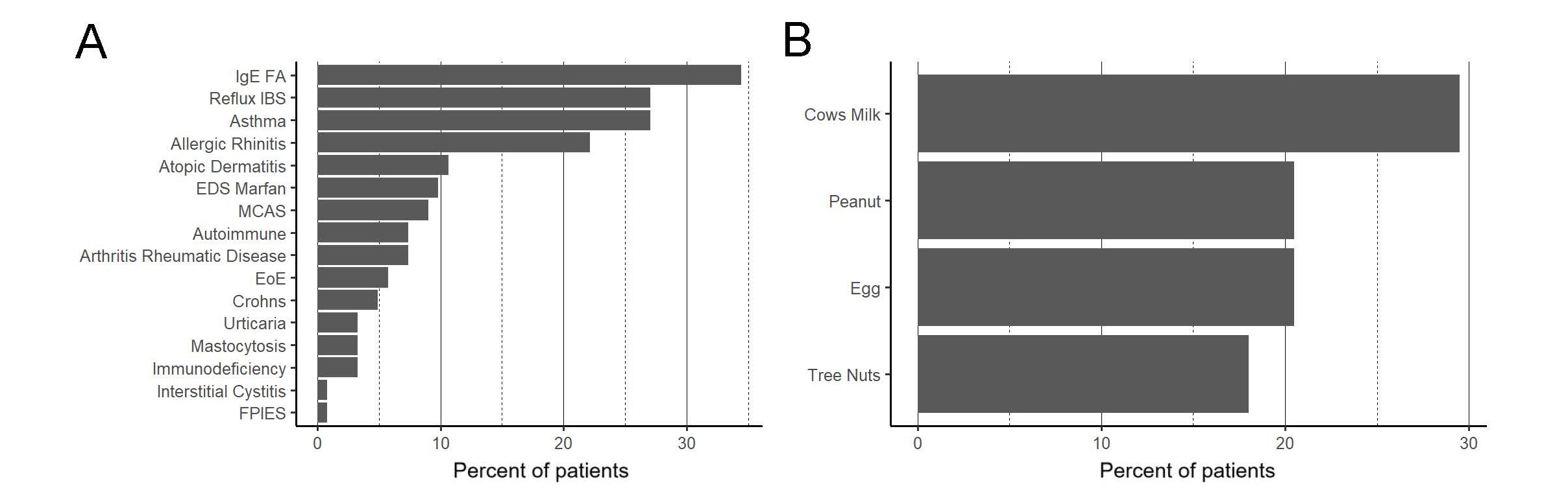
Figure 1. Family history and rates of allergies for our cohort. IgEFA: IgE-Mediated Food Allergy; IBS: Irritable Bowel Syndrome; EDS: Ehlers-Danlos Syndrome; MCAS: Mast Cell Activation Syndrome; EoE: Eosinophilic Esophagitis; FPIES: Food Protein-Induced Enterocolitis Syndrome.
Laboratory test results
Of our 122 patient cohort, 109 had serum tryptase testing. Of these 109, 64 (58.72%) had their highest recorded tryptase under 5.7 ng/mL, 34 (31.19%) with serum tryptase level between 5.7–11.2 ng/mL, 9 (8.26%) with serum tryptase level above 11.2 ng/mL, and 2 (1.83%) with serum tryptase level above 15 (Figure 2).
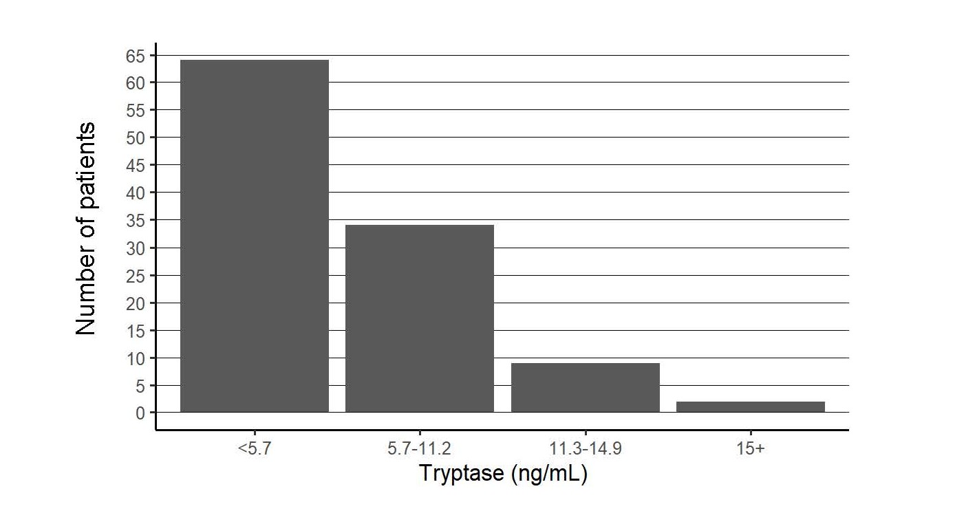
Figure 2. Peak tryptase results for all patients. Results are binned into ranges and plotted as a total count.
Of our cohort, 21 patients (17.2%) had information on tissue CD117+ mast cell immunostaining in some combination of regions of the intestinal mucosa biopsies. 90.48% of stains in the duodenum were elevated (>20 cells per HPF), 75.00% of stains in the stomach antrum were elevated (>20 cells per HPF), and 50.00% of stains in the GE junction were elevated (>20 cells per HPF) (Table 2).
|
Count per HPF (High Power Field) |
Number of Subjects with Peak Count… |
Number of Subjects with Average Count… |
||||
|
Duodenum |
Stomach Antrum |
Gastro- esophageal Junction |
Duodenum |
Stomach Antrum |
Gastro- esophageal Junction |
|
|
≤20 |
2 |
4 |
3 |
2 |
3 |
1 |
|
21 to 49 |
11 |
7 |
1 |
7 |
7 |
0 |
|
≥50 |
8 |
5 |
2 |
5 |
1 |
1 |
Of the 64 (58.72%) patients with a serum tryptase less than 5.7 ng/mL, 11 had mast cell staining. Ten of these patients had tissue CD117+ mast cells over 20 per HPF.
Other results, such as serum histamine, chromogranin A, serotonin, methylmalonic acid, prostaglandin, folate, absolute eosinophils, and urine N-methylhistamine or prostaglandin were too few to be meaningfully analyzed.
Clinical symptoms
In our cohort, all patients have had multi-system symptoms. Among the multi-system symptoms, we found that Gastrointestinal (GI) and cutaneous symptoms were most common (114 subjects, 93.4% and 85 subjects, 69.7%), respectively (Figure 3). The most common GI conditions included irritable bowel syndrome, gastroesophageal reflux, abdominal pain, poor weight gain, constipation, diarrhea, and Small Intestinal Bacterial Overgrowth (SIBO). The most common cutaneous symptoms included flushing, pruritus, urticaria, and angioedema. Seventy-seven (63.6%) had psychiatric/psychological food refusal. Sixty-six (54.1%) had neurological diagnoses, including sleep disturbance and headache. Thirty-four (38.6%) had dysautonomia, including postural orthostatic tachycardia syndrome and temperature sensitivity. Thirty-five (28.9%) had connective tissue disorders, including Ehlers-Danlos syndrome and joint hypermobility. Thirty (24.6%) experienced anaphylaxes. Of the 122 subjects, 22 (18.96%) had eosinophilic esophagitis.
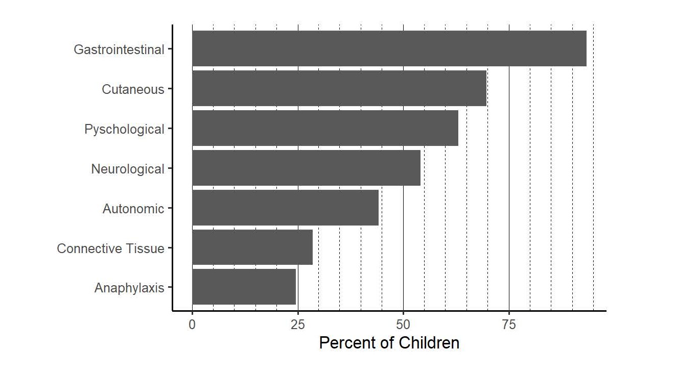
Figure 3. Symptoms subjects experienced starting from the age of symptom onset.
Medication management
In our cohort, the commonly prescribed medications for managing MCAS were antihistamines (H1RA or H2RA), cromolyn, and ketotifen. Out of 122 subjects 116 (95.08%) have demonstrated good clinical response with lessened or resolution of symptoms with some form of combination of these medications (Figure 4). Twenty (16.4%) were managed with antihistamines alone, 33 (27.0%) with antihistamines and cromolyn, 12 (9.8%) with cromolyn alone, 4 (3.3%) with ketotifen alone, and 30 (24.6%) with combined antihistamines, cromolyn and ketotifen. Six (0.05%) subjects did not improve on any of these medications.
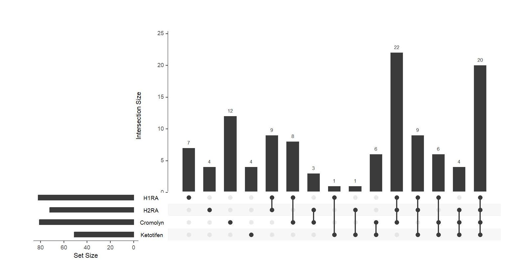
Figure 4. The combination of medications subjects was taken to manage their symptoms with clinical response. Set size (horizontal bars) indicates the total number of patients with each individual condition. Intersection size (vertical bars) indicates the number of patients with each combination of conditions indicated by the dots. H1RA: Histamine Type 1 Receptor Antagonist; H2RA: Histamine Type 2 Receptor Antagonist.
Discussion
To the best of our knowledge, this is the first retrospective chart review conducted on patients with MCAS on a strictly under-18 population. We find that many of these patients do not meet the current diagnostic criteria for MCAS because of the lower serum tryptase levels but have many of the same symptoms as adults with MCAS. Most of this cohort improves on many of the same medications used to treat MCAS in adults. Therefore, we would like to focus our discussion on the diagnostic criteria.
Serum tryptase level is one of the main elements in diagnosing MCAS based on current clinical practice guidelines [3,6,19,20] which is largely based on studies in adults. In our cohort, the tryptase values do not significantly differ between the patients, and many patients (89.91%) have “normal” tryptase values based on the current cut-off of 11.2 ng/mL. MCAS diagnosis is complicated by these findings, as over half of our patients did not meet the tryptase criterion despite fulfilling all the other criteria (response to mast cell directed medications and multi-system clinical symptoms). This raises potentially two possibilities: first, the patients with normal tryptase levels do not actually have MCAS but their clinical symptoms improved with MCAS treatment; or second, the threshold for serum tryptase level in the pediatric MCAS population needs to be lowered. The latter is supported by our findings on pediatric HATS patients (manuscript submitted) and implicated by Castells and colleagues [10], where the tryptase level as low as 6.8 ng/L is significant.
In the six subjects that did not improve on any MCAS medications, two subjects had elevated tryptase and elevated mast cells. Among the other four, two had normal tryptase but elevated mast cells. The last two patients had normal tryptase and no mast cell staining. These results indicate the complex heterogeneity of MCAS in the pediatric population.
It is a clinical dilemma about the accurate diagnosis when we care for these children who present with a multi-system of symptoms related to histamine release, and demonstrating good clinical responses to empiric MCAS treatment, but the serum tryptase levels are below 11.2 ng/mL. A thorough investigation to rule out other medical conditions is crucial for making an accurate diagnosis and to prevent over-medicalization or over diagnosis. At the same time, re-evaluation of the diagnostic criteria in pediatric patients with suspected MCAS, and a careful trial of MCAS treatment (diagnostic therapy) in those patients could provide insights in facilitating the diagnosis, management, and greatly improving the children’s quality of life.
Lastly, MCAS and HATS are likely interrelated conditions, although the precise mechanism of the relationship is still unknown. Clinical symptoms and management for both MCAS and HATS are essentially identical. But, for HATS, the diagnosis is more straightforward because of the identifiable genetic mutation. Whereas it is more challenging for MCAS because serum tryptase level plays a major part in the diagnosis based on current practice guidelines. However, results from our pediatric HATS cohort, the serum tryptase level at 6.8 ng/ml is significant in regard to the presence of HATS mutation. With this, for pediatric MCAS patients, the threshold for serum tryptase level should be lower, between 5.7–6.8 ng/ml based on our results, to prevent underdiagnosis of MCAS in the pediatric population.
The strength of the study includes a large-scale retrospective chart review, detailed analyses of serum tryptase levels and other laboratory tests, the clinical responses to MCAS treatment, and the inclusion of the information on tissue mast cell count. The limitations of the study include single centered study, and the number of study patients is relatively small. A large-scale prospective study with a healthy control group is needed.
Conclusion
In conclusion, we report on the clinical presentations in pediatric patients with suspected MCAS. Our results raised two important questions: first, the criterion for serum tryptase level in pediatric MCAS patients should be redefined, as compared to the current value of 11.2 ng/mL; and second, tissue CD117+ mast cell numbers of GI mucosa biopsies should be considered in diagnosing MCAS in children.
Acknowledgement
The authors would like to thank Dr. Jonathon N. Glickman for his assistance in immunochemical staining of the tissue CD117+ mast cells.
Conflict of Interest
No source of conflicts of interest for this research reported in this manuscript.
Funding
Qian Yuan received personal fee from the Nutricia North America as a consultant. The rest of authors declare no source of funding.
References
2. Hamilton MJ, Hornick JL, Akin C, Castells MC, Greenberger NJ. Mast cell activation syndrome: A newly recognized disorder with systemic clinical manifestations. J Allergy Clin Immunol. 2011;128(1):147–52e2.
3. Valent P, Akin C, Arock M, Brockow K, Butterfield JH, Carter MC, et al. Definitions, criteria and global classification of mast cell disorders with special reference to mast cell activation syndromes: a consensus proposal. Int Arch Allergy Immunol. 2012;157(3):215–25.
4. Lyons JJ. Hereditary Alpha Tryptasemia: Genotyping and Associated Clinical Features. Immunol Allergy Clin North Am. 2018;38(3):483–95.
5. Hamilton MJ. Nonclonal Mast Cell Activation Syndrome: A Growing Body of Evidence. Immunol Allergy Clin North Am. 2018 Aug;38(3):469–81.
6. Lyons JJ, Chovanec J, O’Connell MP, Liu Y, Selb J, Zanotti R, et al. Heritable risk for severe anaphylaxis associated with increased a-tryptase-encoding germline copy number at TPSAB1. J Allergy Clin Immunol 2021;147:622–32.
7. Weinstock LB, Pacc LA, Rezaie A, Afrin LB, Molderings GJ. Mast Cell Activation Syndrome: A Primer for the Gastroenterologist. Dig Dis Sci 2021 Apr;66(4):965–82.
8. Valent P, Hartmann K, Bonadonna P, Gulen T, Brockow K, Alvarez-Twose I, et al. Global Classification of Mast Cell Activation Disorders: An ICD-10-CM-Adjusted Proposal of the ECNM-AIM Consortium. J Allergy Clin Immunol Pract. 2022;10(8):1941–50.
9. Hamilton MJ. Mast Cell Activation Syndrome and Gut Dysfunction: Diagnosis and Management. Curr Gastroenterol Rep. 2024 Apr;26(4):107–14.
10. Castells M, Giannetti MP, Hamilton MJ, Novak P, Pozdnyakova O, Nicoloro-SantaBarbara, et al. Mast cell activation syndrome: Current understanding and research needs. J Allergy Clin Immunol. 2024;154(2):255–63.
11. Afrin LB, Ackerley MB, Bluestein LS, Brewer JH, Brook JB, Buchanan AD, et al. Diagnosis of mast cell activation syndrome: a global "consensus-2". Diagnosis (Berl). 2020 Apr 22;8(2):137–52.
12. Theoharides TC, Valent P, Akin C. Mast cells, mastocytosis, and related disorders. N Engl J Med 2015;373:163–72.
13. Akin C. Mast cell activation disorders. J Allergy Clin Immunol Pract 2014;2: 252–7e1.
14. Metcalfe DD, Baram D, Mekori YA. Mast cells. Physiol Rev. 1997 Oct;77(4):1033–79.
15. Metcalfe DD. Mast cells and mastocytosis. Blood 2008 Aug 15;112(4):946–56.
16. Iding J, Agarwal P, Zhang S, Odin R, Bernstein JA, Zimmermann N. Standardized Quantification of Mast Cells in the Gastrointestinal Tract in Adults. Allergy Clin Immunol Pract 2024 Feb;12(2):472–81.
17. Genta RM, Turner KO, Collins MH, Wechsler JB, Arva NC, Pletneva MA, et al. Quantification of Mucosal Mast Cells in the Gastrointestinal Tract: A Primer for Practicing Pathologists. Arch Pathol Lab Med 2024 Feb 1;148(2):e25–35.
18. Rizvi SA, Oriala C, Thomas J, Li S, Smadi Y. Identifying Mast Cells in Gastrointestinal Biopsies in Pediatric Irritable Bowel Patients. J Pediatr Gastroenterol Nutr 2022 Nov 1;75(5):572–77.
19. Valent P, Akin C, Bonadonna P, Hartmann K, Brockow K, Niedoszytko M, et al. Proposed diagnostic algorithm for patients with suspected mast cell activation syndrome. J Allergy Clin Immunol Pract 2019;7:1125–33e1.
20. Lee E, Picard M. Diagnosis and management of mast cell activation syndrome (MCAS) in Canada: a practical approach. Allergy Asthma Clin Immunol. 2025 Nov 21;21(1):49.
21. Lobbes H, Reynaud Q, Mainbourg S, Lega JC, Durieu I, Durupt S. Dosage de la tryptase : un guide d'utilisation pour le clinicien [Tryptase: A practical guide for the physician]. Rev Med Internet. 2020;41(11):748–55.
22. Sahiner UM, Yavuz ST, Buyuktiryaki B, Cavkaytar O, Arik Yilmaz E, Tuncer A, et al. Serum basal tryptase levels in healthy children: correlation between age and gender. Allergy Asthma Proc. 2014 Sep-Oct;35(5):404–8.
23. Puel M, Rossignol J, Devin C, Madrange M, Diep A, Roland PN, et al. CEREMAST network. Redefining tryptase norms in the pediatric population reveals sex-based differences: Clinical implications. Allergy. 2025 Jan;80(1):335–8.