

Abstract
Cardiac CT angiography (CCTA) has become a mainstream and integral part of many cardiology practices based on a vast base of literature supporting and validating its clinical utility. It is now recommended as a first-line test for the evaluation of chest pain according to the American Heart Association (AHA) and the American College of Cardiology (ACC). As the technology continues to advance, coronary imaging has improved in stride. Unfortunately, despite an arsenal of tests available to detect clinically significant coronary artery disease, many people continue to suffer acute myocardial infarction (MI) and other acute coronary syndromes, leading to significant morbidity and mortality due to unstable coronary artery disease (CAD). These unstable, “vulnerable” or high-risk plaques (HRP) continue to plague cardiologists across the globe. The ability to identify HRP has been a step in the right direction increasing more aggressive therapy. A large body of evidence supporting the utility of CCTA in plaque imaging has emerged and has defined characteristics of HRPs and their relationship to future coronary events. The potential of CCTA plaque imaging exists to not only identify HRP that can lead to more aggressive therapy, but to observe the transformation to stable plaque through follow-up CCTA imaging.
Keywords
Cardiac Computed Tomography Angiography (CCTA), Low attenuation plaque (LAP), Calcified coronary Plaque (CP), Non-calcified coronary plaque (NCP), Artificial intelligence (AI), Plaque Imaging, PCI planning
Abbreviations
AI: Artificial Intelligence; AI-QCT: Artificial Intelligence-guided Quantitative Computed Tomography; AI-QCPA: Artificial Intelligence-guided Quantified Coronary Plaque Analysis; CP: Calcified Plaque; CCTA: Cardiac Computed Tomography Angiography; CAD: Coronary Artery Disease; DLM: Deep-Learning Model; HRP: High-Risk Plaque; IVUS: Intravascular Ultrasound; ICA: Invasive Coronary Angiography; LAP: Low-Attenuation Plaque; MI: Myocardial Infarction; NCP: Non-Calcified Plaque; OCT: Optical Coherence Tomography; PCI: Percutaneous Coronary Intervention; PAV: Plaque Area Volume
Introduction
“The majority of people destined to die suddenly will not have a positive exercise test. The likely reason that they will die suddenly is that only a mild, non-flow-limiting coronary plaque will have been present before the sudden development of an occlusive thrombus [1].” These words from Dr. Stephen Epstein in 1989 still hold true today nearly 37 years later. The goal of creating a world free of MIs remains with numerous advances in imaging, pharmaceutical therapy, and intervention. Despite several risk factor scoring systems, advances in medical therapy and revascularization, CAD remains the leading cause of mortality and morbidity in the world. The prevalence of acute MI in the United States is approximately 7,900,000. There is an annual rate of acute MI of approximately 1.255 million (~785,000 are first time events; ~470,000 are recurrent events) [2]. The underlying cause of many of these events is an unstable, or vulnerable plaque that ruptures leading to complete occlusion of an epicardial coronary artery. These plaques were often thought to be non-flow-limiting with little compromise of the coronary arterial lumen [3]. Current data supports the thought that prior to rupture, plaques grow significantly and are often up to 75% obstructive at the time of rupture [4,5]. The progression of atherosclerotic CAD begins with the accommodation of plaque within the arterial wall that increases the total cross-sectional area of the coronary artery without encroachment on the lumen until a critical point, when the wall can no longer expand and the lumen slowly becomes compromised [6]. The composition of the coronary plaque plays a major role instability. Plaques that are likely to rupture have been previously characterized as having a thin, fibrous cap that is inflamed and overlies a soft lipid-rich core, a thin-cap fibroatheroma (TCFA) [7–10]. High-risk plaques (HRPs) are now characterized as having not only the elements of TCFA but also spotty calcification, positive remodeling and a necrotic core. Identification of these HRPs represents the first step toward being able to treat people with these plaques, and hopefully, prevent future coronary events [11]. One pillar of medical therapy for atherosclerosis is statin therapy. However, up to 75% of people on statin therapy went on to experience major adverse cardiac events in one study [12]. At the root of this remains the HRP. Our ability to identify which patients will experience a plaque rupture remains a challenge.
Background
CCTA has evolved greatly in the last 25 years from the use of electron beam CT and 16-slice multi-detector CT to dual source scanners and 640-slice multidetector CT. The spatial and temporal resolution of modern scanners has greatly reduced the amount of artifact by essentially allowing visualization of the entire heart in one rotation/ one beat. Submillimeter, isovolumic images are now more the norm than the exception with today’s modern scanners. These advances have been critical to the evolution of CCTA and its ability to identify plaque with high sensitivity (~93%), specificity (~92%), and negative predictive value (~94%) compared to intravascular ultrasound (IVUS) while now exposing patients to low amounts of radiation [13]. The identification of coronary plaque has been shown in a study of 23,854 patients to be associated with a 2–3 times higher chance of mortality and an 11-fold higher rate of MACE at 2–3 years [14]. The combination of stenosis evaluation and qualitative extent of coronary atherosclerosis enabled a higher prediction of MACE.
There have been two pathways for CCTA in terms of clinical utility. One is the ischemia pathway. Several studies have demonstrated that CCTA combined with the use of CT fractional flow reserve (FFRct) is accurate when compared to invasive FFR. These studies have also demonstrated that the use of FFRct is associated with less adverse events, reduces costs, and is a better predictor of revascularization and MACE than CCTA alone [15–18]. This body of data was instrumental in establishing a level IA indication for CCTA in the 2021 AHA/ACC chest pain guidelines [19]. The use of FFRct in addition to the CAD-RADS scoring system has reduced the amount of diagnostic invasive coronary angiography and created cost savings of thousands of dollars per patient. The CAD-RADS 2.0 scoring system introduced the concept of classification of patients by plaque volume in addition to stenosis [20]. The focus of this review is on the second pathway of CCTA which is focused on plaque identification, classification and quantification.
Plaque Characterization
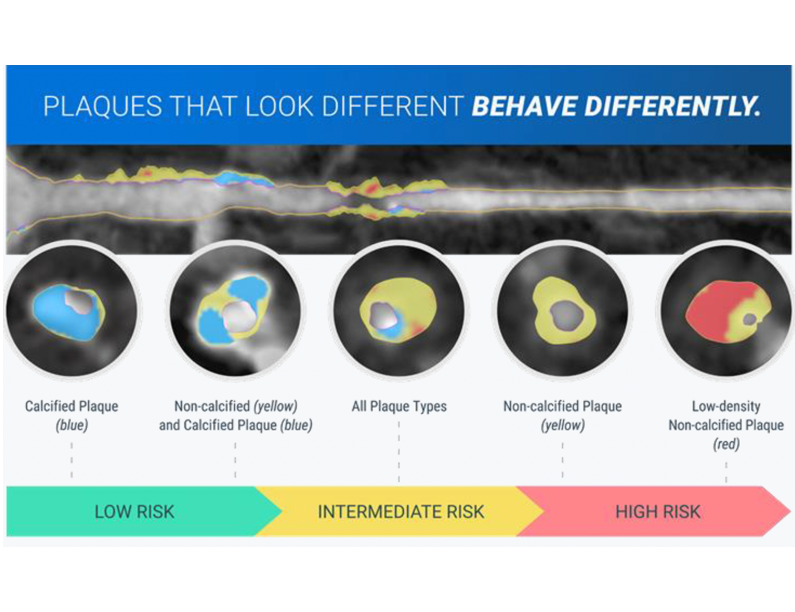
Early investigation of coronary plaque involved comparisons between the Hounsfield units (HU) of plaque on CCTA with IVUS enabled with virtual histology (VH-IVUS). This analysis led to the identification of plaques with a thin cap, necrotic core, and spotty calcification as being high-risk for instability and rupture versus higher HU plaque identified as fibrous and calcified. Coronary plaque by HU can be characterized as follows. Plaque with <30 HU is low-density, non-calcified. Plaque with HU between 30–350 is non-calcified. Finally, plaque with > 350 HU is generally characterized as calcified [21,22]. The question that remains is what plaque progression and what plaque features are important regarding plaque rupture. Quantification of plaque volume is important. If the necrotic core grows within the plaque in volume, it will lead to positive vessel remodeling and thinning of the fibrous cap. In combination with impaired vasomotor capabilities of the vessel, this plaque will be at higher risk for rupture. Attempts to stabilize LRP and prevent plaque rupture involve procalcific reactions. Microcalcifications represent an early stage of this process and are markers of HRP. High rates of microcalcification of coronary plaques has been associated with increased rates of cardiac death and nonfatal MI (p=0.03) [23]. Figure 1 demonstrates the appearance of the types of plaque using CCTA with plaque analysis software.
Artificial Intelligence and Plaque Analysis
AI plaque analysis accuracy
Advances in software and the increasing use of artificial intelligence (AI) have had a profound effect on plaque analysis and have enabled physicians to assess their patients with a personalized approach. AI-guided CCTA analysis helps with workflow as it is less reliant on labor and on experienced readers. It also removes interobserver variability [24]. The use of the Heartflow plaque analysis software, AI-QCPA (an FDA-cleared, AI tool) (Heartflow, Mountain View, CA) versus IVUS demonstrated approximately 95% agreement in the determination of total plaque, calcified plaque (CP), and non-calcified plaque (NCP) volumes respectively in 237 patients at 15 international sites [25]. In a retrospective analysis of the MIAMI study, AI-QCPA demonstrated a high accuracy once again with IVUS for both CP and NCP [26]. The use of this software has been shown to be additive to the information given by stenosis analysis and FFRct and has impacted clinical practice on an individualized basis.
When compared to FFRct, SPECT, QCA, IVUS, NIRS, and OCT, the FDA-cleared machine learning, AI-driven software (AI-QCT) from Cleerly (Cleerly Inc, Denver, CO) has performed well in studies with areas under the curve of 0.86,0.92, 0.96, and 0.97 with the OCT comparison ongoing currently [27]. It has also performed very well versus invasive quantitative coronary analysis (QCA). In a multicenter study of 303 patients comparing AI-QCT with invasive QCA, accuracy, sensitivity, specificity were 86%, 94%, and 82% respectively [28,29]. A couple of interesting studies using deep learning models (DLM) have demonstrated that DLM can estimate coronary vessel lumen and plaque boundaries enabling it to accurately quantify CP, NCP and automatically produce a CAD-RADS score. In a multicenter cohort, DLM showed high accuracy [weighted kappa >0.70 (0.75)] for plaque volume measurements and CAD-RADS classification [30]. In another multicenter study of 1339 patients, unsupervised DLM was used to quantify stenoses and features of HRP on CCTA. There was 93.5% agreement with level 3 CCTA readers on a per-vessel basis and the AUCs for LAP, spotty calcification, and positive remodeling were 0.80, 0.79, and 0.77 respectively [31]. Another interesting study assessed onsite deep learning CT-FFR (cFFRv6) compared to invasive instantaneous wave-free ratio (IFR). CT-FFR used an onsite deep learning algorithm that could be used at the workstation with a result in approximately 10 minutes. The sensitivity, specificity, positive predictive value, and negative predictive value were 89.3%, 68.8%, 83.3%, and 78.6% respectively with accuracy of 81.8% and an AUC of 0.79. CT-FFR had a reasonable correlation with IFR (r~0.37) and was better than CCTA alone vs IFR (AUC 0.52) [32].
AI plaque analysis prognostic value
A retrospective analysis of 4,430 patients from the ADVANCE registry demonstrated that a total plaque volume of >564 mm3 and a total plaque area volume (PAV) of >24.4% were independently associated with significantly higher rates of MACE and late revascularization (p=0.013, p<0.0001 respectively) [33]. In this study, use of AI-QCPA demonstrated that total plaque volume has better predictive value than FFRct for MACE at one year. The (Decisions for Treating Coronary Disease are Changed in Patients Evaluated with Quantified Plaque Analysis) DECODE study demonstrated that the use of AI-QCPA led to a change in the clinical management of 66% of patients when compared to CCTA alone with most of the changes being a more aggressive medical therapy regimen [34]. In the (AI-Derived Plaque Quantification: Coronary CTA and AI-QCPA for determining effective CAD management) DECIDE study, over 50% of patients had their medical regimen changed including 30% of patients with a coronary artery calcium score of 0. This may predict a 15% decrease in cardiovascular risk [35].
AI-QCT allows for significant correlation between PAV and future cardiovascular events. In a retrospective analysis of 303 patients from the CREDENCE (Computed Tomographic Evaluation of Atherosclerotic DEterminaNts of Myocardial IsChEmia) trial, plaque burden was demonstrated to correlate with prognosis. The study proposed a clinically useful CCTA coronary atherosclerosis staging system (Table 1) in which plaque volume and percent atheroma correlate with stenosis severity on QCA and coronary ischemia on invasive FFR. Higher plaque burden by either PAV or TPV was associated with more extensive disease. Stage 1 represented predominately nonobstructive disease while Stage 3 represented a large proportion of multivessel disease. Increasing plaque burden was also associated with more ischemia with the stage 1 group being 70-85% non-ischemic while the stage 3 group demonstrated ~ 70% ischemia. This suggests a dose-response relationship between plaque burden and disease severity with increasing plaque burden being associated with more coronary artery involvement and more ischemia. In this study an increase of >1% atheroma per year was associated with a worse prognosis [36–38]. The use of AI-QCT in a group of 536 patients who were staged similarly revealed higher rates of all-cause mortality, nonfatal myocardial infarction, nonfatal stroke and coronary revascularization with increased PAV. Additionally, PAV data was able to better predict future ASCVD risk [39]. The use of AI-QCT for ischemia has also been demonstrated to be very useful in predicting the likelihood of ischemia compared to other modalities. In studies, patients with positive AI-QCT ischemia have worse survival compared to those with a negative result (HR 7.24 for a cardiac event at 8 years).
|
Stage |
Plaque Volume in mm3 |
Percentage of atheroma |
|
0 |
0 |
0% |
|
1 |
1–250 |
1–5% |
|
2 |
251–750 |
6–15% |
|
3 |
>750 |
>15% |
Clinical Study Data
The identification of low-density NCP as a high-risk feature has been central to the ability to predict future cardiac events. In the ICONIC (Incident COroNary Syndromes Identified by Computed Tomography) trial, low density NCP volume was the strongest predictor of future acute coronary syndromes [40]. This was true even when 2/3 of the future culprit lesions had less than 50% stenosis on CCTA. Of note, the more calcified plaques were associated with lower rates of acute coronary syndrome. Although the overall rates of cardiac events were low in the PROMISE (Prospective Multicenter Imaging Study for Evaluation of Chest pain) trial, low density-NCP was identified as being a strong predictor of MI [41,42]. There are many studies that demonstrate the importance of plaque analysis and cardiac events. Because this is not meant to be an exhaustive review of all CCTA studies, the following studies were chosen for review.
SCOT-HEART (Scottish Computed Tomography of the Heart)
The SCOT-HEART family of studies has provided many insights into the utility of CCTA to evaluate chest pain patients. The initial trial demonstrated a significant reduction in MI in the CCTA cohort versus standard of care [43]. The presence of low-attenuation plaque (LAP) increased the risk of fatal and nonfatal MI on retrospective analysis of the CCTA scans performed in the original trial. The study examined 1697 patients. After a median follow-up of 4.7 years, 37 MIs occurred. LAP, coronary artery calcification (CAC), and the presence of obstructive CAD were all higher in the patient subgroup that experienced fatal and nonfatal MI. LAP was the strongest predictor irrespective of CAC or obstructive CAD. LAP was more predictive than peri-coronary adipose tissue for prediction of MI. Patients who had a LAP burden of >4% (MI patients had 7.4% compared to 4.1% in the non-MI group) had 5 times the likelihood of a future MI (HR 4.65, p<0.01) [44,45].
An interesting subsequent study looked at whether AI based quantitative image analysis techniques like radiomics could be used to evaluate plaques by phenotype. The concept is that various morphologic plaque patterns can be characterized using mathematical algorithms and machine-learning. In this study, a total of 15 eigen radiomic features were associated with MI [46]. Eight of these had a strong correlation when added to clinical risk factors, Agatston calcium score, presence of obstructive coronary disease, and quantitative plaque burden (total plaque, CP, and NCP) (Figure 2). These radiomic phenotypes provided better prediction of future MI (nonfatal and fatal) than conventional metrics and were strongest in predicting events in the longer-term follow-up group (>5 years). This could potentially help identify people at risk for future MI and could be a helpful in identifying patients who would benefit from more intensive therapy earlier in their disease process. Analysis such as this may allow for a more personalized approach to CAD diagnosis with CCTA in the future.
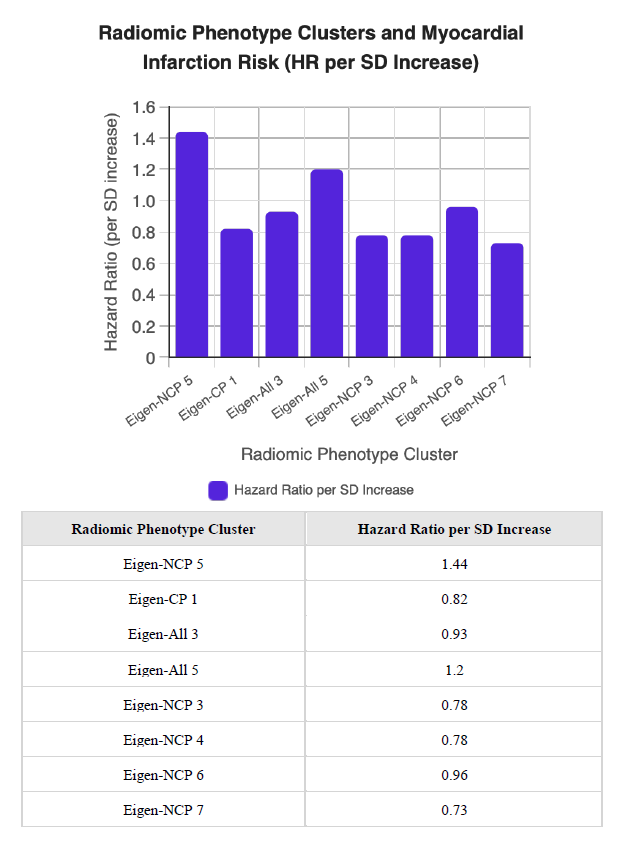
Figure 2. The chart below displays the association between eight key radiomic phenotype clusters and the risk of fatal or nonfatal myocardial infarction, as measured by hazard ratio (HR) per standard deviation (SD) increase. Clusters with HR above 1 indicate increased risk, while those below 1 indicate decreased risk.
PARADIGM
The PARADIGM study examined the effect of statin therapy on plaque progression using serial CCTA studies. There were 1255 eligible patients for analysis. Four hundred seventy-four patients with 1079 lesions were taking statins. Seven hundred eighty-one patients with 2196 lesions were taking statin therapy. High-risk plaque (HRP) was defined as plaque having at least two of the following characteristics: positive arterial remodeling, LAP (< 30 HU), or spotty calcification (<3 mm in any direction within the plaque). The annualized incidence of HRP, positive remodeling, spotty calcification, and LAP were all lower in the group taking statins (0.9% vs 1.6%; 5.2% vs 7.2%; 0.2% vs 0.5%; 0.8% vs 1.0%, all p<0.001). This was basically a 35% reduction in HRP at between 3–4 years on statins. Interestingly, the progression of PAV was slower (p=0.002), and there was an increase in calcified PAV (p<0.001) in the statin group [47]. This is clinically important because it demonstrates that HRP can be converted to lower risk plaque using statins. These plaque findings were independent of severity of stenosis. The plaques essentially became more stable even though the degree of coronary stenosis did not change significantly.
CREDENCE
CREDENCE established the importance of an anatomic approach using novel volumetric and compositional plaque measures beyond stenosis, versus functional assessment for identifying ischemic segments, compared to invasive FFR. In this multi-center derivation–validation cohort, 612 patients with signs and symptoms of ischemia, referred for nonemergent invasive angiography with invasive FFR, completed coronary computed tomographic angiography (CCTA) quantification of atherosclerotic plaque and FFR by CT (FFR–CT) and semiquantitative scoring of rest and stress perfusion by cardiac MR, PET or SPECT. The primary endpoint was invasive FFR 0.8 or less. Hemodynamically significant stenosis was present in 26.5% of 1727 vessels. Image quality for CCTA was acceptable in 99% of patients [48].
Prevalence of CAD (greater than 50% stenosis) on a per vessel basis was 58% by CCTA, with abnormal FFR CT on nearly half. High risk plaque features were common. Positive remodeling (at least 1.1) was identified in 70.2% of patients (N = 430), LAP was identified in 14.1% of patients (N = 76), spotty calcification identified in 15.4% of patients (N= 94). Moderate concordance between invasive FFR and FFR–CT (K = 0.45) was observed. 91% of normal FFR-CT had normal invasive FFR while only 51% of abnormal FFR-CT had abnormal invasive FFR.
AUC for CCTA stenosis severity was 0.82 with significant predictors including percentage of noncalcified atheroma volume, lumen volume, number of lesions with high-risk plaque and number of lesions with greater than 30% stenosis. Adding atherosclerotic plaque significantly improved AUC to 0.88 (P less than 0.001). Addition of FFR–CT to atherosclerotic plaque and stenosis did not improve discrimination with an ROC curve (AUC = 0.88). The AUC curve for stress MPI including SRS and SDS within specific vascular territories with 0.69, which did not change with addition of LVEF (AUC 0.69) or with exercise electrocardiogram findings (AUC 0.70) [15,36]. LAP, positive remodeling greater than 1.1, napkin ring sign or spotty calcification, in addition to lumen volume, percentage of noncalcified atheroma volume, and number of lesions with greater than 30% stenosis were strongly associated with positive invasive FFR and represent instability of lipid rich plaque with similar values in the derivation of the patient cohort [36].
Key takeaways from CREDENCE are that anatomic features, namely noncalcified plaque volume, presence of high-risk plaque and vessel size are independently associated with invasive FFR, independent of stenosis severity with a stronger link to invasive FFR than physiologic assessments of myocardial perfusion. Notably FFR CT did not significantly improve discrimination of normal versus abnormal invasive FFR.
Coronary CT plaque analysis in percutaneous coronary intervention planning
CCTA has been increasingly used in coronary PCI planning, given its significant diagnostic and planning value [49]. Figure 3 shows the currently available information that can be determined by CCTA including vessel size, plaque quantification and characterization, as well as determination of the presence of ischemia. Coronary CTA can provide accurate diagnostic value for the evaluation of ischemia while also helping determine focal disease that would benefit from PCI, as well as evaluate residual physiologic disease burden after intervention [50]. In the case of chronic total occlusion (CTO), CCTA planning is invaluable in cases with ambiguous proximal or distal caps, ambiguous vessel course, or non-visualized distal vessel. It allows determination of vessel course at different preset angulations that can be used during the procedure, with or without co-registration with the imaging system. Multiple CT-based scoring systems have been developed to predict successful CTO crossing [51]. CCTA has additional applications in determining the optimal angle for evaluating aorto-ostial lesions and in planning diagnosis and treatment in prior-CABG patients [52].
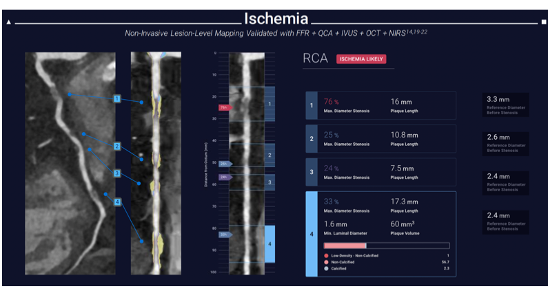
Figure 3. Plaque analysis with ischemia evaluation. Representative report after evaluation with AI-QCT. Descriptions are given regarding the type and extent of coronary plaque assessed from the CCTA images. There is also an ischemia evaluation included that states whether ischemia is likely or not likely based on the AI algorithm used to assess ischemia based on plaque characteristics. Courtesy of Cleerly, Inc.
Recently, there have been significant advancements in the plaque analysis algorithms in CCTA. The plaque analysis algorithms have improved the use of CCTA for pre-planning PCI in general, and CTO PCI specifically [53]. Pre-procedure identification of plaque composition improves the efficiency and safety of the procedure by enabling early selection of the necessary tools. To identify plaque composition, the operator views the vessel in reconstructed MPR images (straight and curved) to determine lesion length, then reviews the cross sections to assess the extent of disease and plaque composition throughout the vessel course.
Coronary CTA can improve success rates for coronary CTO PCI, especially in more complex cases with J-CTO 2 or higher [54]. AI tools are also showing promise for accurate, fully automated CTO PCI planning [55]. The specific role of plaque analysis compared with the overall CTA 3D reconstruction and co-registration has not been specifically studied. However, plaque analysis has been increasingly used in CTO PCI planning due to its numerous practical applications. The initial step of plaque analysis includes the evaluation of the CTO in the reconstructed MPR images. This allows evaluation of CTO length, an important predictor of CTO wire crossing. In the original J-CTO score, a CTO length ≥20 mm predicted failure of antegrade wiring within 30 minutes [56], whereas in the CT-based K-CTO score, a CTO length ≥15 mm predicted the same.
A specific use for plaque analysis in CTO planning is distinguishing between soft plaques. CT-Plaque analysis of the CTO body is a superior planning tool compared with coronary angiography. The evaluation of the extent of coronary calcification by coronary angiography is significantly limited. Coronary CTA can identify the extent, length, thickness, density, and arc of calcification. In longer lesions with severe calcification (>270°) and higher density (>637 Hounsfield units) [57], the operator has information allowing for evaluation of different plaque modification techniques (atherectomy, IVL, specialty balloons). This can improve both procedural efficiency and safety by avoiding potential complications such as balloon-induced dissection and perforation. The presence of calcification is an important predictor of antegrade crossing failure. In the K-CTO score, calcification of ≥180° with cross-sectional area (CSA) ≥50% was an important predictor of failure to cross antegrade within 30 minutes, adding 1 point to the difficulty score. When calcifications are central (100% CSA), also called full moon calcification, this adds 2 points to the difficulty score.
The practical application of these findings is real as it can help offer procedural guidance. When evaluating a CTO body that is shorter with predominantly soft plaque, antegrade wiring should be attempted first. On the other hand, a longer CTO body with heavy calcification, especially if any segment shows full-moon calcification, makes attempts at true-to-true wiring pointless; the operator should start with early dissection-reentry techniques.
Challenges for AI-CCTA
Several important challenges remain for the continuing widespread use of AI-CCTA. These can be broken down into a few categories. Workflow is a challenge. There is a need for standardized acquisition protocols and measurement techniques. The turnaround time for current AI plaque assessments is several hours, which is why the idea of onsite CT-FFR capability is intriguing. This would reduce turnaround time from hours to minutes [32]. Standardized reporting and the ability to incorporate into the electronic health record are also a challenge currently. From a technology standpoint, there should be consensus on what the minimum is for scanner technology as this would reduce the number of vessels/scans that are not interpretable by current AI plaque technology. Another technical challenge is plaque quantification and characterization. Agreement on how plaques are measured and described is needed (for example, plaques causing MACE are not only LAP). The ability to better characterize plaque by reducing artifact from calcium with photon counting is needed on a wider scale. However, this leads to the next challenge, which is economics. The scalability of high-end CCTA with AI plaque assessment and photon counting is greatly affected by the cost associated with these technologies. It is currently not able to be placed in many hospitals due to a lack of capital. Finally, as in many cardiovascular research studies, CCTA studies lack representation of many groups in the population and would benefit from greater diversity in enrollment.
Conclusion
In the span of a couple decades, CCTA has emerged as a first-line option for ischemia evaluation. The evolution of scanners has made submillimeter, isovolumic images widespread. This has allowed for improved plaque imaging. The evolution of data has taken us from assessment of risk based on coronary calcification to a more elegant assessment of plaque. Identification and quantification of LAP have led to a better understanding of HRP and plaque rupture which has led to a focus on more aggressive medical therapy with statins. Serial CCTA may prove to be very useful in terms of reducing MACE, namely fatal and nonfatal MI by allowing clinicians to visualize plaque that has transformed from plaque with high-risk features to stable plaque that is fibrotic or calcific. Additionally, the use of CCTA as a road map for planning coronary interventions, including complex procedures like CTO has been shown to be very useful. Identifying the type of plaque and being able to plan how to modify calcified plaque will make these procedures safer. The future will likely involve the use of CCTA and AI and/or deep learning plaque analysis further upstream to truly make an impact on future cardiac events.
References
2. AstraZeneca. Morbidity and Mortality 2012 Chart Book on Cardiovascular, Lung, and Blood Diseases. London: AstraZeneca; 2012. Available from: https://www.astrazeneca.com/content/dam/az/media-centre-docs/article_files/articles-2015/Documents/2012_ChartBook_508.pdf
3. Ambrose JA, Tannenbaum MA, Alexopoulos D, Hjemdahl-Monsen CE, Leavy J, Weiss M, et al. Angiographic progression of coronary artery disease and the development of myocardial infarction. J Am Coll Cardiol. 1988 Jul;12(1):56–62.
4. Narula J, Nakano M, Virmani R, Kolodgie FD, Petersen R, Newcomb R, et al. Histopathologic characteristics of atherosclerotic coronary disease and implications of the findings for the invasive and noninvasive detection of vulnerable plaques. J Am Coll Cardiol. 2013 Mar 12;61(10):1041–51.
5. Stone GW, Maehara A, Lansky AJ, de Bruyne B, Cristea E, Mintz GS, et al. A prospective natural-history study of coronary atherosclerosis. N Engl J Med. 2011 Jan 20;364(3):226–35.
6. Glagov S, Weisenberg E, Zarins CK, Stankunavicius R, Kolettis GJ. Compensatory enlargement of human atherosclerotic coronary arteries. N Engl J Med. 1987 May 28;316(22):1371–5.
7. Libby P. The interface of atherosclerosis and thrombosis: basic mechanisms. Vasc Med. 1998;3(3):225–9.
8. Falk E, Shah PK, Fuster V. Coronary plaque disruption. Circulation. 1995 Aug 1;92(3):657–71.
9. Virmani R, Kolodgie FD, Burke AP, Farb A, Schwartz SM. Lessons from sudden coronary death: a comprehensive morphological classification scheme for atherosclerotic lesions. Arterioscler Thromb Vasc Biol. 2000 May;20(5):1262–75.
10. Dalager-Pedersen S, Ravn HB, Falk E. Atherosclerosis and acute coronary events. Am J Cardiol. 1998 Nov 26;82(10B):37T–40T.
11. Ahmadi A, Argulian E, Leipsic J, Newby DE, Narula J. From Subclinical Atherosclerosis to Plaque Progression and Acute Coronary Events: JACC State-of-the-Art Review. J Am Coll Cardiol. 2019 Sep 24;74(12):1608–17.
12. Libby P. The forgotten majority: unfinished business in cardiovascular risk reduction. J Am Coll Cardiol. 2005 Oct 4;46(7):1225–8.
13. Fischer C, Hulten E, Belur P, Smith R, Voros S, Villines TC. Coronary CT angiography versus intravascular ultrasound for estimation of coronary stenosis and atherosclerotic plaque burden: a meta-analysis. J Cardiovasc Comput Tomogr. 2013 Jul-Aug;7(4):256–66.
14. Hadamitzky M, Achenbach S, Al-Mallah M, Berman D, Budoff M, Cademartiri F, et al. Optimized prognostic score for coronary computed tomographic angiography: results from the CONFIRM registry (COronary CT Angiography EvaluatioN For Clinical Outcomes: An InteRnational Multicenter Registry). J Am Coll Cardiol. 2013 Jul 30;62(5):468–76.
15. Driessen RS, Danad I, Stuijfzand WJ, Raijmakers PG, Schumacher SP, van Diemen PA, et al. Comparison of Coronary Computed Tomography Angiography, Fractional Flow Reserve, and Perfusion Imaging for Ischemia Diagnosis. J Am Coll Cardiol. 2019 Jan 22;73(2):161–73.
16. Patel MR, Nørgaard BL, Fairbairn TA, Nieman K, Akasaka T, Berman DS, et al. 1-Year Impact on Medical Practice and Clinical Outcomes of FFRCT: The ADVANCE Registry. JACC Cardiovasc Imaging. 2020 Jan;13(1 Pt 1):97–105.
17. Douglas PS, De Bruyne B, Pontone G, Patel MR, Norgaard BL, Byrne RA, et al. 1-Year Outcomes of FFRCT-Guided Care in Patients With Suspected Coronary Disease: The PLATFORM Study. J Am Coll Cardiol. 2016 Aug 2;68(5):435–45.
18. Ferencik M, Mayrhofer T, Bittner DO, Emami H, Puchner SB, Lu MT, et al. Use of High-Risk Coronary Atherosclerotic Plaque Detection for Risk Stratification of Patients With Stable Chest Pain: A Secondary Analysis of the PROMISE Randomized Clinical Trial. JAMA Cardiol. 2018 Feb 1;3(2):144–52.
19. Gulati M, Levy PD, Mukherjee D, Amsterdam E, Bhatt DL, Birtcher KK, et al. 2021 AHA/ACC/ASE/CHEST/SAEM/SCCT/SCMR Guideline for the Evaluation and Diagnosis of Chest Pain: A Report of the American College of Cardiology/American Heart Association Joint Committee on Clinical Practice Guidelines. Circulation. 2021 Nov 30;144(22):e368–454.
20. Cury RC, Leipsic J, Abbara S, Achenbach S, Berman D, Bittencourt M, et al. CAD-RADS™ 2.0 - 2022 Coronary Artery Disease-Reporting and Data System: An Expert Consensus Document of the Society of Cardiovascular Computed Tomography (SCCT), the American College of Cardiology (ACC), the American College of Radiology (ACR), and the North America Society of Cardiovascular Imaging (NASCI). J Cardiovasc Comput Tomogr. 2022 Nov-Dec;16(6):536–57.
21. de Graaf MA, Broersen A, Kitslaar PH, Roos CJ, Dijkstra J, Lelieveldt BP, et al. Automatic quantification and characterization of coronary atherosclerosis with computed tomography coronary angiography: cross-correlation with intravascular ultrasound virtual histology. Int J Cardiovasc Imaging. 2013 Jun;29(5):1177–90.
22. van Velzen JE, Schuijf JD, de Graaf FR, Nucifora G, Pundziute G, Jukema JW, et al. Plaque type and composition as evaluated non-invasively by MSCT angiography and invasively by VH IVUS in relation to the degree of stenosis. Heart. 2009 Dec;95(24):1990–6.
23. Moss A, Daghem M, Tzolos E, Meah MN, Wang KL, Bularga A, et al. Coronary Atherosclerotic Plaque Activity and Future Coronary Events. JAMA Cardiol. 2023 Aug 1;8(8):755–64.
24. Jonas RA, Weerakoon S, Fisher R, Griffin WF, Kumar V, Rahban H, et al. Interobserver variability among expert readers quantifying plaque volume and plaque characteristics on coronary CT angiography: a CLARIFY trial sub-study. Clin Imaging. 2022 Nov;91:19–25.
25. Narula J, Stuckey TD, Nakazawa G, Ahmadi A, Matsumura M, Petersen K, et al. Prospective deep learning-based quantitative assessment of coronary plaque by computed tomography angiography compared with intravascular ultrasound: the REVEALPLAQUE study. Eur Heart J Cardiovasc Imaging. 2024 Aug 26;25(9):1287–95.
26. Ihdayhid AR, Tzimas G, Peterson K, Ng N, Mirza S, Maehara A,et al. Diagnostic Performance of AI-enabled Plaque Quantification from Coronary CT Angiography Compared with Intravascular Ultrasound. Radiol Cardiothorac Imaging. 2024 Dec;6(6):e230312.
27. Nurmohamed NS, Danad I, Jukema RA, de Winter RW, de Groot RJ, Driessen RS, et al. Development and Validation of a Quantitative Coronary CT Angiography Model for Diagnosis of Vessel-Specific Coronary Ischemia. JACC Cardiovasc Imaging. 2024 Aug;17(8):894–906.
28. Griffin WF, Choi AD, Riess JS, Marques H, Chang HJ, Choi JH, et al. AI Evaluation of Stenosis on Coronary CTA, Comparison With Quantitative Coronary Angiography and Fractional Flow Reserve: A CREDENCE Trial Substudy. JACC Cardiovasc Imaging. 2023 Feb;16(2):193–205.
29. Bernardo R, Nurmohamed NS, Bom MJ, Jukema R, de Winter RW, Sprengers R, et al. Diagnostic accuracy in coronary CT angiography analysis: artificial intelligence versus human assessment. Open Heart. 2025 Jan 11;12(1):e003115.
30. Herten VRLM, Hampe N, Takx RAP, Franssen KJ, Wang Y, Sucha D, et al. Automatic Coronary Artery Plaque Quantification and CAD-RADS Prediction Using Mesh Priors. IEEE Trans Med Imaging. 2024 Apr;43(4):1272–83.
31. Ihdayhid AR, Sehly A, He A, Joyner J, Flack J, Konstantopoulos J, et al. Coronary Artery Stenosis and High-Risk Plaque Assessed With an Unsupervised Fully Automated Deep Learning Technique. JACC Adv. 2024 Mar 6;3(9):100861.
32. Roshan MP, Gigliotti GV, Gonzalez J, Cury RA, Lamy C, Sayegh K, et al. Novel deep learning CCTA-FFR for detecting functionally significant coronary stenosis: Comparison with iFR. J Cardiovasc Comput Tomogr. 2026 Mar-Apr;20(2):132–9.
33. Dundas J, Leipsic J, Fairbairn T, Ng N, Sussman V, Guez I, et al. Interaction of AI-Enabled Quantitative Coronary Plaque Volumes on Coronary CT Angiography, FFRCT, and Clinical Outcomes: A Retrospective Analysis of the ADVANCE Registry. Circ Cardiovasc Imaging. 2024 Mar;17(3):e016143.
34. Rinehart S, Raible SJ, Ng N, Mullen S, Huey W, Rogers C, et al. Utility of Artificial Intelligence Plaque Quantification: Results of the DECODE Study. J Soc Cardiovasc Angiogr Interv. 2024 Mar 26;3(3Part B):101296.
35. Rinehart S, Blankstein R, McCarthy CP, Januzzi JL, Scherer M, O'Neal WT, et al. Guiding Preventive Care Strategies for Patients With Atherosclerotic Plaque on Coronary CTA: Primary Outcomes of the DECIDE Registry. JACC Cardiovasc Imaging. 2026 Jun;19(6):706–16.
36. Stuijfzand WJ, van Rosendael AR, Lin FY, Chang HJ, van den Hoogen IJ, Gianni U, et al. Stress Myocardial Perfusion Imaging vs Coronary Computed Tomographic Angiography for Diagnosis of Invasive Vessel-Specific Coronary Physiology: Predictive Modeling Results From the Computed Tomographic Evaluation of Atherosclerotic Determinants of Myocardial Ischemia (CREDENCE) Trial. JAMA Cardiol. 2020 Dec 1;5(12):1338–48.
37. Min JK, Chang HJ, Andreini D, Pontone G, Guglielmo M, Bax JJ, et al. Coronary CTA plaque volume severity stages according to invasive coronary angiography and FFR. J Cardiovasc Comput Tomogr. 2022 Sep-Oct;16(5):415–22.
38. Motoyama S, Ito H, Sarai M, Kondo T, Kawai H, Nagahara Y, et al. Plaque Characterization by Coronary Computed Tomography Angiography and the Likelihood of Acute Coronary Events in Mid-Term Follow-Up. J Am Coll Cardiol. 2015 Jul 28;66(4):337–46.
39. Nurmohamed NS, Bom MJ, Jukema RA, de Groot RJ, Driessen RS, van Diemen PA, et al. AI-Guided Quantitative Plaque Staging Predicts Long-Term Cardiovascular Outcomes in Patients at Risk for Atherosclerotic CVD. JACC Cardiovasc Imaging. 2024 Mar;17(3):269–80.
40. Chang HJ, Lin FY, Lee SE, Andreini D, Bax J, Cademartiri F, et al. Coronary Atherosclerotic Precursors of Acute Coronary Syndromes. J Am Coll Cardiol. 2018 Jun 5;71(22):2511–22.
41. Douglas PS, Hoffmann U, Patel MR, Mark DB, Al-Khalidi HR, Cavanaugh B, et al. Outcomes of anatomical versus functional testing for coronary artery disease. N Engl J Med. 2015 Apr 2;372(14):1291–300.
42. Taron J, Foldyna B, Mayrhofer T, Osborne MT, Meyersohn N, Bittner DO, et al. Risk Stratification With the Use of Coronary Computed Tomographic Angiography in Patients With Nonobstructive Coronary Artery Disease. JACC Cardiovasc Imaging. 2021 Nov;14(11):2186–95.
43. SCOT-HEART Investigators; Newby DE, Adamson PD, Berry C, Boon NA, Dweck MR, et al. Coronary CT Angiography and 5-Year Risk of Myocardial Infarction. N Engl J Med. 2018 Sep 6;379(10):924–33.
44. Tzolos E, Williams MC, McElhinney P, Lin A, Grodecki K, Flores Tomasino G, et al. Pericoronary Adipose Tissue Attenuation, Low-Attenuation Plaque Burden, and 5-Year Risk of Myocardial Infarction. JACC Cardiovasc Imaging. 2022 Jun;15(6):1078–88.
45. Williams MC, Kwiecinski J, Doris M, McElhinney P, D'Souza MS, Cadet S, et al. Low-Attenuation Noncalcified Plaque on Coronary Computed Tomography Angiography Predicts Myocardial Infarction: Results From the Multicenter SCOT-HEART Trial (Scottish Computed Tomography of the HEART). Circulation. 2020 May 5;141(18):1452–62.
46. Kolossváry M, Lin A, Kwiecinski J, Cadet S, Slomka PJ, Newby DE, et al. Coronary Plaque Radiomic Phenotypes Predict Fatal or Nonfatal Myocardial Infarction: Analysis of the SCOT-HEART Trial. JACC Cardiovasc Imaging. 2025 Mar;18(3):308–19.
47. Lee SE, Chang HJ, Sung JM, Park HB, Heo R, Rizvi A, et al. Effects of Statins on Coronary Atherosclerotic Plaques: The PARADIGM Study. JACC Cardiovasc Imaging. 2018 Oct;11(10):1475–84.
48. Danad I, Raijmakers PG, Driessen RS, Leipsic J, Raju R, Naoum C, et al. Comparison of Coronary CT Angiography, SPECT, PET, and Hybrid Imaging for Diagnosis of Ischemic Heart Disease Determined by Fractional Flow Reserve. JAMA Cardiol. 2017 Oct 1;2(10):1100–7.
49. Sandoval Y, Leipsic JA, Collet C, Ali ZA, Azzalini L, Barbato E, et al. Coronary Computed Tomography Angiography to Guide Percutaneous Coronary Intervention: Expert Opinion from a SCAI/SCCT Roundtable. J Soc Cardiovasc Angiogr Interv. 2025 May 1;4(6):103664.
50. Sonck J, Nagumo S, Norgaard BL, Otake H, Ko B, Zhang J, et al. Clinical Validation of a Virtual Planner for Coronary Interventions Based on Coronary CT Angiography. JACC Cardiovasc Imaging. 2022 Jul;15(7):1242–55.
51. Opolski MP, Achenbach S, Schuhbäck A, Rolf A, Möllmann H, Nef H, et al. Coronary computed tomographic prediction rule for time-efficient guidewire crossing through chronic total occlusion: insights from the CT-RECTOR multicenter registry (Computed Tomography Registry of Chronic Total Occlusion Revascularization). JACC Cardiovasc Interv. 2015 Feb;8(2):257–67.
52. Jones DA, Beirne AM, Kelham M, Rathod KS, Andiapen M, Wynne L, et al. Computed Tomography Cardiac Angiography Before Invasive Coronary Angiography in Patients With Previous Bypass Surgery: The BYPASS-CTCA Trial. Circulation. 2023 Oct 31;148(18):1371–80.
53. Carvalho PEP, Jalli S, Ser OS, Cheng V, Cavalcante JL, Lesser J, et al. Artificial intelligence-guided quantitative coronary CT angiography (AI-QCT) automated detection and occlusion length estimation of chronic total occlusions. J Cardiovasc Comput Tomogr. 2025 Nov-Dec;19(6):677–83.
54. Hong SJ, Kim BK, Cho I, Kim HY, Rha SW, Lee SH, et al. Effect of Coronary CTA on Chronic Total Occlusion Percutaneous Coronary Intervention: A Randomized Trial. JACC Cardiovasc Imaging. 2021 Oct;14(10):1993–2004.
55. Morino Y, Abe M, Morimoto T, Kimura T, Hayashi Y, Muramatsu T, et al. Predicting successful guidewire crossing through chronic total occlusion of native coronary lesions within 30 minutes: the J-CTO (Multicenter CTO Registry in Japan) score as a difficulty grading and time assessment tool. JACC Cardiovasc Interv. 2011 Feb;4(2):213–21.
56. Yu CW, Lee HJ, Suh J, Lee NH, Park SM, Park TK, et al. Coronary Computed Tomography Angiography Predicts Guidewire Crossing and Success of Percutaneous Intervention for Chronic Total Occlusion: Korean Multicenter CTO CT Registry Score as a Tool for Assessing Difficulty in Chronic Total Occlusion Percutaneous Coronary Intervention. Circ Cardiovasc Imaging. 2017 Apr;10(4):e005800.
57. Kurogi K, Ishii M, Nagatomo T, Tokai T, Kaichi R, Takae M, et al. Mean density of computed tomography for predicting rotational atherectomy during percutaneous coronary intervention. J Cardiovasc Comput Tomogr. 2023 Mar-Apr;17(2):120–9.