

Abstract
Nature recently listed 20 major T cell milestones since the early breakthroughs of Drs. Jacques Miller and Max Cooper. However, to make the story more complete, several key discoveries may need to be mentioned. One of the major missing episodes is the discovery of the bone marrow in mice as the origin of T and B cells, revealed by Dr. Henry Claman and his colleagues. The complete absence of the critical role of B cells in the final differentiation of T cells into memory lineages could be a major oversight in modern immunology. In this short essay, I will try to summarize some of the published evidence showing that T cell differentiation into memory cells is triggered only by B cells in germinal centers after priming at infection sites for CD8 T cells and in the T cell zone for CD4 T cells in the spleen and draining lymph nodes. Based on the analysis, T cells need two steps to develop into long-lived memory cells, and a new T cell lineage model is presented.
Keywords
B cell, T cell, Dendritic cell, Germinal center, Follicular T helper cells, Memory T cell, BCL6
Introduction
A collection of major milestones in T cells was recently published in Nature (https://www.nature.com/immersive/d42859-022-00032-7/index.html) since the discoveries and characterization of two major players in adaptive immunity, T cells and B cells, by Drs. Jacques Miller [1], Max Cooper [2] in the 1960s. However, the discovery of the mammalian (mouse) bone marrow as the primordial lymphoid lymphatic cell reservoir (including T cells, B cells, and other immune cells ) by Claman and his colleagues actually opened up modern immunology [3]. Literally, if not all, at least most of the milestones listed were produced from the mouse model. However, this essay only focuses on one of the two most critical sides of the relationship between B cells and T cells: how B cells help T cells to differentiate into memory cells, since how T cells help B cells was one of the main themes of the listed milestones. An overwhelming bias of research in the field of immunology is how T cells determine the functional proliferation and differentiation of B cells in animals. It took almost half a century for people to elucidate the detailed underlying mechanism of how T cells help B cells in the germinal centers (GCs) of the spleen and local lymph nodes through a special lineage of T cells, the follicular T helper cells (Tfh) [4–6]. Due to the lack of a B cell-specific maturation organ or apparatus like the Thymus for T cells, how B cells mutually help T cells (T cell proliferation and differentiation) has been overlooked or severely underappreciated in the field of immunology, although groundbreaking discoveries were made even before people revealed how Tfh cells guide the maturation process of B cells in GCs.
B cells were Found to be Antigen-Presenting Cells
Marrack and Kappler were pioneers in showing that B cells express both Ir (MHC-II) [7] and MHC-I [8] and that hybridoma B cells present antigens to hybridoma T cells [9,10], while Chestnut and Grey showed that normal B cells present antigens to T cells as efficiently as to that of macrophages at the same time [11,12].
Depletion of B cells Impairs T cell Proliferation in Spleen and Lymph Nodes
Ron and Segal were the first to report that B cell depletion by anti-µ antibody treatment in mice results in a defect in antigen-reactive T-cell proliferation [13,14]. Segal also demonstrated that splenic B cells function as antigen-presenting cells to prime T cells and convert T cells into effector T cells [15]. Lanzavecchia was the first to show that antigens taken up by B cells are processed within B cells and subsequently presented by B cells to T cells via the MHC molecules [16]. This was later confirmed by Janeway and Ron, who demonstrated that B cells are the antigen-presenting cells required to initiate T cells in the local lymph nodes [17,18]. Kupfer directly observed how B cells present antigens to T cells using immunofluorescence microscopy [19]. Overall, the essential role of B cells as antigen-presenting cells to drive T cell activation in the spleen and lymph nodes was established in the 1980s.
T cells Fail to Differentiate into Memory T cells without B cells
In the early 1990s, it was reported that a continuous supply of antigens was necessary or essential for the generation of both memory T helper cells and memory cytotoxic T cells [20,21]. It was shown that B-cell depletion leads to the generation of short-lived CD8 memory T cells as well as exhausted CD8 T cells. Shortly thereafter, it was reported that depletion of B cells could lead to the failure to generate not only memory CD8 T cells but also memory CD4 T cells, conferring exhausted characteristics to both CD4 and CD8 T cells in µMT/µMT mice [22]. Shortly thereafter, it was again confirmed that B cells play a critical role in the development of CD4 memory T cells [23,24]. These results demonstrated that B cells are essential for the development of long-lasting memory T cells.
Germinal Centers are the Place Where B cells Help T cells to Differentiate into Memory T cells
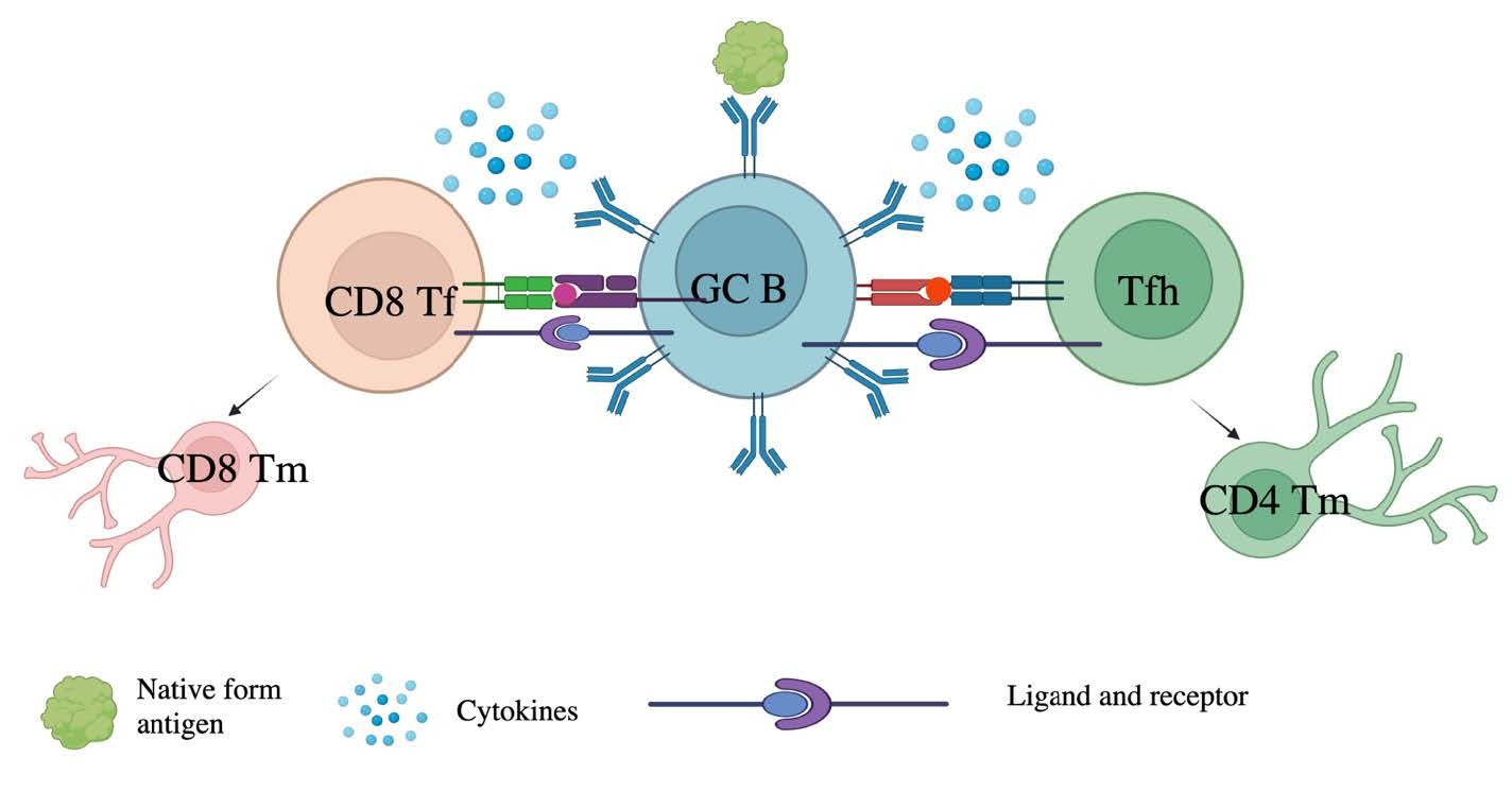
An emerging question is where B cells present antigens to T cells to help T cell differentiation. A fundamental discovery from the BCL-6-deficient mice showed that deletion of BCL-6 not only leads to the complete loss of germinal center formation but also to the failure of B cell and T cell differentiation into memory populations [25,26]. Another excellent finding from Jenkins’ group is that antigen-specific T cells migrate to the B cell zone and interact with B cells to form germinal centers shortly after antigen exposure [27]. This suggests that GCs are the main, if not the only, site for B cells to communicate with T cells. Tokuhisa’s group later showed that BCL-6 is not only essential for the development of CD8 memory T cells [28,29] but also for the development of the CD4 memory T cells [30]. A later report from Jenkins’ group showed that BCL-6 is essential for the development of all memory T cells [31], further supporting that GCs are the primary site for T cell differentiation based on the only GC-expressing feature of BCL-6. Later, follicular T cells were shown to directly support B cell differentiation in the GCs [4–6]. These results demonstrate that B cells, as the only T cell antigen-presenting cells in the GCs, are essential for memory T cell formation and that T cells promote the B cell high-affinity maturation through somatic hypermutation in a mutually beneficial scenario (Figure 1). Another surprising piece of evidence in support of this speculation is that even a single B cell clone expressing a specific antibody to a lymphocytic choriomeningitis virus (LCMV) antigen on the surface of B cell-transgenic mice on top of B cell-deficient mice could lead to normal development and differentiation of CD4 memory T cells [32].
Dendritic Cells Prime T cells in the T cell Zone, While B cells Take Over after Priming
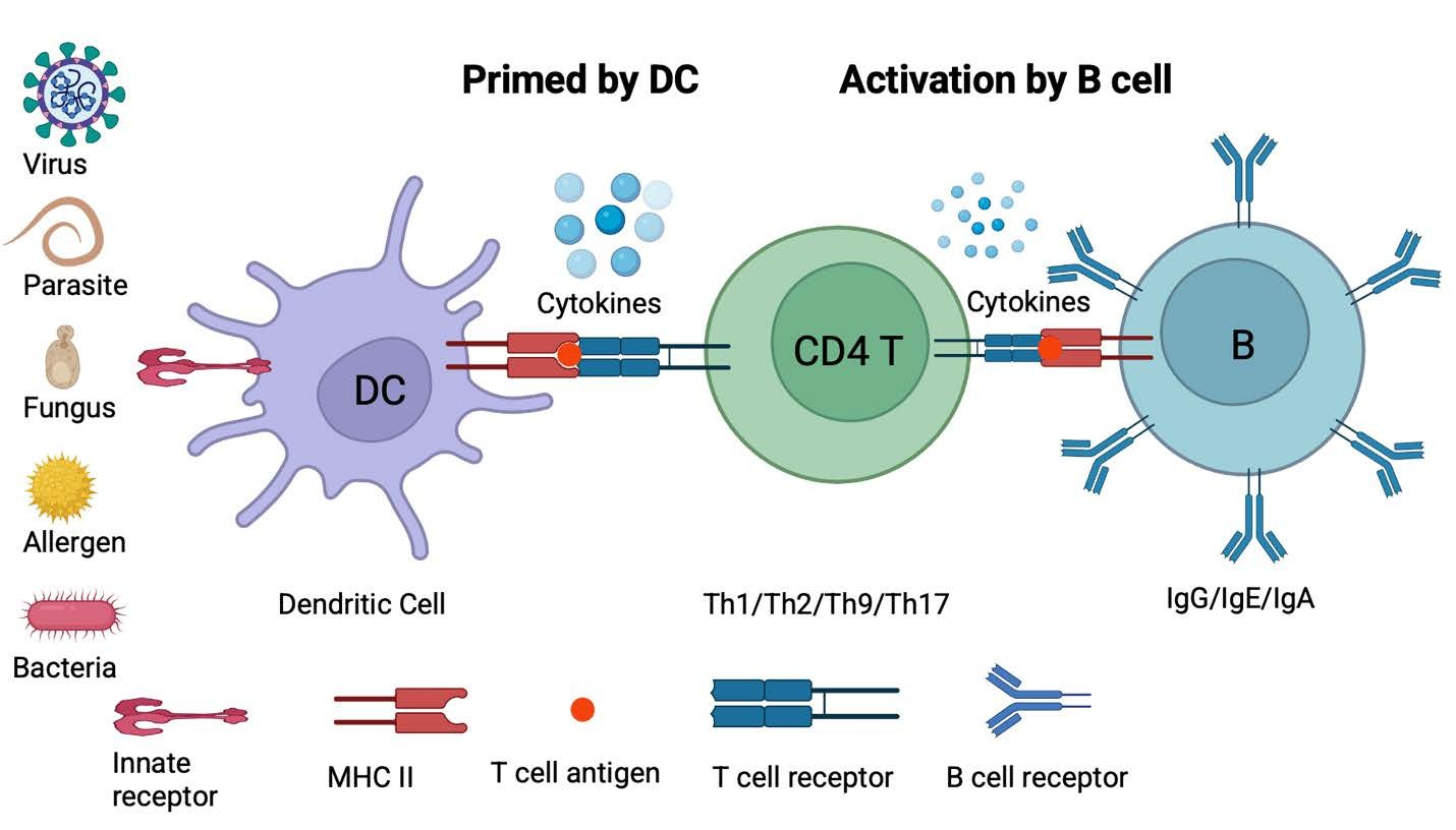
A common view in the immunology field is that dendritic cells (DCs) are the dominant antigen-presenting cells for T cells to proliferate and differentiate into both effector T cells and memory T cells. A seminal discovery from Jenkins’ group showed that DCs are required to prime CD4 T cells in the lymph nodes, but after priming by DCs, B cells take over from DCs to become the dominant, if not sole, antigen-presenting cells in the lymph nodes [27,33]. This suggests that DCs are only required for early CD4 T cell priming in the T cell zone of the lymph nodes during pathogen attack, while B cells are the actual players to support T cell differentiation in GCs.
B cells are Essential for Proper T cell Differentiation in Humans
Interestingly, when anti-CD20 antibodies became therapeutic agents for the treatment of cancer and autoimmune diseases, it was found that patients treated with anti-CD20 antibodies became vulnerable to viral [34] and bacterial [35] infections with defects in memory T cells. Furthermore, patients with B-cell depletion were unable to mount normal humoral and cellular immune responses following SARS-CoV-2 vaccination [36–38]. A more striking observation was that anti-CD20-treated patients failed to clear SARS-CoV-2 [39]. The intriguing fact is that SARS-CoV-2-specific effector and memory T cells are properly produced, but without normal clearance capacity similar to that of exhausted T cells [39]. These results strongly support the concept that T cells without B cells could not properly develop into effector or memory T cells.
A Two-Step Process, First Primed by Infected Cells (for CD8 T cells) or DCs (for CD4 T cells) and Second Activated by B cells, is Needed for Memory T cell Differentiation
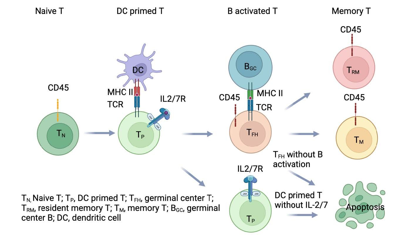
Based on the above analysis of all published results, we can confidently conclude that there are two steps for CD4 T cell activation and differentiation. First, dendritic cells will present antigens to T cells and prime CD4 T cells in the T cell zone. However, CD8 T cells are usually primed at the site of infection and migrate to the T cell zone via the lymphatic circulation system (detail will be analyzed in another essay). Second, both primed CD4 T and CD8 T cells will migrate to the B cell zone and work together with B cells (Figure 2). CD4 T cells will help B cell maturation in germinal centers. Conversely, B cells are the major antigen-presenting cells that drive the T cell maturation process, which most likely occurs in the germinal centers of the spleen and local lymph nodes. It is likely that the feedback signals from germinal center B cells during the matching or pairing between B and T cells in the light zone of GCs trigger T cell activation (second time) and subsequent differentiation into memory T cells (Figure 1), suggesting that these GC-experienced T cells give rise to memory T cells. Without the help of B cells, T cells either remaining in the T cell zone or migrating into the B cell zone after priming will eventually differentiate into exhausted T cells or undergo apoptotic cell death after the resolution of infection. Furthermore, it may remind us not only that current developmental pathways or lineage classifications of memory T cells have caveats but also that current markers assigned to define different types of effector or memory T cells are questionable. Most importantly, it suggests that both follicular CD4 and CD8 cells are halfway along their journey to develop into memory T cells starting from the primed stage. After a second match with germinal center B cells, all these successfully B cell-matched T cells will differentiate into long-lived memory T cells, either memory CD8 or memory CD4 T cells. In this regard, we conclude that germinal center follicular T cells are the essential intermediate T cell lineage for the final long-lived memory T cells for both CD4 T cells (Figure 3) and CD8 T cells (not shown, similar to the CD4 T cell pathway, but CD8 T cells are first primed at infection sites by infected cells or macrophages, but not DC cells in the T cell zone).
Acknowledgments
I would like to thank National Jewish Health for their support; Philippa Marrack, John Kappler, Marc K. Jenkins, and John Cambier for valuable suggestions; Yongmei Jiang, Cheng-Yuan Zhang, Dong-Xiao Li, Peng Sun, Yun-Xia Jiang, Shijun Jiang, and Chengmei Li for private financial support, Alex Zhang for editing. It is partially supported by an NIH grant GM135421 (G.Z.) and NB Life Laboratory LLC, Colorado, USA. Figures 1–3 are generated by the BioRender program.
Contributions
GZ conceived the concept and wrote up the manuscript.
Conflict of Interest
G.Z. holds equity in NB Life Laboratory LLC, Colorado, USA.
References
2. Cooper MD, Peterson RD, Good RA. Delineation of the thymic and bursal lymphoid systems in the chicken. Nature. 1965 Jan 9;205:143–6.
3. Claman HN, Chaperon EA, Triplett RF. Thymus-marrow cell combinations. Synergism in antibody production. Proc Soc Exp Biol Med. 1966 Aug-Sep;122(4):1167–71.
4. Johnston RJ, Poholek AC, DiToro D, Yusuf I, Eto D, Barnett B, et al. Bcl6 and Blimp-1 are reciprocal and antagonistic regulators of T follicular helper cell differentiation. Science. 2009 Aug 21;325(5943):1006–10.
5. Nurieva RI, Chung Y, Martinez GJ, Yang XO, Tanaka S, Matskevitch TD, et al. Bcl6 mediates the development of T follicular helper cells. Science. 2009 Aug 21;325(5943):1001–5.
6. Yu D, Rao S, Tsai LM, Lee SK, He Y, Sutcliffe EL, et al. The transcriptional repressor Bcl-6 directs T follicular helper cell lineage commitment. Immunity. 2009 Sep 18;31(3):457–68.
7. Marrack P, Kappler JW. The role of H-2-linked genes in helper T-cell function. III. Expression of immune response genes for trinitrophenyl conjugates of poly-L(Tyr, Glu)-poly-D,L-Ala--poly-L-Lys in B cells and macrophages. J Exp Med. 1978 Jun 1;147(6):1596–610.
8. Marrack P, Kappler JW. The role of H-2-linked genes in helper T cell function. VII. Expression of I region and immune response genes by B cells in bystander help assays. J Exp Med. 1980 Nov 1;152(5):1274–88.
9. Kappler J, White J, Wegmann D, Mustain E, Marrack P. Antigen presentation by Ia+ B cell hybridomas to H-2-restricted T cell hybridomas. Proc Natl Acad Sci U S A. 1982 Jun;79(11):3604–7.
10. Walker E, Warner NL, Chesnut R, Kappler J, Marrack P. Antigen-specific. I region-restricted interactions in vitro between tumor cell lines and T cell hybridomas. J Immunol. 1982 May;128(5):2164–9.
11. Chesnut RW, Colon SM, Grey HM. Antigen presentation by normal B cells, B cell tumors, and macrophages: functional and biochemical comparison. J Immunol. 1982 Apr;128(4):1764–8.
12. Chesnut RW, Grey HM. Studies on the capacity of B cells to serve as antigen-presenting cells. J Immunol. 1981 Mar;126(3):1075–9.
13. Ron Y, De Baetselier P, Gordon J, Feldman M, Segal S. Defective induction of antigen-reactive proliferating T cells in B cell-deprived mice. Eur J Immunol. 1981 Dec;11(12):964–8.
14. Ron Y, De Baetselier P, Tzehoval E, Gordon J, Feldman M, Segal S. Defective induction of antigen-reactive proliferating T cells in B cell-deprived mice. II. Anti-mu treatment affects the initiation and recruitment of T cells. Eur J Immunol. 1983 Feb;13(2):167–71.
15. Tzehoval E, De Baetselier P, Ron Y, Tartakovsky B, Feldman M, Segal S. Splenic B cells function as immunogenic antigen-presenting cells for the induction of effector T cells. Eur J Immunol. 1983 Feb;13(2):89–94.
16. Lanzavecchia A. Antigen-specific interaction between T and B cells. Nature. 1985 Apr 11-17;314(6011):537–9.
17. Janeway CA Jr, Ron J, Katz ME. The B cell is the initiating antigen-presenting cell in peripheral lymph nodes. J Immunol. 1987 Feb 15;138(4):1051–5.
18. Ron Y, Sprent J. T cell priming in vivo: a major role for B cells in presenting antigen to T cells in lymph nodes. J Immunol. 1987 May 1;138(9):2848–56.
19. Kupfer A, Swain SL, Janeway CA Jr, Singer SJ. The specific direct interaction of helper T cells and antigen-presenting B cells. Proc Natl Acad Sci U S A. 1986 Aug;83(16):6080–3.
20. Gray D, Matzinger P. T cell memory is short-lived in the absence of antigen. J Exp Med. 1991 Nov 1;174(5):969–74.
21. Oehen S, Waldner H, Kündig TM, Hengartner H, Zinkernagel RM. Antivirally protective cytotoxic T cell memory to lymphocytic choriomeningitis virus is governed by persisting antigen. J Exp Med. 1992 Nov 1;176(5):1273–81.
22. Homann D, Tishon A, Berger DP, Weigle WO, von Herrath MG, Oldstone MB. Evidence for an underlying CD4 helper and CD8 T-cell defect in B-cell-deficient mice: failure to clear persistent virus infection after adoptive immunotherapy with virus-specific memory cells from muMT/muMT mice. J Virol. 1998 Nov;72(11):9208–16.
23. Linton PJ, Harbertson J, Bradley LM. A critical role for B cells in the development of memory CD4 cells. J Immunol. 2000 Nov 15;165(10):5558–65.
24. van Essen D, Dullforce P, Gray D. Role of B cells in maintaining helper T-cell memory. Philos Trans R Soc Lond B Biol Sci. 2000 Mar 29;355(1395):351–5.
25. Dent AL, Shaffer AL, Yu X, Allman D, Staudt LM. Control of inflammation, cytokine expression, and germinal center formation by BCL-6. Science. 1997 Apr 25;276(5312):589–92.
26. Ye BH, Cattoretti G, Shen Q, Zhang J, Hawe N, de Waard R, et al. The BCL-6 proto-oncogene controls germinal-centre formation and Th2-type inflammation. Nat Genet. 1997 Jun;16(2):161–70.
27. Garside P, Ingulli E, Merica RR, Johnson JG, Noelle RJ, Jenkins MK. Visualization of specific B and T lymphocyte interactions in the lymph node. Science. 1998 Jul 3;281(5373):96–9.
28. Ichii H, Sakamoto A, Hatano M, Okada S, Toyama H, Taki S, et al. Role for Bcl-6 in the generation and maintenance of memory CD8+ T cells. Nat Immunol. 2002 Jun;3(6):558–63.
29. Ichii H, Sakamoto A, Kuroda Y, Tokuhisa T. Bcl6 acts as an amplifier for the generation and proliferative capacity of central memory CD8+ T cells. J Immunol. 2004 Jul 15;173(2):883–91.
30. Ichii H, Sakamoto A, Arima M, Hatano M, Kuroda Y, Tokuhisa T. Bcl6 is essential for the generation of long-term memory CD4+ T cells. Int Immunol. 2007 Apr;19(4):427–33.
31. Pepper M, Pagán AJ, Igyártó BZ, Taylor JJ, Jenkins MK. Opposing signals from the Bcl6 transcription factor and the interleukin-2 receptor generate T helper 1 central and effector memory cells. Immunity. 2011 Oct 28;35(4):583–95.
32. Whitmire JK, Asano MS, Kaech SM, Sarkar S, Hannum LG, Shlomchik MJ, et al. Requirement of B cells for generating CD4+ T cell memory. J Immunol. 2009 Feb 15;182(4):1868–76.
33. Itano AA, McSorley SJ, Reinhardt RL, Ehst BD, Ingulli E, Rudensky AY, et al. Distinct dendritic cell populations sequentially present antigen to CD4 T cells and stimulate different aspects of cell-mediated immunity. Immunity. 2003 Jul;19(1):47–57.
34. Misumi I, Whitmire JK. B cell depletion curtails CD4+ T cell memory and reduces protection against disseminating virus infection. J Immunol. 2014 Feb 15;192(4):1597–608.
35. Elsegeiny W, Eddens T, Chen K, Kolls JK. Anti-CD20 antibody therapy and susceptibility to Pneumocystis pneumonia. Infect Immun. 2015 May;83(5):2043–52.
36. Apostolidis SA, Kakara M, Painter MM, Goel RR, Mathew D, Lenzi K, et al. Cellular and humoral immune responses following SARS-CoV-2 mRNA vaccination in patients with multiple sclerosis on anti-CD20 therapy. Nat Med. 2021 Nov;27(11):1990–2001.
37. Katz JD, Bouley AJ, Jungquist RM, Douglas EA, O'Shea IL, Lathi ES. Humoral and T-cell responses to SARS-CoV-2 vaccination in multiple sclerosis patients treated with ocrelizumab. Mult Scler Relat Disord. 2022 Jan;57:103382.
38. Liebers N, Speer C, Benning L, Bruch PM, Kraemer I, Meissner J, et al. Humoral and cellular responses after COVID-19 vaccination in anti-CD20-treated lymphoma patients. Blood. 2022 Jan 6;139(1):142–7.
39. Cords L, Knapp M, Woost R, Schulte S, Kummer S, Ackermann C, et al. High and Sustained Ex Vivo Frequency but Altered Phenotype of SARS-CoV-2-Specific CD4+ T-Cells in an Anti-CD20-Treated Patient with Prolonged COVID-19. Viruses. 2022 Jun 10;14(6):1265.