Abstract
This manuscript reviews key advancements in muscle biology, focusing on molecular mechanisms underlying muscle plasticity and regeneration. We explore the roles of myokines, particularly interleukin-6 (IL-6), and the mechanistic target of rapamycin (mTOR) signaling pathway in muscle adaptation and repair. Additionally, we examine the therapeutic potential of nutrition, stem cells, and regenerative medicine, including extracellular vesicles (EVs) and gene therapy, to address age-related muscle loss and injuries. Future research directions and clinical applications are discussed to inform ongoing inquiries in this field.
Introduction
The last several years have seen an impressive breakthrough in muscle biology, particularly due to the emergence of new technologies that have improved our knowledge of muscle plasticity, regeneration, and pathophysiology. The complexity of how muscle fibers adapt to a variety of stimuli, such as exercise, injury, and disease, are reasons why these processes should be studied in detail. This discussion commentary addresses current developments in the field such as the development of muscle stem cells, the signaling pathways, regenerative medicine as well as new challenges to researchers.
Plasticity of Muscle: Introduction
Muscle plasticity refers to the ability of skeletal muscle to adapt to different stimuli, such as mechanical load, nutritional intake, and metabolic demands. This adaptability is crucial for maintaining muscle health and functionality. Recent research highlights the role of external factors, including exercise, nutrition, and hormonal influences, in shaping muscle composition and performance [1,2].
Exercise-Induced Muscle Adaptations
The primary stimulus to muscle plasticity has been exercise and the most recent findings have clarified the molecular pathways to these changes. Resistance training has been reported to induce significant alterations in muscle fiber type, hypertrophy, and strength, with training frequency of 2–3 times a week, intensity at 70–85% of one-repetition maximum (1RM), and volume of 3–5 sets of 6–12 repetitions recommended. The interactions between myokines, including interleukin-6 (IL-6), and myostatin, and muscle adaptation are becoming a focus of attention. An example is that IL-6 is secreted by contracting muscle and was found to maintain and grow muscle, as well as regulate the metabolism and inflammation of muscles.
A recent study in Cell Metabolism demonstrated the effects of IL-6 on satellite cell activation and muscle repair after injury, which can be used as a therapeutic target of age-related muscle loss [3]. Notably, the rapamycin (mTOR) (mechanistic target of rapamycin) pathway has become the central actor, connecting the availability of amino acids with that of muscle protein synthesis. Muscle diseases are linked to disruption in mTOR signaling, suggesting that a comprehension of this pathway can inform the development of strategies to counter muscle atrophy. A leucine intake of 2-3 g/day (0.03 g/kg body weight) post-exercise is suggested for optimal results [4].
Nutrition’s Influence on Muscle Health
Nutrition is an important consideration in muscle health and performance adaptations. Recent results highlight the significance of certain ingredients in the nutrition, including amino acids, especially leucine and their effects on muscle recovery and hypertrophy. Leucine is a strong stimulator of the mTOR pathway and studies have shown that when it is supplemented along with resistance training, it can significantly increase the increase in muscle mass and strength [5].
In addition, scientists are currently examining how dietary habits, including ketogenic diets and intermittent fasting, affect muscle metabolism. In 2025, a meta-analysis study suggested that ketogenic diets might be able to maintain muscle mass when losing weight but might negatively affect high-intensity performance. These results highlight how complex nutritional interventions are and how they require personalized solutions depending on the desired outcome: muscle gain, weight loss, and endurance [6].
The Stem Cells in Muscles: The Secret of Healing
The regeneration of muscles is a dynamic process that mainly includes muscle stem cells (satellite cells, SCs). Recent developments have illuminated the regulation and role of SCs in muscle repair, especially when an injury or disease is encountered [7].
Dynamics and Quiescence of Satellite Cell Dynamics
Satellite cells are dormant in the adult muscle tissue waiting to be activated either by injury or stress. Recent studies have clarified what signals are used to sustain this dormant state, with one of them being the transcription factor, Pax7 [8]. In a study published in 2024, SCs capable of regenerating tissues when exposed to manipulative manipulations of the inflammatory microenvironment have been shown to be reactivated, thereby increasing the regenerative potential of quiescent SCs in regenerative therapies [9].
In addition, age-related changes in SC function and quantity play a major role in sarcopenia, or the loss of muscle mass and strength associated with aging. The prevalence of sarcopenia is estimated to affect approximately 30–50% of adults over 65 years, representing a significant economic burden with costs exceeding $18 billion annually [10]. Approaches aimed at replenishing the satellite cell pool through pharmacological or genetic means could provide therapeutic avenues to counteract muscle degeneration in aging populations.
Innovations in Regenerative Medicine
The regenerative medicine drive has resulted in new approaches to improve muscle recovery and healing. Among potential options are extracellular vesicles (EVs) of mesenchymal stem cells (MSCs). The EVs have been demonstrated to have bioactive molecules, which can regulate inflammation, repair muscle, and stimulate stem cell activity. A recent study revealed that MSC-derived EVs positively impacted muscle regeneration in a mouse model of injury by increasing SC proliferation and differentiation [11]. Human randomized controlled trials (RCTs) on MSC-EVs and CRISPR clinical trials are essential for demonstrating safety and efficacy in humans.
Gene therapy is also promising, especially when it comes to genetic muscular disorders like Duchenne muscular dystrophy (DMD). An innovative strategy based on CRISPR/Cas9 technology has arisen, which offers specific corrections to the dystrophin genes, which restore their functionality in models of DMD. Further progress in methods of delivery of gene therapies is essential to make these findings applicable to clinical practice.
Muscle Fiber Type and Metabolism: New Perspectives
There are several types of fibers made up of skeletal muscle and each has different metabolism and functional characteristics. Recent developments in single-cell transcriptomics have made it possible to more subtly define muscle fiber populations, showing that muscle tissue is heterogeneous.
Fiber-Type Plasticity and Metabolism
Training regime, diet and genetic background influence the plasticity of muscle fiber types. High-throughput sequencing studies have revealed new biomarkers related to fiber-type transition, especially the transition between fast-twitch and slow-twitch fibers in the presence of endurance training [1].
More recent research has also focused on metabolic changes associated with various types of fiber. The fast-twitch muscles are mainly glycolytic, and the slow-twitch muscles are oxidative. It is essential to understand what causes these changes in metabolism to optimize training programs in athletic groups and to create interventions aimed at metabolic pathologies [2].
New Clues to the Stillness of Fiber
The injury of muscle elicits an integrated process including satellite cells, inflammatory cells, and myogenic precursors resulting in the regeneration of fibers. One of the latest publications emphasized the role of metabolic stress after injuries in stimulating a change in the fiber-type composition to fatigue-resistant fibers and improving functional recovery. These insights are essential for developing rehabilitation protocols for injured populations and enhancing outcomes in clinical settings, particularly for individuals recovering from surgeries or sports-related injuries [12].
New Problems in Muscle Biology
Although muscle biology has advanced, there are still numerous issues that should be studied and be addressed with novel approaches.
Translational Research Gaps
Among the main obstacles is the ability to convert the results of preclinical trials into clinical treatments. Although animal models have been used to develop promising interventions, these interventions are frequently challenged when attempting to translate them into human contexts due to safety, efficacy, and regulatory approval concerns. The key to closing this gap is the involvement of scientists, clinicians, and regulatory agencies to enable access to novel treatments.
Understanding Muscle Aging
The issue of muscle aging is a big challenge in clinical settings especially as life expectancy increases. Further studies are needed to decipher the complicated biology of aging muscles, which are the molecular processes that lead to sarcopenia. Exploring the interaction between muscle metabolism, SC functioning, and systemic (inflammation and hormonal changes) factors will help in designing effective interventions in the elderly population [13]
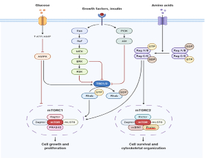
Figure 1. mTOR signaling pathway.
The Role of the Microbiome
Newer data suggests the material role of the gut microbiome in muscle health and performance. The impact of the microbiome on metabolism, inflammation, and nutrient absorption may have far-reaching effects on muscle development and maintenance [14]. But the study is still in its infancy and the mechanisms that are involved in these interactions need to be understood in order to be able to devise future therapeutic approaches.
Conclusion
The developments in muscle biology have enhanced our understanding of the intricate processes governing muscle plasticity, regeneration, and disease. Integrating advanced methodologies, such as single-cell genomics, along with innovative treatment strategies and an appreciation for the interactions of external factors like diet and exercise, offers new opportunities for improving muscle health.
However, significant challenges remain, particularly regarding translational research, muscle aging, and the role of the microbiome. Continued interdisciplinary collaboration and technological advancements will be key to overcoming these obstacles and unlocking innovative treatments that enhance muscle health across diverse populations.
Statements and Declarations
Funding
The authors declare that no funds, grants, or other support were received during the preparation of this manuscript.
Competing interests
The authors have no relevant financial or non-financial interests to disclose.
Author contributions
The work reported in this study has been carried out by the author, Prerna Mehta, who has also independently written this manuscript. This contains its development and planning, sample acquisition and testing and writing and finalizing the paper for publication.
Compliance with ethical standards
In compliance with the standards of publishing the results of this research, the author, Prerna Mehta, hereby states that there is no conflict of interest to report. This work did not include human subjects and animals, hence, no informed consent, or animal welfare statements were required. Guidelines to ethical best practice have been followed in the preparation of this manuscript.
References
2. Lloyd EM, Pinniger GJ, Murphy RM, Grounds MD. Slow or fast: Implications of myofibre type and associated differences for manifestation of neuromuscular disorders. Acta Physiol (Oxf). 2023 Aug;238(4):e14012.
3. Nara H, Watanabe R. Anti-Inflammatory Effect of Muscle-Derived Interleukin-6 and Its Involvement in Lipid Metabolism. Int J Mol Sci. 2021 Sep 13;22(18):9889.
4. Merchant RA, Chan YH, Anbarasan D, Seetharaman S, Au L, Nachammai V, et al. Impact of exercise and leucine-enriched protein supplementation on physical function, body composition, and inflammation in pre-frail older adults: a quasi-experimental study. Front Med (Lausanne). 2023 Aug 14;10:1204198.
5. Rehman SU, Ali R, Zhang H, Zafar MH, Wang M. Research progress in the role and mechanism of Leucine in regulating animal growth and development. Front Physiol. 2023 Nov 17;14:1252089.
6. Dy?ka D, Rodze? ?, Rodze? M, Pacholak-Klimas A, Ede G, Sethi S, et al. Ketogenic Diets for Body Weight Loss: A Comparison with Other Diets. Nutrients. 2025 Mar 10;17(6):965.
7. Careccia G, Mangiavini L, Cirillo F. Regulation of Satellite Cells Functions during Skeletal Muscle Regeneration: A Critical Step in Physiological and Pathological Conditions. Int J Mol Sci. 2023 Dec 29;25(1):512.
8. Sincennes MC, Brun CE, Lin AYT, Rosembert T, Datzkiw D, Saber J, et al. Acetylation of PAX7 controls muscle stem cell self-renewal and differentiation potential in mice. Nat Commun. 2021 May 31;12(1):3253.
9. Sadiq IZ, Abubakar FS, Katsayal BS, Ibrahim B, Adamu A, Usman MA, et al. Stem cells in regenerative medicine: Unlocking therapeutic potential through stem cell therapy, 3D bioprinting, gene editing, and drug discovery. Biomedical Engineering Advances. 2025 Jun 1;9:100172.
10. Goates S, Du K, Arensberg MB, Gaillard T, Guralnik J, Pereira SL. Economic Impact of Hospitalizations in US Adults with Sarcopenia. J Frailty Aging. 2019;8(2):93–9.
11. Aziziyan F, Asl SS, Mahdipour M, Fard RN, Sheykhhasan M. Mesenchymal stem cell-derived extracellular vesicles in musculoskeletal regeneration: mechanisms, applications, and future prospects. Stem Cell Res Ther. 2026 Jan 3;17(1):66.
12. Kaczmarek A, Kaczmarek M, Cia?owicz M, Clemente FM, Wola?ski P, Badicu G, et al. The Role of Satellite Cells in Skeletal Muscle Regeneration-The Effect of Exercise and Age. Biology (Basel). 2021 Oct 18;10(10):1056.
13. Mehta P. Revolutionizing therapeutics: Exploring novel biotechnological methods for disease management and treatment. Biophilia Insights. 2025 Jun 7;3(1).
14. Mehta P. Wearable Technology Revolution: Improving Health Monitoring and Well-Being. Medinformatics. 2025 Nov 18.