

Abstract
N-acetylcysteine (NAC) is an exogenous antioxidant drug against oxidative tissue injury. The current study aims to investigate the ameliorating role of NAC on cisplatin-induced nephrotoxicity and kidney injury biomarkers in male rats using an immunohistochemical study. The study was carried out on 50 male rats divided into two main groups and injected intraperitoneally. The control group consisted of 30 rats (GPI) and included three subgroups: GPIa received saline, GPIb received 500 mg/kg/day NAC, and GPIc received a single dose of 5 mg/kg CP. The treated group (20 rats, GPII) included two subgroups: GPIIa, administered CP after receiving NAC, and GPIIb, administered CP before receiving NAC. Fixed formalin-embedded paraffin sections were stained by hematoxylin and eosin to study the histopathological changes in rat kidney; silver methenamine stain was used to assess the thickness of the basement membrane, and immunoexpression of TNF-α and KIM-1 was assessed using an immunohistochemical protocol. Finally, we evaluated the urinary space, basement membrane thickness, and intensity of staining by image analysis software (ImageJ). The histopathology after 5 days of rats being administered CP revealed marked degeneration of kidney tissue, characterized by dilation of renal tubules, necrosis and pyknosis of tubular epithelial cells, thickening of the basement membrane, and atrophy of glomeruli, along with a significant increase in urinary space (p<0.001). An improvement of renal tubules and urinary space was observed in animals that received NAC before and after the administered CP. An increase in TNF-α and KIM-1 immunoexpression in the CP (GPIc) was observed, along with a significant increase in integrated optical density (IOD) (p≤0.05). However, a significant decrease of both biomarkers was seen in group CP after receiving NAC (p≤0.05). Therefore, receiving NAC leads to improved kidney tissue and alleviated kidney biomarkers. Thereby, receiving NAC before the CP intake may be useful for ameliorating the CP toxicity.
Keywords
Cisplatin, N-acetylcysteine, Kidney tissue, Immunohistochemical staining, TNF-α, KIM-1, Rats
Abbreviations
ABC: Avidin Biotin Complex; AKI: Acute Kidney Injury; CKD: Chronic Kidney Disease; DAB: 3,3-diaminobenzidin; HRP: Horse Radish Protein; PBS: Phosphate Buffer Saline; PASM: Periodic Acid-Silver Methenamine Stain; IOD: Integrated Optical Density; KIM-1: Kidney Injury Molecule-1; TNF-α: Tumor Necrosis Factor-α
Introduction
Chemotherapy drugs work on altering cell metabolism and cellular enzymes that may have the potential to interfere with some critical cellular processes [1], including programmed cell death, DNA damage and replication, drug resistance, and immune system reactivity [2]. Their anticancer properties might be eliminated by altering their chemical potency structure [3]. Cisplatin (CP) has been widely used in chemotherapy against cancers of the lung [4], head and neck, esophagus, stomach, colon, bladder, cervix, uterus, and most other advanced cancers such as cancers of the breast, pancreas, liver, kidney, prostate, glioblastomas, metastatic melanomas, and peritoneal or pleural mesotheliomas [5]. The anticancer activity of cisplatin depends on its binding to DNA, which induces the formation of inter-and intra-strand cross-links, causing defects in the DNA templates and arresting DNA synthesis and replication [6]. Also, cisplatin induces oxidative stress by generating Reactive Oxygen Species (ROS) and depleting cellular antioxidants, which leads to cellular dysfunction, activates inflammatory pathways, and causes death [7]. The disturbance of cellular antioxidants is producing chemokines and pro-inflammatory cytokines that recruit immune cells and amplify the inflammatory response of tissue injury and impair tissue repair [8,9]. N-Acetyl Cysteine (NAC) is a compound derived from the amino acid L-cysteine. It is an exogenous antioxidant drug that may protect against oxidative tissue injury [10]. It is able to ameliorate platinum deposition in the kidney, oliguria, proteinuria, and Blood Urea Nitrogen (BUN) concentration induced by cisplatin administration [11]. It was able to suppress the renal failure observed in a patient who was exposed to a cisplatin overdose [12]. The biomarkers are endogenous molecules that identify the physiological or pathophysiological processes distributed in different parts of the nephron and control the pharmacological responses [13]. The tumor necrosis factor-α (TNF-α) plays a pathogenic role in stimulating cytokine. As a pro-inflammatory cytokine, it increases the expression of inflammatory cytokines and chemokines. The chemokines expression in the kidney produces acute renal failure [14]. TNF-α was produced by a broad range of tissue and cells, including immune cells [15] and resident renal cells such as glomerular [16], tubular epithelial cells [17], and endothelial cells [18]. The TNF-α receptors expressed in glomerular (endothelium, mesangial and epithelial) and tubular cells [19]. On the other hand, the Kidney Injury Molecule-1 (KIM-1) is a transmembrane tubular protein that was undetectable in normal kidneys. It is expressed on epithelial cells of renal proximal tubule and has important role in renal tubulointerstitial damage [11], but it was markedly induced in renal injury including Acute Kidney Injury (AKI) and Chronic Kidney Disease (CKD) [20]. KIM-1 is associated with renal fibrosis and inflammation. It is upregulated in renal disease, indicating that it can be used as a non-invasive renal biomarker [21,22]. This study amid to evaluate the protective role of NAC against Cp induced nephrotoxicity using immunohistochemical assessment of renal biomarkers.
Materials and Methods
Experimental design
This study was carried out on 50 male Sprague Dawley rats weighing 100–120 g, obtained from the animal facility of Theodor Bilharz Institute, Cairo, Egypt. The animals were housed in plastic cages under standard laboratory conditions and had free access to food and water. All animal procedures were performed in accordance with the ARRIVE guidelines and the ICLAS (International Council for Laboratory Animal Science) Ethical Guideline for Researchers for the care and use of laboratory animals. The study was approved by the Institutional Animal Ethics Committee of Alexandria University (ALEXU-IACUC), Alexandria, Egypt.
The animals were divided into two main groups: Group I (GPI) consisted of 30 rats served as the control group and divided into three subgroups (10 rats for each). They were injected Intraperitoneally (IP) as follows: GPIa with normal saline, GPIb with NAC (500 mg/kg/day) for ten days [23], and GPIc with a single dose of 5 mg/kg cisplatin [24]. Group II (GPII) consisted of 20 rats, which served as the experimental group, divided into two subgroups (10 rats for each). GPIIa: Rats injected with NAC as in GPIb, on day 10, they were administered a single dose of 5 mg/kg cisplatin. GPIIb: Rats injected with a single dose of 5 mg/kg cisplatin on the 5th day received NAC (500 mg/kg/day) for 10 days.
Sample preparations
Animals were sacrificed after five days of CP and ten days of NAC administration. The rats were fasted overnight and sacrificed under anesthesia by inhalation of 3% isoflurane, with minimal suffering to the animals. The kidneys were removed, weighted and fixed in 10% neutral buffered formalin, dehydrated in ascending series of ethyl alcohol, cleared in xylene, and embedded in paraffin wax for processing the paraffin blocks. Then the sections of 5 μm were cut and processed for the following staining methods:
Haematoxylin and eosin stain
Sections were brought down to distilled water, stained with Mayer’s haematoxylin for 7 Min, eosin for 3 Min and processed to be mounted with Canada balsam [25]. Then the staining sections for all groups were examined under the light microscope and photographed. These microphotographs were stored in the computer for measuring the urinary space.
Periodic Acid-Silver Methenamine stain (PASM)
Periodic acid-silver methenamine stain (Sigma Aldrich 1.00820) was used to study the thickness of the basement membrane of the renal tubules and Bowman’s capsule. Aldehydes produced from the oxidized carbohydrate reduced the silver ions to metallic silver. Paraffin sections were brought down to distilled water, rinsed in periodic acid for 15 Min at 60°C, washed in distilled water. Sections were stained with methenamine silver solution for 1 hr at 60°C, washed in distilled water, treated with sodium thiosulphate, counter stained with light green for 1 min, and mounted with Canada balsam [26]. They were examined under the light microscope, photographed by digital camera and saved on the computer.
Immunohistochemical staining
Immunohistochemical staining of TNF-α and KIM-1proteins was carried out using the labeling streptavidin biotin complex (A universal kit (SABC–HRP) reagent and DAB stain) from Thermo Fisher–USA [27]. Five µm-thick paraffin sections were placed on coated glass slides. Sections were deparaffinized, rehydrated, and rinsed in distilled water. Slides were placed on a slide holder and submerged in large amount of antigen retrieval (citrate buffer pH 6) in an oven at 95°C for 20 min. Slides were removed from the oven to room temperature and allowed to cool for 30 Min. A circle was drawn on the slide around the tissue with a hydrophobic barrier pen. Slides were washed in Phosphate Buffer Saline (PBS) twice 5 min each. Endogenous peroxides activity was quenched using 3.0% H2O2 in PBS. Slides were washed in PBS twice 5 min each. Slides were incubated with serum blocking reagent for 30 min to block nonspecific binding. Slides were washed in pieces of tissue paper. Slides were incubated with primary antibodies of TNF-α and KIM-1over night at 4O°C in the humidified chamber. Negative control slides were incubated without adding primary antibody. Then Slides were allowed to reach the room temperature and then washed in PBS twice 5 min each. Slides incubated with conjugated ABC-HRP reagent for 30 min in humid chamber at room temperature, then washed in PBS twice 5 min each. Working solution of DAB was prepared and applied to tissue sections, and the reaction was monitored as the chromogenic reaction turned the epitope sites brown. Next step was preceded when the intensity of the signal is appropriate for imaging. Slides were rinsed in distilled water, stained with hematoxylin as nuclear counter stain, and then rinsed in running tap-water until the appropriate color of the hematoxylin appeared. Finally, Sections were dehydrated, cleared, and mounted. The (TNF-α, KIM-1) expressions are indicated by dark brown stain labeling to the epithelial cells membrane and cytoplasm. Negative control slides were stained without adding primary antibodies (TNF-α, KIM-1 proteins), and the blue color was an indicator to the negative. The semi quantitative evaluation of the immunohistochemical results of TNF-α and KIM-1 were done according to (+1 weak, +2 moderate, +3 strong, +4 intense).
Image analysis
Measurement of urinary space
The digitized image analysis identified the measurement of the urinary space for 10 images for the two groups. The image was used under the X40 objective lens, a maximal and a minimal pixel length in each field allow the measure performed on the 10 images of each group saved on the hard drive before measure. The 10 renal corpuscles from the cortex of each image were chosen randomly under a light microscope. The average score across the whole image should be taken to produce the width of urinary space (the area between the visceral and parietal epithelium of Bowman's capsular) using computer image analysis software (Image J) [28].
Integrated optical density (IOD)
The measurement of the Integrated Optical Density (IOD) of immunostaining density of TNF and KIM expression based on Gray-level acquisition, the analysis data was carried out by reading 10 fixed areas in one of 10 images for each group. The study used the X40 objective (actuallyX400 with Bar 50). The images were performed on and saved on the hard drive of computer to be analyzed. The maximal and minimal pixel intensity of the gray value in the field measured by pixel. The average score across the whole image should be taken. The mean values of each reaction were based on the mean of pixel number. The IOD based on Gray-level transition probabilities in digitized images upon degree of visible gray value from dark to light (0 up to 250) [29].
Statistical analysis
Data were expressed as mean ± SEM. Comparison of mean was done by the student’s t-test (One-Way ANOVA). Values of P <0.05 were considered statistically significant; evaluation was conducted with SPSS software.
Results
Histopathological findings
The examination of kidney sections stained with H & E of control group (GPI), saline GPIa, and NAC GPIb showed normal histological structure of renal tubules and glomeruli (Figures 1a and 1b). Cp GPIc After five days of single dose injection showed disintegration of renal tubular epithelial cells as desquamation, widespread necrosis, pyknosis, and karyolysis nuclei were observed. Proteinaceous casts in the tubular lumen, congested and atrophied glomeruli were noticed (Figures 1c and 1d). Treated group (GPII); rats administered cisplatin post receiving NAC for 10 days GPIIa, the kidney sections appeared with mild improvement of renal tubules, which have regularly arranged cuboidal epithelial cells of rounded nuclei (Figure 1e). While GPIIb of animals administered cisplatin before receiving NAC for ten days showed necrotic tubular epithelial cells with disintegrated and sloughing cytoplasm, the nuclei appeared with irregularly arranged and condensed chromatin at the periphery of the tubules with few pyknosis and karyolysis nuclei (Figure 1f).
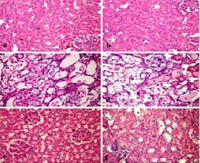
Figure 1. Paraffin section photomicrograph of rat kidney stained with H&E, bar = 50 µm, (a): GPIa showing normal kidney architecture of proximal tubules (→), and glomerulus (►). (b): GPIb showing normal histological Cal kidney tissue. (c & d): GPIc showing damage and disorganization of kidney tubules and intratubular casts (C), a pyknosis (p), and karyolysis (k) nuclei was seen. (e): GPIIa showing reorganized renal tubules (→) and glomeruli (►). (f): GPIIb showing dilatation of some tubular epithelial cells (*) and atrophied glomerulus (arrow) with infiltrated inflammatory cells.
Periodic acid-silver methenamine stain (PASM)
The Periodic acid-silver methenamine stain (PASM) was used for demonstrating the basement membrane of renal tubules and Bowman’s capsule. GPIa and GPIb, showed condensed silver stain of the renal tubular basement membrane and mild stain intensity of the Bowman’s capsules wall (Figures 2a and 2b). GPIc showed an increased thickness of the basement membrane and Bowman’s capsule wall mostly of atrophied glomeruli after 5 days of CP administration (Figures 2c and 2d). In the treated subgroups, the animals administered CP after 10 days of receiving NAC (GPIIa) showed a thin deposition of silver stain of the renal tubules, basement membrane, and brush border, and a mild thickness of Bowman’s capsule wall (Figure 2e). The animals administered CP for five days before receiving NAC for ten days (GPIIb) showed dark brown color deposits in most renal tubule basement membranes and thickness of Bowman’s capsule wall, which is decreased in density compared to the CP administration (Figure 2f).
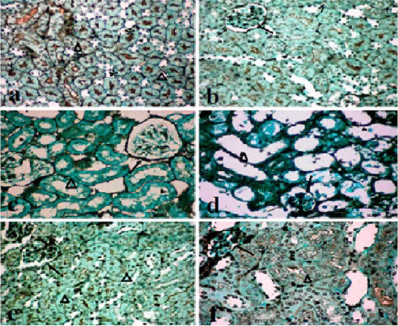
Figure 2. Paraffin section photomicrograph of rat kidney stained by PASM, bar = 50 µm, (a): GPIa showing condensed silver stain in brush border (*), and thin shadow in the basement membrane of renal tubules (▲). (b): GPIb showing Bowman’s capsule wall (bc), and thin basement membrane of renal tubules (▲). (c & d): GPIc showing glomeruli with thickness capsule wall (→), and basement membrane in most renal tubules. (e) GPIIa and (f): GPIIb showing thin shadow of silver stain in the renal tubule basement membranes and mild thickness of Bowman’s capsule.
Immunohistochemical findings
TNF-α immunoexpression
The TNF-α expression appeared as a dark brown stain labeling the epithelial cells of the injured area of renal tubules and glomeruli. Negative control slides were stained without adding primary antibody (TNF-α protein), and the blue color was an indicator of the negative control. Kidney sections of GPIa and GPIb showed negative TNF-α immunorexpression within the renal tubules (Figures 3a and 3b). Post five days of cisplatin administration, GPIc showed strong TNF-α immunoexpression with an intense positive stain in the epithelial cells of the necrotic and glomerulus mesangial cells (Figures 3c and 3d). GPIIa showed a decrease of TNF-α immunoexpression in regenerative epithelial cells of the renal tubules (Figure 3e). While GPIIb demonstrated a mild decrease of TNF-α expression compared to GPIc, which appeared as a strong stain in most injured renal tubules and glomeruli (Figure 3f).
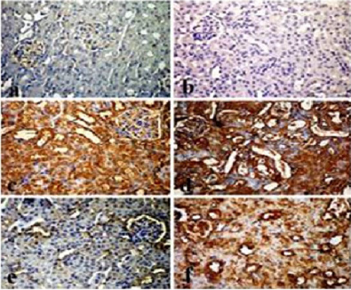
Figure 3. Photomicrograph of FFPE Immunostaining by TNF-α (ABC & DAB Stain, Bar = 50 µm), (a): GPIa showing weak immunoexpression of TNF-α in the membranes of few renal tubular cells and mesangial cells in the glomerulus. (b): GPIb showing negative TNF-α immunoexpression in renal tubules. (c & d): GPIc showing strong to intense TNF-α immunorexpression (*) within the destructive renal tubules and atrophied glomerulus. (e): GPIIa showing moderate positive stain in most renal tubule cell membranes and glomeruli. (f): GPIIb showing increased in the TNF-α expression compared to (e).
KIM-1 immunoexpression
The KIM-1 immunoexpression detected in Formalin-fixed Paraffin sections (FFPE) appeared as a brown color labeling the epithelial cell membrane and cytoplasm of the renal tubules, while the blue color indicated a negative control reaction. Photomicrographs of the rat kidney control group section of GPIa and GPIb showed weak immunostaining in the cell membrane of renal tubules and mesangial cells of glomeruli (Figures 4a and 4b). After 5 days of CP administration, GPIc showed an intense KIM-1 positive immunostaining in most cells of the renal tubules and Bowman’s capsules. However, there is a strong expression in the mesangial cells of glomeruli and the cytoplasm of degenerative renal tubular cells (Figures 4c and 4d). GPIIa rats administered CP after receiving NAC for 10 days showed moderate positive KIM-1 immunostaining (Figure 4e), which decreased compared to GPIc. At GPIIb, administering CP before receiving NAC for 10 days showed a mild decrease of the KIM-1 expression; it appeared as strong positive immunostaining in the renal tubular epithelia cell membrane and mesangial cells of glomeruli (Figure 4f).
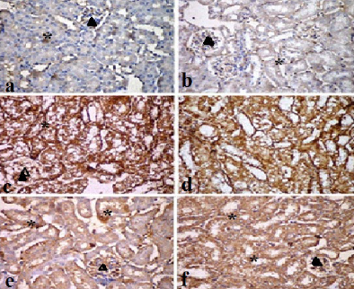
Figure 4. Photomicrograph of FFPE of KIM-1 immunostaining by EABC & DAB stain (Bar = 50 µm). (a): GPIa showing weak immunoexpression of KIM-1 in the membranes of a few renal tubular cells and mesangial cells in the glomerulus. (b) GPIb showing weak KIM-1 immunoexpression detected in renal tubules (*) and few mesangial cells in glomerulus (▲). (c & d): GPIc showing an intense KIM-1 immunorexpression with the destructive renal tubular epithelial cell membrane and cytoplasm (*) and atrophied glomerulus (▲). (e): GPIIa showing decreased KIM-1 immunoexpression, appearing as moderately positive in most renal tubular epithelial cell membranes and glomeruli. (f): GPIIb showing increased KIM-1 expression as a strong positive KIM-1 immunostaining density in injured renal tubules (*) and atrophied glomeruli (▲).
Digital imaging findings
Measurement of urinary space
Table 1 Distribute the mean values of the width of urinary spaces, which were 2.68 ± 0.10, 2.74 ± 0.12, and 4.41 ± 0.13 for GPI; GPIa, GPIb, and CP administration after 5 days (GPIc), and 2.79 ± 0.13 and 3.94 ± 0.13 for GPII. A significant difference was noticed in the urinary spaces of subgroup rats administered CP for 5 days and in the CP before receiving NAC for 10 days, but there was no statistically significant difference between the urinary spaces in saline, NAC, and rats administered CP after receiving NAC for 10 days.
Integrated Optical Density (IOD)
The image analysis identified the Integrated Optical Density (IOD) of stain density and calculated the relative density of quantitative immunohistochemical stains of both antibodies, TNF-α and KIM-1, by algorithmic calibrations. An IOD in digitized images was calibrated from dark to light (180 down to 70 by pixel). It was illustrated in Table 1 and Figure 5.
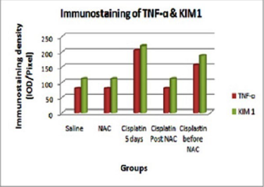
Figure 5. The bar graph represents the quantitative immunoexpression of both markers in rats' kidney tissues.
IOD of TNF-α immunostaining
Table 1 shows the mean ± SD values of the TNF-α immunostaining density IOD for control subgroups GPI; GPIa, GPIb, and CP administration after 5 days (GPIc) were 79.68 ± 0.55, 79.73 ± 0.84, and 205.53 ± 0.82, respectively. A significant difference was noticed between the IOD in the cisplatin-administered group, but there was no statistically significant difference between the IOD in the saline and NAC subgroups. The mean values of the IOD for treated GP II (CP after receiving NAC and CP before receiving NAC for 10 days) were 80.02 ± 0.91 and 205.04 ± 5.54, respectively. A significant difference was noticed between the two subgroups.
IOD of KIM-1 immunostaining
Table 1 shows the distribution of the mean ± SD values of the Kim1 immunostaining density IOD for control subgroups; saline GPIa, NAC GPIb, and CP GPIc after 5 days were 111.71 ± 0.63, 111.77 ± 0.77, and 218.81 ± 0.88, respectively. A highly significant increase was noticed in the cisplatin-administered group (P<0.001) compared to the NAC and saline subgroups. But there was no statistically significant difference between the two subgroups. The mean values of the IOD for GPII (CP after receiving NAC and CP before receiving NAC for 10 days) were 111.88 ± 0.48 and 187.40 ± 1.06, respectively. A significant difference increase was noticed between the two subgroups (P<0.001).
|
Parameters ► Groups▼ |
Urinary space µm |
IOD of TNF-α pixel |
IOD of KIM 1 pixel |
|
GPIa Saline |
2.68d ± 0.10 |
79.73d ± 0.84 |
111.71a ± 0.63 |
|
GPIb NAC |
2.74d ± 0.12 |
79.73d ± 0.97 |
111.77d ± 0.77 |
|
GPIc Cisplatin 5 days |
4.41a ± 0.13 |
205.53a ± 0.82 |
218.81a ± 0.88 |
|
GPIIa Cisplatin post 10 days NAC |
2.79d ± 0.13 |
80.02d ± 0.91 |
111.88d ± 0.48 |
|
GPIIb Cisplatin before 10 days NAC |
3.94b ± 0.13 |
156.80b ± 1.02 |
187.40b ± 1.06 |
|
F |
110.136 |
3441.339 |
17230.796* |
|
P |
<0.001 |
<0.001 |
<0.001 |
|
F: F test (ANOVA) Different superscripts in the same column statically significant *: Statistically significant at p ≤0.05. a, b, c Significantly different from the control value (d) at P ≤0.05. |
|||
Discussion
Cisplatin (CP) is a potent chemotherapeutic agent used to kill cancer cells [30] and is widely used as an adjuvant therapy for most cancer cases [31]. A study explained that the CP mainly accumulates in the renal epithelial cells induced nephrotoxicity [32]. The CP increased production of reactive oxygen and nitrogen species, resulting in significant damage to cell structure and function, including DNA breaks, lipid peroxidation, protein nitration, enzyme dysfunction [33]. Haase et al. [34], claimed that the histopathological examination of the kidney is the gold standard method to detect renal damage. This study focused on kidney injury induced by a single dose of cyclophosphamide before and after receiving N-acetylcysteine. The histopathological results revealed a single dose of CP induces dilatation of the renal tubules with a thickened epithelial lining of the renal tubules and loss of brush border, necrotic and pyknotic epithelial tubular cells and an increase in atrophied glomeruli was observed. The finding illustrated the cell membrane destruction, tubular dysfunction through production of the Reactive Oxygen Species (ROS). Many investigators reported that CP treatment exhibited reduced glomerular filtration rate [7,35]. The accumulation of glycoproteins around renal glomeruli and tubular basement membrane was due to increased collagen deposition and could explain the renal damage [36]. An intense Methenamine Periodic Acid Silver (MPAS) stain indicated a marked thickening of both the tubular and glomerular basement membrane [37]. Our results clarified that MPAS reaction verified the thickness of the renal tubules and Bowman's capsule wall. So, the thickness of the glomerular and tubular basement membranes leads to cellular damage, and progressive loss of kidney filtration function.
N-Acetylcysteine (NAC), a thiol antioxidant prevented and reduced CP nephrotoxicity [38,39]. Our histopathological results revealed an improvement of kidney architecture in groups receiving NAC before and after CP intake. Bowman's space (urinary space), the area between the visceral and parietal epithelium of Bowman's capsule revealed a statistically significant difference (p <0.001) in its width after the 5th day of administering a single dose of CP and administering CP before receiving the NAC group, but there was no significant difference between the urinary spaces in the groups receiving saline, NAC, and administered CP post receiving NAC. This finding illustrated the role of the NAC for reducing the atrophy of glomeruli numbers and the recovery of some glomeruli and regulation of renal tubules. The same result was documented: that the CP administration post receiving NAC, some glomeruli seemed to have disappeared, and the remaining glomeruli often appeared to have enlarged periglomerular space [39]. Also, a recovering of glomerular organization and a reduction of urinary spaces was noticed after receiving NAC [40]. Many investigators explained that NAC is a powerful antioxidant [41,42] and has a protective effect against kidney damage caused by free radicals and reverses the anti-apoptotic pathway induced by CP administration [43,44]. This finding illustrated the positive role of the NAC on slowing progression and managing kidney health. Also, methenamine silver stain clarified a thin shadow stain in most of the renal tubular basement membrane and brush border. This finding revealed the efficacy of NAC against the CP injury. Receiving NAC before or after CP administration could reduce the inhibition of membranous physiological function, as well as reduce the urinary space width.
Furthermore, the main objective of the present work was to detect the localization and immunoexpression of TNF-α in the kidney tissue. The change in the levels of pro-inflammatory cytokines such as TNF-α has crucial roles in kidney tissue, including immune cells, mesangial cells, glomeruli, tubular epithelial cells, and endothelial cells [45,46]. The present immunohistochemical analysis of TNF-α revealed a weak expression in renal tubules and glomeruli in the control saline and NAC subgroups but elevated in the CP group as a strong brown positive stain which may contribute to renal injury. Whereas, in the group administered CP after receiving NAC for ten days, there was a marked reduction in TNF-α expression of most regenerated renal epithelial cells compared with the CP-administered group. These results were confirmed by a study that reported that the urine TNF-α was undetectable in saline-treated mice and was up regulated in mice injected with CP for 24–48 hr and sustained to 72 hr [47]. This is evident in serum: the TNF-α concentration increased gradually after CP injection and markedly peaked after three days [48]. Otherwise, the group of CP before receiving NAC revealed a mild reduction of TNF-α expression in most renal tubular cells and glomeruli as compared with the CP group. This reduction of TNF-α expression supports the role of the NAC in inhibiting the inflammatory cytokines induced by CP nephrotoxicity. This result documented that NAC, a potent anti-inflammatory and antioxidant and could be inhibiting TNF-α biosynthesis [49].
KIM-1 is a sensitive and accurate molecular biochemical marker for early kidney damage detection of CP-induced nephrotoxicity. The present results of the immunohistochemical staining revealed a weak staining in the control saline and NAC subgroups, but 5 days after cisplatin administration, the labeling of KIM-1 was more widely distributed in the cytoplasm at the apical surface of the epithelial cells of the renal tubules, glomerular mesangial cells, and endothelial cells. Elevated KIM-1 levels were also found in human renal diseases associated with renal fibrosis, inflammation, and dysfunction [21,50]. The high KIM-1 expression was due to increased kidney injury induced by the CP. KIM 1 is undetectable in normal renal tissue but highly expressed in kidney injury, it was expressed at high levels. The elevated KIM-1 expression was considered an early indicator of acute kidney injury over the conventional biomarker [51]. CP complex activated signaling pathways, such as KIM-1 expression in kidney tissue injury, contribute to a decline in glomerular filtration rate [22]. This finding is in accordance with low expression of KIM-1 immunostaining in epithelial cells of the rats’ renal tubules administered CP after receiving NAC. Whereas a strong positive immunostaining of KIM-1 demonstrated in the renal tubular epithelial cell membranes and mesangial cells of the glomeruli in rats administered CP before receiving NAC. Similarly, KIM-1 expression is up regulated in the kidneys of rats administered CP only [52].
In addition, the quantitative elevation of the immunostaining density through the digital software analysis called the Integrated Optical Density (IOD) confirmed the immunoexpression of both markers and revealed the effectiveness of NAC treatment in preventing nephrotoxicity. The immunostaining density of TNF-α and KIM-1 revealed a significant increase (p≤0.05) of the IOD in the CP administration and CP before receiving NAC groups. But there was no statistically significant difference in the IOD in groups of saline, NAC, and CP post receiving NAC. So, the image analysis can be used to evaluate an immunohistochemical expression for producing a guideline score and insight into the role of the different markers on tumorigenesis.
Conclusion
The findings from this study was concluded that cisplatin increased the expression of both kidney biomarkers, TNF-α & KIM-1, while animals receiving NAC before and after CP administration decreased them. It could be attributed to the regaining of kidney structure in animals receiving NAC, leading to the alleviation of both markers. The immunohistochemistry technique revealed that both markers are more sensitive and specific to detect the renal damage. Also, the efficacy of the NAC supplement before CP exhibited anti-inflammatory properties through the improvement of kidney histological architecture and immune responses to oxidative stress through ameliorating the biomarker signal complex of TNF-α and KIM-1 expression.
Acknowledgments
The authors wish to thank the Cooperative of the Medical Center of Technology Research, Alexandria University, Egypt, for providing the animal care and immunohistochemical techniques.
Funding
The current work did not receive any research funding from any organization.
Ethics Approval
The protocol has been performed in accordance with relevant guidelines and regulations and peer-reviewed by a specialized scientific reviewers committee of the Medical Research Institute, Alexandria University. It was approved by the Institutional Animal Ethics Committee of Alexandria University (ALEXU-IACUC), Alexandria Egypt.
Consent for Publication
The co-author's consent to the publication.
Conflicts of Interest
None to declare.
Disclosure
The authors have nothing to report.
Data Availability
The dataset is available from the corresponding author on reasonable request.
Authors Contributions
Abdelazim Ahmed Ismail: Conceptualization, study design, and substantive revision of the manuscript. Afaf Mousad Amin: Conceptualization, main supervision, drafting, and revision of the manuscript. Amani Hussein Kazem: Interpretation of histopathological results and drafting of the manuscript. Noura Abdel-Kader Matar: Animal care, histopathology and histochemical procedures, and interpretation and drafting of the results. Mona Abdel-Hamed Yehia: Interpretation of immunohistochemistry data, software execution, drafting of the work, and revision of the manuscript.
References
2. Delou JMA, Souza ASO, Souza LCM, Borges HL. Highlights in Resistance Mechanism Pathways for Combination Therapy. Cells. 2019 Aug 30;8(9):101.
3. Anand U, Dey A, Chandel AKS, Sanyal R, Mishra A, Pandey DK, et al. Cancer chemotherapy and beyond: Current status, drug candidates, associated risks and progress in targeted therapeutics. Genes Dis. 2022 Mar 18;10(4):1367–401.
4. Pabla N, Dong Z. Cisplatin nephrotoxicity: mechanisms and renoprotective strategies. Kidney Int. 2008 May;73(9):994–1007.
5. Boulikas T, Vougiouka M. Recent clinical trials using cisplatin, carboplatin and their combination chemotherapy drugs (review). Oncol Rep. 2004 Mar;11(3):559–95.
6. Mohammed OA, Abdel-Reheim MA, Saleh LA, Alamri MMS, Alfaifi J, Adam MIE, et al. Alvespimycin Exhibits Potential Anti-TGF-β Signaling in the Setting of a Proteasome Activator in Rats with Bleomycin-Induced Pulmonary Fibrosis: A Promising Novel Approach. Pharmaceuticals (Basel). 2023 Aug 9;16(8):1123.
7. Nasr M, Cavalu S, Saber S, Youssef ME, Abdelhamid AM, Elagamy HI, et al. Canagliflozin-loaded chitosan-hyaluronic acid microspheres modulate AMPK/NF-κB/NLRP3 axis: A new paradigm in the rectal therapy of ulcerative colitis. Biomed Pharmacother. 2022 Sep;153:113409.
8. Saber S, Yahya G, Gobba NA, Sharaf H, Alshaman R, Alattar A, et al. The Supportive Role of NSC328382, a P2X7R Antagonist, in Enhancing the Inhibitory Effect of CRID3 on NLRP3 Inflammasome Activation in Rats with Dextran Sodium Sulfate-Induced Colitis. J Inflamm Res. 2021 Jul 21;14:3443–63.
9. Elmorsy EA, Saber S, Hamad RS, Abdel-Reheim MA, El-Kott AF, AlShehri MA, et al. Advances in understanding cisplatin-induced toxicity: Molecular mechanisms and protective strategies. Eur J Pharm Sci. 2024 Dec 1;203:106939.
10. Nayak DU, Karmen C, Frishman WH, Vakili BA. Antioxidant vitamins and enzymatic and synthetic oxygen-derived free radical scavengers in the prevention and treatment of cardiovascular disease. Heart Dis. 2001 Jan-Feb;3(1):28–45.
11. Appenroth D, Winnefeld K, Schröter H, Rost M. Beneficial effect of acetylcysteine on cisplatin nephrotoxicity in rats. J Appl Toxicol. 1993 May-Jun;13(3):189–92.
12. Luo J, Tsuji T, Yasuda H, Sun Y, Fujigaki Y, Hishida A. The molecular mechanisms of the attenuation of cisplatin-induced acute renal failure by N-acetylcysteine in rats. Nephrol Dial Transplant. 2008 Jul;23(7):2198–205.
13. Zhang WR, Parikh CR. Biomarkers of Acute and Chronic Kidney Disease. Annu Rev Physiol. 2019 Feb 10;81:309–33.
14. Ramesh G, Reeves WB. TNF-alpha mediates chemokine and cytokine expression and renal injury in cisplatin nephrotoxicity. J Clin Invest. 2002 Sep;110(6):835–42.
15. Ramesh G, Reeves WB. TNFR2-mediated apoptosis and necrosis in cisplatin-induced acute renal failure. Am J Physiol Renal Physiol. 2003 Oct;285(4):F610–8.
16. Baud L, Oudinet JP, Bens M, Noe L, Peraldi MN, Rondeau E, et al. Production of tumor necrosis factor by rat mesangial cells in response to bacterial lipopolysaccharide. Kidney Int. 1989 May;35(5):1111–8
17. Neale TJ, Rüger BM, Macaulay H, Dunbar PR, Hasan Q, Bourke A, et al. Tumor necrosis factor-alpha is expressed by glomerular visceral epithelial cells in human membranous nephropathy. Am J Pathol. 1995 Jun;146(6):1444–54.
18. Ramesh G, Reeves WB. p38 MAP kinase inhibition ameliorates cisplatin nephrotoxicity in mice. Am J Physiol Renal Physiol. 2005 Jul;289(1):F166–74.
19. Albaugh G, Kann B, Strande L, Vemulapalli P, Hewitt C, Alexander JB. Nicotine induces endothelial TNF-alpha expression, which mediates growth retardation in vitro. J Surg Res. 2001 Aug;99(2):381–4
20. Baud L, Ardaillou R. Tumor necrosis factor in renal injury. Miner Electrolyte Metab. 1995;21(4–5):336–41.
21. Kin Tekce B, Tekce H, Aktas G, Sit M. Evaluation of the urinary kidney injury molecule-1 levels in patients with diabetic nephropathy. Clin Invest Med. 2014 Dec 1;37(6):E377–83.
22. Van Timmeren MM, van den Heuvel MC, Bailly V, Bakker SJ, van Goor H, Stegeman CA. Tubular kidney injury molecule-1 (KIM-1) in human renal disease. J Pathol. 2007 Jun;212(2):209–17.
23. Shadnia S, Dasgar M, Taghikhani S, Mohammadirad A, Khorasani R, Abdollahi M. Protective effects of alpha-tocopherol and N-acetyl-cysteine on diazinon-induced oxidative stress and acetylcholinesterase inhibition in rats. Toxicol Mech Methods. 2007;17:109–15.
24. Jonker JW, Schinkel AH. Pharmacological and physiological functions of the polyspecific organic cation transporters: OCT1, 2, and 3 (SLC22A1-3). J Pharmacol Exp Ther. 2004 Jan;308(1):2–9.
25. Bancroft DJ, Gamble M. Theory and Practice of Histological Technique. 5th ed. London: Churchill Livingstone; 2002. pp.116–7.
26. Cathro HP, Shen SS, Truong LD. Diagnostic histochemistry in medical diseases of the kidney. Semin Diagn Pathol. 2018 Nov;35(6):360–9.
27. Salguero FJ, Mekonnen T, Ruiz-Villamor E, Sánchez-Cordón PJ, Gómez-Villamandos JC. Detection of monokines in paraffin-embedded tissues of pigs using polyclonal antibodies. Vet Res. 2001 Nov-Dec;32(6):601–9.
28. Abdu S, Ali A, Ansari S. Cytotoxic effect of ochratoxin A on the renal corpuscles of rat kidney: could ochratoxin A cause kidney failure? Histol Histopathol. 2011 May;26(5):543–9.
29. Matos LL, Stabenow E, Tavares MR, Ferraz AR, Capelozzi VL, Pinhal MA. Immunohistochemistry quantification by a digital computer-assisted method compared to semiquantitative analysis. Clinics (Sao Paulo). 2006 Oct;61(5):417–24.
30. Hajdu SI. A note from history: landmarks in history of cancer, part 1. Cancer. 2011 Mar 1;117(5):1097–102.
31. Galluzzi L, Senovilla L, Vitale I, Michels J, Martins I, Kepp O, et al. Molecular mechanisms of cisplatin resistance. Oncogene. 2012 Apr 12;31(15):1869–3.
32. Santos NA, Bezerra CS, Martins NM, Curti C, Bianchi ML, Santos AC. Hydroxyl radical scavenger ameliorates cisplatin-induced nephrotoxicity by preventing oxidative stress, redox state unbalance, impairment of energetic metabolism and apoptosis in rat kidney mitochondria. Cancer Chemother Pharmacol. 2008 Jan;61(1):145–55.
33. Huang Q, Dunn RT 2nd, Jayadev S, DiSorbo O, Pack FD, Farr SB, et al. Assessment of cisplatin-induced nephrotoxicity by microarray technology. Toxicol Sci. 2001 Oct;63(2):196–207.
34. Haase M, Mertens PR. Urinary biomarkers--silver bullets to faster drug development and nephron protection. Nephrol Dial Transplant. 2010 Oct;25(10):3167–9.
35. Chirino YI, Trujillo J, Sánchez-González DJ, Martínez-Martínez CM, Cruz C, Bobadilla NA, et al. Selective iNOS inhibition reduces renal damage induced by cisplatin. Toxicol Lett. 2008 Jan 4;176(1):48–57.
36. Palipoch S, Punsawad C. Biochemical and histological study of rat liver and kidney injury induced by Cisplatin. J Toxicol Pathol. 2013 Sep;26(3):293–9.
37. Ghaly EN, Gergis SW, Aziz JN, Yassa HD, Hassan HA. Role of mesenchymal stem cell therapy in cisplatin induced nephrotoxicity in adult Albino rats: Ultrastructural & Biochemical study. Act Med Int. 2014 Jul 1;1(2): 57–66.
38. Deneke SM. Thiol-based antioxidants. Curr Top Cell Regul. 2000;36:151–80.
39. Wang And X, Guo Z. The role of sulfur in platinum anticancer chemotherapy. Anticancer Agents Med Chem. 2007 Jan;7(1):19–34.
40. Conesa EL, Valero F, Nadal JC, Fenoy FJ, López B, Arregui B, et al. N-acetyl-L-cysteine improves renal medullary hypoperfusion in acute renal failure. Am J Physiol Regul Integr Comp Physiol. 2001 Sep;281(3):R730–7.
41. Sorbello M, Morello G, Parrinello L, Molino C, Rinzivillo D, Pappalardo R, et al. Effect of N-acetyl-cysteine (NAC) added to fenoldopam or dopamine on end-tidal carbon dioxide and mean arterial pressure at time of renal artery declamping during cadaveric kidney transplantation. Transplant Proc. 2010 May;42(4):1056–60.
42. Wu YJ, Muldoon LL, Neuwelt EA. The chemoprotective agent N-acetylcysteine blocks cisplatin-induced apoptosis through caspase signaling pathway. J Pharmacol Exp Ther. 2005 Feb;312(2):424–31.
43. Tarladacalisir YT, Kanter M, Uygun M. Protective effects of vitamin C on cisplatin-induced renal damage: a light and electron microscopic study. Ren Fail. 2008;30(1):1–8.
44. Cermik H, Taslipinar MY, Aydin I, Agilli M, Aydin FN, Ucar F, et al. The relationship between N-acetylcysteine, hyperbaric oxygen, and inflammation in a rat model of acetaminophen-induced nephrotoxicity. Inflammation. 2013 Oct;36(5):1145–52.
45. Lu CY, Hartono J, Senitko M, Chen J. The inflammatory response to ischemic acute kidney injury: a result of the 'right stuff' in the 'wrong place'? Curr Opin Nephrol Hypertens. 2007 Mar;16(2):83–9.
46. Zhang B, Ramesh G, Norbury CC, Reeves WB. Cisplatin-induced nephrotoxicity is mediated by tumor necrosis factor-alpha produced by renal parenchymal cells. Kidney Int. 2007 Jul;72(1):37–44.
47. Ramesh G, Kimball SR, Jefferson LS, Reeves WB. Endotoxin and cisplatin synergistically stimulate TNF-alpha production by renal epithelial cells. Am J Physiol Renal Physiol. 2007 Feb;292(2):F812–9.
48. Miyawaki Y, Ueki M, Ueno M, Asaga T, Tokuda M, Shirakami G. D-allose ameliorates cisplatin-induced nephrotoxicity in mice. Tohoku J Exp Med. 2012 Nov;228(3):215–21.
49. Shalby AB, Assaf N, Ahmed HH. Possible mechanisms for N-acetyl cysteine and taurine in ameliorating acute renal failure induced by cisplatin in rats. Toxicol Mech Methods. 2011 Sep;21(7):538–46.
50. Vaidya VS, Ferguson MA, Bonventre JV. Biomarkers of acute kidney injury. Annu Rev Pharmacol Toxicol. 2008;48:463–93.
51. Hoffmann D, Adler M, Vaidya VS, Rached E, Mulrane L, Gallagher WM, et al. Performance of novel kidney biomarkers in preclinical toxicity studies. Toxicol Sci. 2010 Jul;116(1):8–22.
52. Tonomura Y, Tsuchiya N, Torii M, Uehara T. Evaluation of the usefulness of urinary biomarkers for nephrotoxicity in rats. Toxicology. 2010 Jun 29;273(1–3):53–9