Abstract
Background: Pleomorphic rhabdomyosarcoma (PRMS) is a rare and aggressive soft tissue sarcoma that typically occurs in the extremities of older adults. Involvement of the splenic hilum is extremely rare and has not been previously reported in the literature.
Case presentation: We present the case of an 81-year-old female with a remote history of high-grade serous carcinoma of the ovary who presented with new-onset vaginal bleeding. Imaging revealed a mass in the left upper quadrant at the interface of the spleen and pancreatic tail. Surgical resection revealed a lesion at the splenic hilum, and final pathology confirmed pleomorphic rhabdomyosarcoma. The postoperative course was uncomplicated, aside from a transient pancreatic leak managed with a drain.
Conclusion: To our knowledge, this case represents the first presentation of PRMS at the splenic hilum, confounded by patients' history of prior ovarian malignancy. Accurate histopathologic diagnosis and timely surgical management are critical in such rare and diagnostically challenging cases.
Keywords
Pleomorphic rhabdomyosarcoma, Soft tissue sarcoma, Splenic Hilum, Ovarian cancer, Exploratory laparotomy, Multidisciplinary approach, Postoperative surveillance
Introduction
Pleomorphic rhabdomyosarcoma (PRMS) is the least common subtype of rhabdomyosarcoma, occurring almost exclusively in adults, with a predilection for the deep soft tissues of the extremities [1–3]. Visceral involvement, especially of the upper abdominal organs, is exceedingly rare. To our knowledge, there are no reported cases of PRMS originating from the splenic hilum. This case is further complicated by the patient’s history of high-grade serous ovarian carcinoma, which raised initial suspicion for recurrent gynecologic malignancy. We report the first known case of PRMS localized to the splenic hilum. Written patient consent was obtained.
Case Presentation
An 81-year-old female with a significant oncologic history of high-grade serous carcinoma of the ovary, status post total abdominal hysterectomy, bilateral salpingo-oophorectomy, omentectomy, and adjuvant chemotherapy three years prior, presented with new-onset vaginal bleeding. Physical exam was unremarkable. Laboratory studies were unremarkable; tumor markers were not elevated.
Imaging
Contrast-enhanced CT of the abdomen and pelvis demonstrated a complex left upper quadrant mass, centered between the tail of the pancreas and the spleen. The lesion appeared to arise exophyticAlly from the pancreas and was described as either a complex cystic lesion or a solid mass (Figure 1). There was no other evidence of metastatic disease.
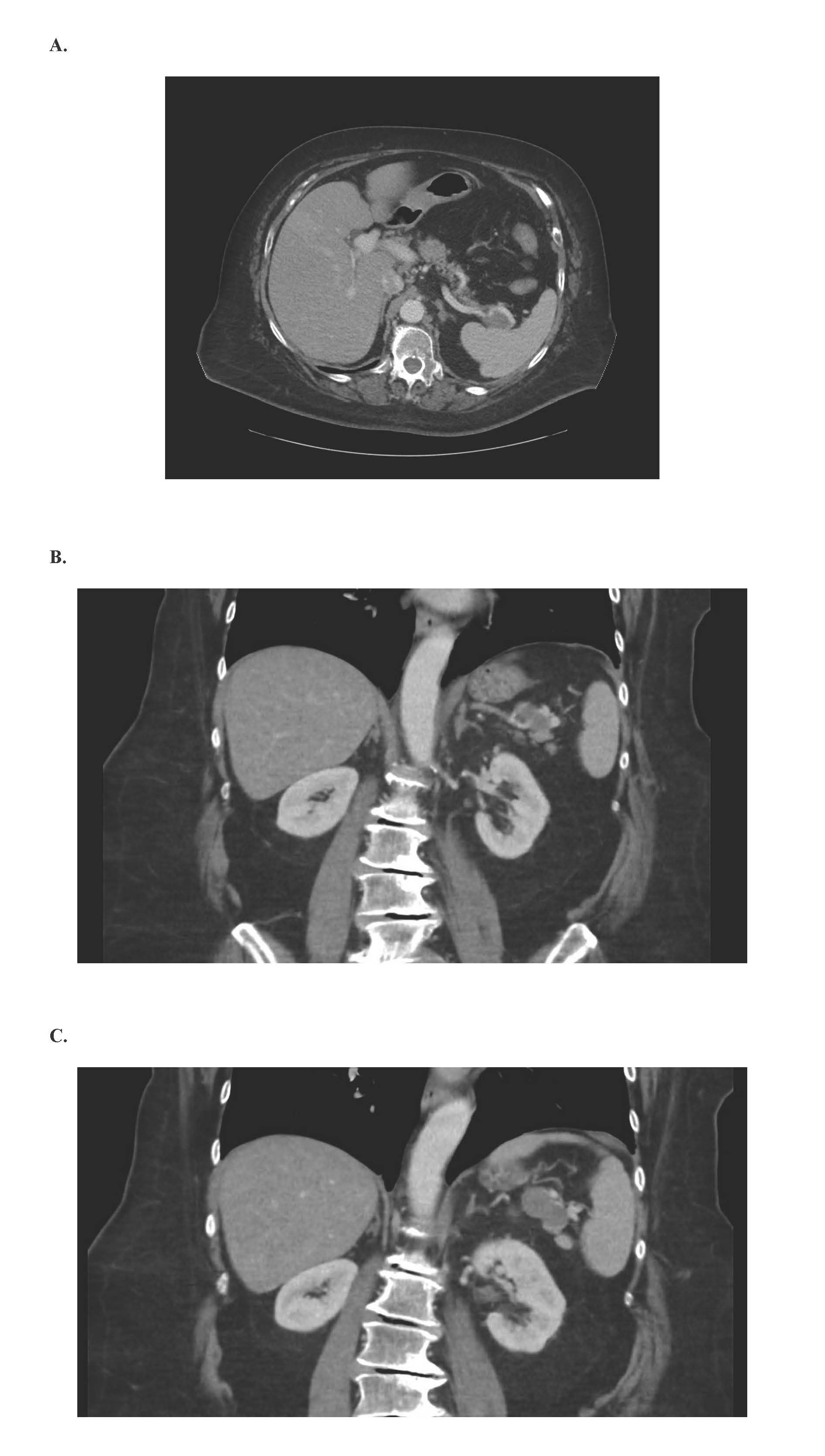
Figure 1. CT images.
Surgical findings
Exploratory laparotomy revealed a well-circumscribed, firm mass at the splenic hilum, intimately associated with the tail of the pancreas. A splenectomy with en bloc resection of the mass and distal pancreatectomy was performed (Figure 2). No gross peritoneal disease or evidence of recurrent ovarian cancer was identified intraoperatively.
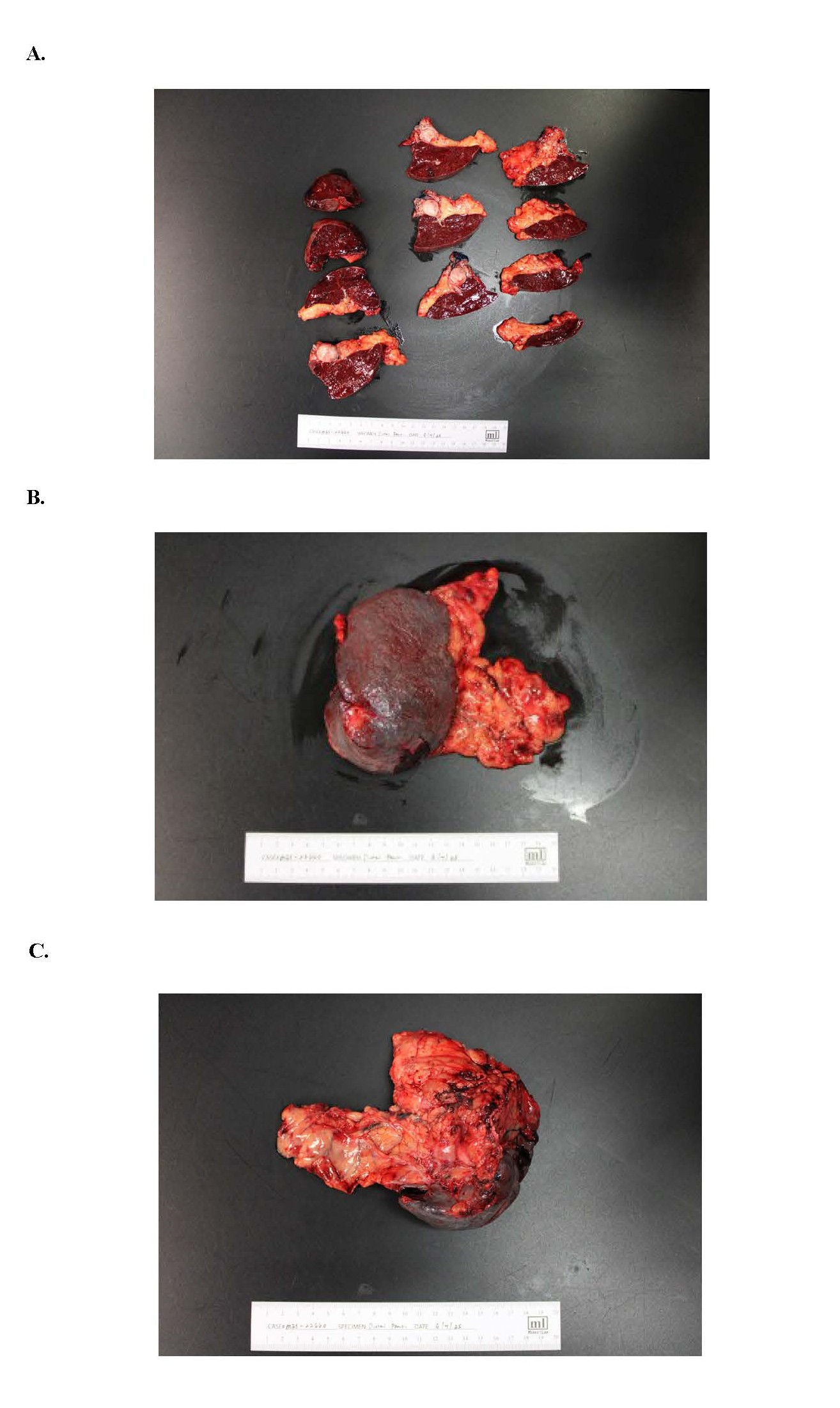
Figure 2. Gross pathology.
Pathology
The mass was centered at the splenic hilum, abutting but not clearly invading the pancreatic parenchyma. Hematoxylin and eosin (H&E) stain demonstrated pleomorphism, and the presence of rhabdom oblasts (Figures 3A and 3B). Immunohistochemistry demonstrated that tumor cells to be strongly and diffusely positive for vimentin and Desmin as well as focally positive for Myogenin (Figure 3C) consistent with pleomorphic rhabdomyosarcoma. Margins were negative for tumor. No ovarian cancer recurrence was identified.
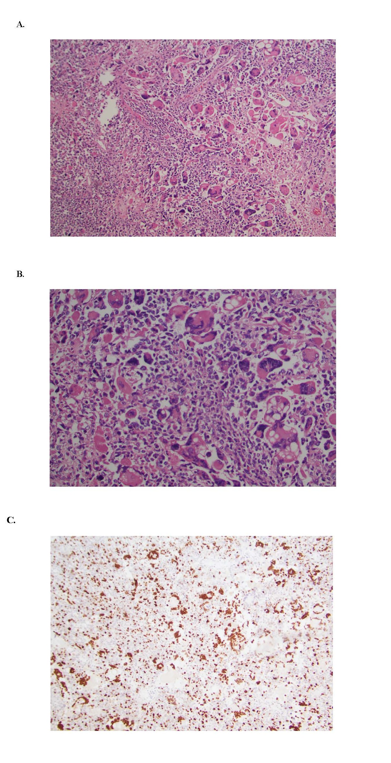
Figure 3. Pathology Showing (A) Hematoxylin and Eosin (H&E) Stain At 100X, (B) H&E Stain At 200X, and (C) Myogenin Stain-100x.
Postoperative course
The patient’s immediate recovery was uncomplicated. Drain output was minimal, but a postoperative pancreatic leak was suspected based on elevated drain amylase (385 U/L vs serum 34 U/L). The patient was discharged on postoperative day 3 with the drain in place. The leak resolved spontaneously, and the drain was removed on postoperative day 5 in clinic.
Oncology plan
After multidisciplinary discussion, the patient was recommended for surveillance given the rarity of the tumor, lack of established adjuvant guidelines, and the patient's age and performance status. Follow-up CT imaging at three-month intervals was planned.
Discussion
Pleomorphic rhabdomyosarcoma is a rare high-grade sarcoma comprising less than 5% of all rhabdomyosarcomas, most often occurring in the extremities of adults over age fifty. The pathogenesis remains unclear, and unlike embryonal or alveolar subtypes, PRMS typically lacks known translocations or fusion genes. Visceral presentations are rare, and involvement of the splenic hilum is unprecedented in current literature.
This case highlights several diagnostic challenges. First, the location between the pancreas and spleen with a cystic appearance raised differential considerations including pancreatic neoplasm, lymphoma, metastatic carcinoma, or even a benign pseudocyst. Second, the patient’s history of ovarian cancer confounded the preoperative differential.
Histologic diagnosis is essential, as PRMS can morphologically mimic other high-grade sarcomas or carcinomas. The presence of rhabdomyoblastic differentiation confirmed by immunohistochemical positivity for desmin, and myogenin is diagnostic.
There is no standard adjuvant treatment protocol for PRMS due to its rarity. Complete surgical resection with negative margins is the cornerstone of therapy. The role of chemotherapy or radiation remains unclear but may be considered on a case-by-case basis. In elderly patients or those with low tumor burden and good margins, observation is often reasonable.
Conclusion
This case describes the first presentation of pleomorphic rhabdomyosarcoma at the splenic hilum. It underscores the importance of considering rare primary sarcomas in the differential diagnosis of upper abdominal masses and highlights the critical role of surgical resection and histologic confirmation. Multidisciplinary decision-making is key to guiding postoperative surveillance or adjuvant treatment in such unusual presentations.
Conflicts of Interest
The authors have no conflicts of interest to disclose.
Funding Statement
None.
Acknowledgements
The authors thank Dr. Jenny Knight and Dr. Luminita Rezeanu, Department of Pathology, University of South Carolina – School of Medicine Greenville, for assistance with histopathologic review and imaging acquisition.
References
2. Goldblum JR, Folpe AL, Weiss SW. Enzinger and Weiss's Soft Tissue Tumors. 2020;19:652–96.
3. Deb PQ, Chokshi RJ, Li S, Suster DI. Pleomorphic Rhabdomyosarcoma: A Systematic Review with Outcome Analysis and Report of a Rare Abdominal Wall Lesion. Int J Surg Pathol. 2023 Aug;31(5):548–56.