

Abstract
Polycystic Ovary Syndrome (PCOS) is a heterogeneous endocrine–metabolic disorder and one of the leading causes of anovulatory infertility. Although women with PCOS are often managed as a relatively homogeneous population during assisted reproductive treatment, the four Rotterdam phenotypes differ substantially in endocrine function, metabolic profile, ovarian reserve, follicular microenvironment, ovarian responsiveness, and reproductive potential. These biological differences may contribute to the considerable variability observed in In Vitro Fertilization (IVF) outcomes and treatment-related complications. This review examines the clinical significance of PCOS phenotypes in assisted reproduction by integrating current evidence on endocrine and metabolic heterogeneity, ovarian response to stimulation, oocyte competence, embryo development, implantation, cumulative live birth rates, and pregnancy-related outcomes. Overall, hyperandrogenic phenotypes demonstrate greater ovarian responsiveness and a higher risk of Ovarian Hyperstimulation Syndrome (OHSS), whereas non-hyperandrogenic phenotypes are generally associated with more favorable reproductive outcomes. These findings suggest that phenotypic classification provides clinically meaningful information beyond conventional predictors such as age, ovarian reserve markers, body mass index, and previous treatment response. Incorporating phenotype-specific characteristics into IVF planning may improve risk stratification, protocol selection, OHSS prevention, and patient counseling. Future integration of phenotype-based classification with emerging molecular biomarkers and artificial intelligence–assisted predictive models may further advance precision reproductive medicine. Overall, PCOS phenotypes should be regarded not merely as diagnostic categories but as clinically relevant indicators of reproductive behavior that can support safer, more individualized, and more effective IVF management.
Keywords
Polycystic ovary syndrome, PCOS phenotypes, In vitro fertilization, Phenotype-guided IVF, Personalized reproductive medicine
Introduction
Polycystic Ovary Syndrome (PCOS) is one of the most common endocrine disorders affecting women of reproductive age and remains a leading cause of anovulatory infertility worldwide. Depending on the diagnostic criteria applied, its prevalence ranges from approximately 5%–21%, reflecting variations in both diagnostic definitions and population characteristics [1,2]. Beyond reproductive dysfunction, PCOS is increasingly recognized as a complex multisystem disorder characterized by hyperandrogenism, ovulatory dysfunction, insulin resistance, obesity, chronic low-grade inflammation, and metabolic abnormalities that may persist throughout a woman's lifespan [3,4].
Infertility is among the most clinically significant manifestations of PCOS. Although chronic anovulation has traditionally been considered the principal cause of subfertility, growing evidence suggests that reproductive dysfunction extends beyond ovulatory disturbances alone. Altered folliculogenesis, abnormalities within the follicular microenvironment, oxidative stress, mitochondrial dysfunction, endocrine imbalance, and impaired endometrial receptivity may collectively compromise fertility potential [5,6]. Consequently, a substantial proportion of women with PCOS ultimately require Assisted Reproductive Technologies (ART), particularly In Vitro Fertilization (IVF), to achieve pregnancy [7,8].
Despite significant advances in ovarian stimulation protocols and laboratory techniques, IVF outcomes remain highly variable among women with PCOS. Considerable differences have been reported in ovarian responsiveness, oocyte competence, embryo development, implantation rates, Cumulative Live Birth Rates (CLBR), and pregnancy-related complications following IVF treatment [9–11]. Furthermore, while some patients exhibit excessive ovarian responses and an increased risk of OHSS others demonstrate more moderate treatment responses despite seemingly adequate ovarian reserve parameters [12,13]. These observations suggest that clinically relevant biological differences exist within the PCOS population.
The introduction of the Rotterdam criteria in 2003 expanded the diagnostic spectrum of PCOS and established four phenotypic categories: Phenotype A (hyperandrogenism + ovulatory dysfunction + polycystic ovarian morphology), Phenotype B (hyperandrogenism + ovulatory dysfunction), Phenotype C (hyperandrogenism + polycystic ovarian morphology), and Phenotype D (ovulatory dysfunction + polycystic ovarian morphology) (Rotterdam ESHRE/ASRM Consensus, 2004). Although initially developed as diagnostic classifications, these phenotypes are increasingly recognized as biologically distinct subgroups characterized by differences in endocrine function, metabolic burden, ovarian reserve characteristics, and reproductive potential [4,12].
Recent studies suggest that phenotype-specific differences may influence key determinants of IVF success, including ovarian response to stimulation, embryo development, implantation potential, live birth outcomes, and treatment-related risks [9,11,13]. Emerging evidence suggests that reproductive outcomes may differ across PCOS phenotypes, raising interest in the potential role of phenotypic classification in assisted reproductive treatment [10,12]. These findings have generated increasing interest in the potential role of phenotypic classification as a tool for individualized reproductive management.
Therefore, the aim of this review is to examine the biological and clinical significance of PCOS phenotypes in the context of assisted reproduction and to evaluate their potential value as predictors of IVF outcomes. By integrating current evidence regarding endocrine and metabolic heterogeneity, ovarian response patterns, embryo development, pregnancy outcomes, and treatment-related risks, this review proposes a phenotype-guided framework for personalized reproductive medicine in women with PCOS.
Why PCOS Phenotypes Matter in IVF
PCOS has traditionally been approached as a single clinical disorder. However, accumulating evidence indicates that women diagnosed with PCOS exhibit substantial variability in endocrine function, metabolic status, ovarian reserve characteristics, and reproductive potential. This biological diversity has important implications for fertility treatment, particularly in the context of assisted reproductive technologies such as in IVF [3,14].
The introduction of the Rotterdam criteria expanded the clinical spectrum of PCOS and led to the recognition of four major phenotypes. Although originally established as diagnostic classifications, these phenotypes differ considerably in hormonal profiles, ovarian reserve markers, metabolic characteristics, inflammatory status, and reproductive behavior [12]. Consequently, phenotypic classification may provide clinically relevant information beyond diagnosis alone and help explain the variability observed in IVF outcomes.
One of the most important phenotype-related differences concerns ovarian responsiveness during controlled ovarian stimulation. Hyperandrogenic phenotypes, particularly Phenotype A, are frequently characterized by elevated Anti-Müllerian Hormone (AMH) levels, increased Antral Follicle Count (AFC), and enhanced ovarian sensitivity to gonadotropins. While these characteristics may increase oocyte yield, they also substantially increase the risk of OHSS, one of the most significant complications of IVF treatment [12,13,15]. In contrast, other phenotypes may exhibit more balanced ovarian responses and lower treatment-related risks.
Phenotype-specific differences extend beyond ovarian response alone. Hyperandrogenism and insulin resistance have been associated with impaired follicular development, altered granulosa cell function, oxidative stress, and disturbances within the follicular microenvironment, all of which may influence oocyte competence and embryo development [5,7]. These biological differences may ultimately affect fertilization rates, embryo quality, implantation success, and live birth outcomes.
Recent studies have further demonstrated variability in CLBR and pregnancy-related complications among PCOS phenotypes. Classical hyperandrogenic phenotypes generally appear to demonstrate diminished reproductive potential, whereas non-hyperandrogenic phenotypes have been associated with improved reproductive prognosis and higher CLBR [9,10]. Such findings suggest that reproductive prognosis may differ substantially even among women sharing the same overall diagnosis of PCOS.
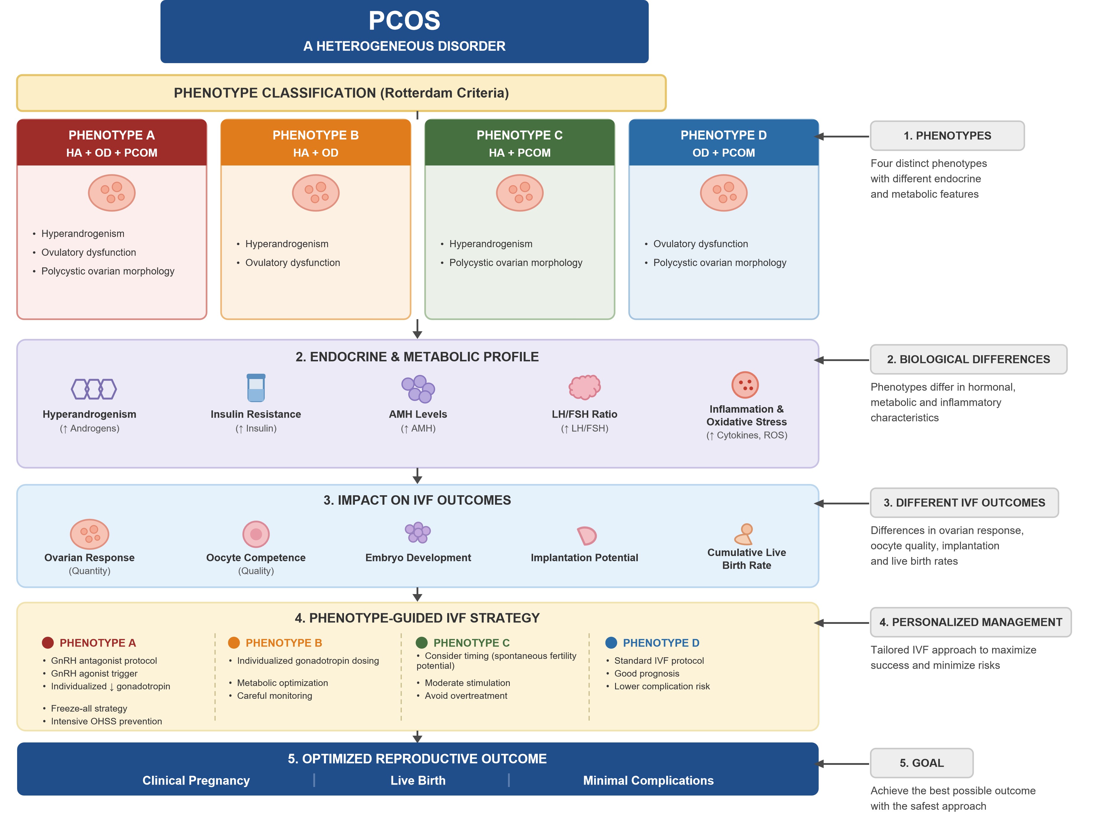
From a clinical perspective, phenotypic classification may contribute to more individualized IVF management. Incorporating phenotype-specific endocrine and metabolic characteristics into treatment planning may improve risk stratification, facilitate selection of appropriate stimulation protocols, optimize OHSS prevention strategies, and support more personalized patient counselling. Taken together, current evidence supports the concept that PCOS phenotypes may serve as clinically relevant predictors of ovarian response, reproductive success, and treatment-related complications. A proposed phenotype-based framework for personalized IVF management is presented in Figure 1 [11,12].
Biological Basis of Phenotype-Specific Reproductive Dysfunction
Endocrine and metabolic heterogeneity across PCOS phenotypes
PCOS encompasses distinct endocrine and metabolic phenotypes characterized by differences in hormonal regulation, metabolic dysfunction, inflammatory status, and reproductive potential. Although the Rotterdam criteria classify PCOS according to the presence or absence of hyperandrogenism, ovulatory dysfunction, and Polycystic Ovarian Morphology (PCOM), increasing evidence indicates that these phenotypes differ substantially in their underlying biological characteristics [12,14].
Hyperandrogenism represents one of the principal determinants of phenotype-specific reproductive dysfunction. Phenotypes A and B, characterized by both hyperandrogenism and ovulatory dysfunction, generally exhibit the most pronounced endocrine abnormalities. Elevated androgen concentrations impair follicular maturation, alter granulosa cell function, and disrupt ovarian steroidogenesis, contributing to chronic anovulation and reduced fertility potential [3]. In contrast, Phenotype D lacks significant hyperandrogenism and is typically associated with a milder endocrine profile and enhanced reproductive performance [9,10].
Insulin resistance is another major contributor to phenotypic variability. Hyperinsulinemia enhances ovarian androgen production through synergistic interactions with Luteinizing Hormone (LH) while simultaneously reducing hepatic Sex Hormone-Binding Globulin (SHBG) synthesis, resulting in increased circulating free androgen levels [16,17]. These metabolic disturbances are particularly evident in classical hyperandrogenic phenotypes and have been linked to impaired follicular development, altered endometrial receptivity, and adverse pregnancy outcomes [14].
AMH has emerged as one of the most informative biomarkers of phenotype-specific ovarian function. Elevated AMH concentrations are strongly associated with increased AFC, ovarian reserve characteristics, and responsiveness to controlled ovarian stimulation [5,18]. Phenotype A generally exhibits the highest AMH concentrations and ovarian reserve markers, whereas lower AMH levels have been reported in the other PCOS phenotypes, with an overall decreasing pattern of A > D > C > B [12,15,19]. The proposed contribution of elevated AMH to impaired follicular development and follicular arrest is illustrated in Figure 2. Nevertheless, phenotype-related differences in AMH should not be interpreted in isolation, as AMH concentrations and the broader phenotypic expression of PCOS may also be influenced by patient-related and metabolic factors. Elevated AMH levels, when considered alongside ultrasonographic ovarian morphology and the presence of hyperandrogenism identified by clinical and/or biochemical findings, may provide supportive evidence for the diagnosis of PCOS [20]. However, AMH should not be considered a stand-alone biomarker for PCOS diagnosis or phenotypic assessment and should be interpreted within the broader clinical and diagnostic context [21].
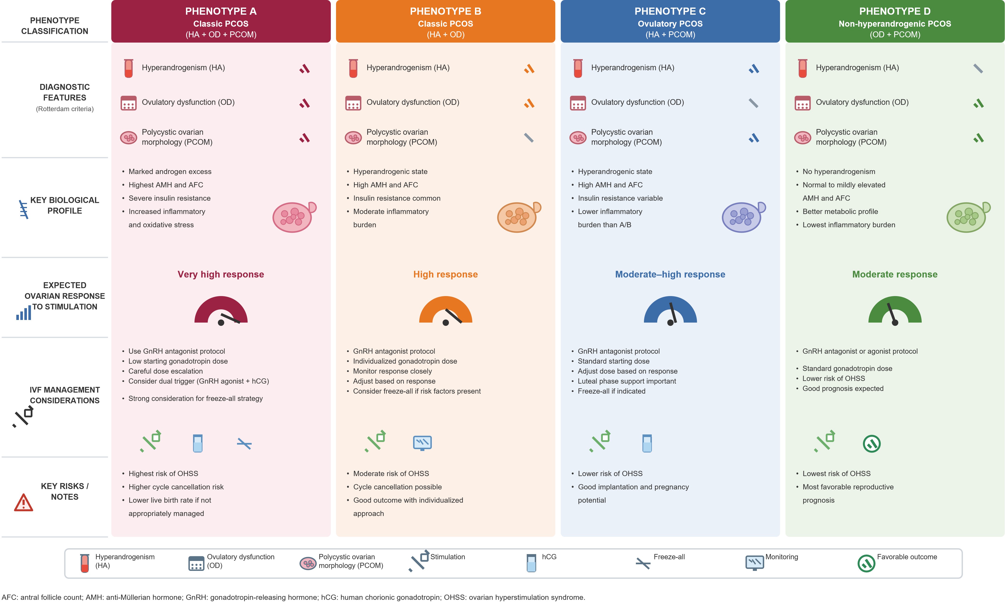
Figure 2. Phenotype-specific clinical characteristics and IVF considerations in women with Polycystic Ovary Syndrome (PCOS). The figure summarizes the endocrine and metabolic pathways involved in the pathogenesis of PCOS. Abnormal Gonadotropin-Releasing Hormone (GnRH) pulsatility leads to an increased Luteinizing Hormone (LH)/Follicle-Stimulating Hormone (FSH) ratio, promoting ovarian androgen production and ovulatory dysfunction. Insulin resistance and compensatory hyperinsulinemia further exacerbate hyperandrogenism through synergistic effects on ovarian steroidogenesis and reduced hepatic Sex Hormone-Binding Globulin (SHBG) synthesis. The interaction of these pathways contributes to follicular arrest, chronic anovulation, and reproductive dysfunction. The severity of these mechanisms varies among PCOS phenotypes and may influence fertility outcomes and response to IVF treatment.
Elevated AMH may predict a greater oocyte yield but is also associated with an increased risk of excessive ovarian response and OHSS, with serum AMH demonstrating greater predictive performance than conventional clinical parameters such as age and BMI [13]. In clinical practice, AMH should therefore be interpreted alongside complementary predictors of ovarian response, including age, AFC, BMI, and, when available, the ovarian response observed during previous stimulation cycles [22].
Follicular microenvironment and mitochondrial competence
Follicular Fluid (FF), which fills the antral cavity of growing ovarian follicles, provides a critical microenvironment for oocyte development. The composition of follicular fluid in women with PCOS has been shown to exhibit alterations in the levels of hormones, growth factors, cytokines, metabolites, and oxidative stress markers. In particular, concentrations of fatty acids, insulin, estrogen, and pregnenolone are markedly increased, whereas progesterone (P4) levels are reduced. Elevated Free Fatty Acid (FFA) levels are closely associated with the inflammatory state of the follicular microenvironment, while miRNAs present in follicular fluid are thought to modulate steroidogenesis. Furthermore, Heparin-Binding Epidermal Growth Factor (EGF)-like growth factor (HB-EGF), which is abundant in the follicular fluid of patients with PCOS, together with increased Arachidonic Acid (AA) levels, may contribute to the induction of mitochondrial dysfunction. Collectively, these alterations create a disrupted follicular microenvironment in PCOS that may adversely affect oocyte maturation [23,24].
Among these alterations, dysregulation of the inflammatory milieu appears to be a prominent feature of the follicular microenvironment in PCOS. In the follicular fluid (FF) of women with PCOS, increased levels of pro-inflammatory cytokines, including Interleukin (IL)-6, IL-8, IL-18, IL-1β, and Tumor Necrosis Factor-alpha (TNF-α), have been reported, whereas levels of the anti-inflammatory cytokine IL-10 have been found to be reduced. This imbalance between pro- and anti-inflammatory mediators contributes to the establishment of a pro-inflammatory follicular microenvironment. Hyperandrogenic phenotypes appear to exhibit a more pronounced inflammatory profile, potentially compromising reproductive potential [23,25]. Such an altered inflammatory milieu may adversely affect oocyte quality and preimplantation embryo development, thereby potentially compromising IVF outcomes in women with PCOS [24,26,27].
Chronic low-grade inflammation and oxidative stress further contribute to phenotype-specific differences in reproductive function [7]. Among PCOS phenotypes, the highest Oxidative Stress Index (OSI) values have been reported in phenotypes D and B. Increased oxidative stress may compromise DNA integrity, thereby adversely affecting oocyte development and maturation [28].
Excessive ROS generation within the altered follicular microenvironment may impair mitochondrial homeostasis and compromise oocyte competence [7]. Such oxidative stress may lead to alterations in Mitochondrial DNA (mtDNA) copy number, structural mitochondrial damage, and impaired mitochondrial function. A strong association between reduced mtDNA copy number and PCOS has been demonstrated, with decreased mtDNA copy number being linked to abnormal follicular development and metabolic disturbances in women with PCOS. Moreover, mtDNA copy number has been proposed as a potentially important biomarker for assessing oocyte maturation and fertilization potential [29].
Gut microbiome and metabolic–endocrine interactions
In PCOS, gut microbiota dysbiosis has been reported, characterized by reduced alpha diversity and alterations in the abundance of specific bacterial taxa associated with metabolic disturbances [30,31]. This reduction in alpha diversity has also been associated with obesity in several studies, and gut microbiota dysbiosis observed in PCOS appears to share certain compositional features with obesity-associated microbial alterations [30]. In addition, alterations in the gut microbiota of women with PCOS have been proposed to be associated with metabolic parameters, sex hormone levels, and mediators of the gut–brain axis [32].
Alterations in the gut microbiome may contribute to the development of PCOS; however, current evidence has not yet established a definitive causal relationship between gut microbiota alterations and PCOS [33]. Furthermore, whether distinct PCOS phenotypes are characterized by specific gut microbiota profiles remains unclear, highlighting an important gap in our understanding of phenotype-specific biological heterogeneity in PCOS [30].
Environmental endocrine disruptors
Exposure to endocrine-disrupting chemicals and environmental toxicants during critical periods of development, particularly when prolonged, may predispose individuals to the development of PCOS and associated metabolic disturbances [3]. Chemicals present in pesticides, plastics, household cleaning products, and cosmetics may disrupt hormonal balance and thereby increase the risk of PCOS. Endocrine-Disrupting Chemicals (EDCs) commonly encountered in daily life, such as Bisphenol A (BPA) and phthalates, may interfere with estrogen and androgen receptor signaling, thereby disrupting hormonal homeostasis. These compounds have also been shown to interfere with normal ovarian steroidogenesis and the regulation of the Hypothalamic–Pituitary–Gonadal (HPG) axis [10,35]. However, whether the endocrine and metabolic effects of environmental toxicants differ across PCOS phenotypes remains insufficiently understood.
Taken together, interactions among hyperandrogenism, insulin resistance, AMH dysregulation, inflammation, oxidative stress, alterations in the gut microbiome, and environmental exposures may contribute to the biological heterogeneity observed across PCOS phenotypes. These interconnected endocrine, metabolic, and environmental mechanisms are summarized in Figure 3, while the principal biological and reproductive characteristics of the four PCOS phenotypes are presented in Table 1.
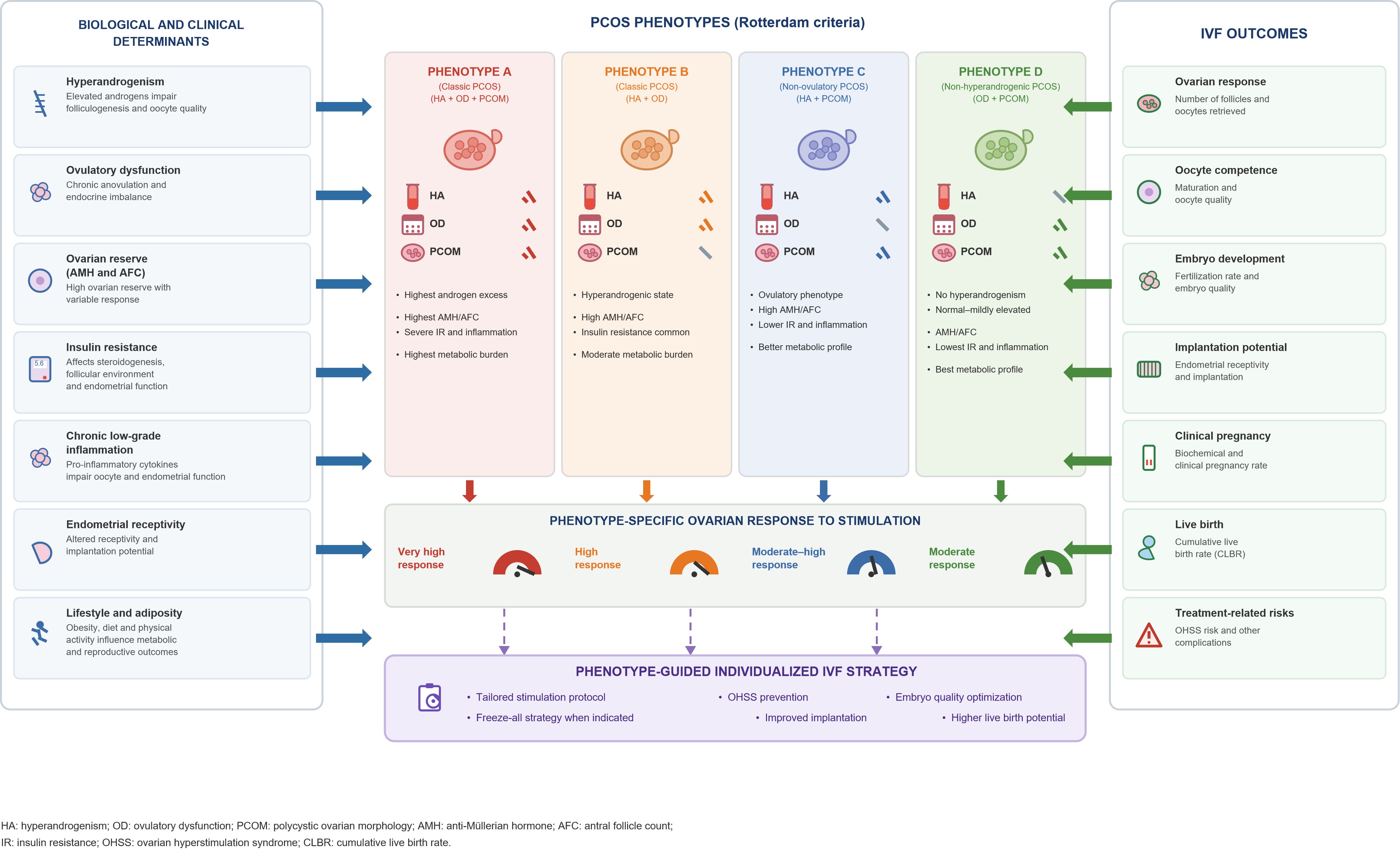
Figure 3. AMH-mediated follicular arrest and impaired folliculogenesis in Polycystic Ovary Syndrome (PCOS). This schematic compares normal follicular development with the disrupted folliculogenesis observed in PCOS. Elevated Anti-Müllerian Hormone (AMH) levels inhibit Follicle-Stimulating Hormone (FSH)-dependent follicular recruitment and suppress aromatase activity, resulting in impaired estradiol production and failure of dominant follicle selection. Consequently, multiple follicles remain arrested at preantral and small antral stages, leading to anovulation and the characteristic polycystic ovarian morphology. These alterations may contribute to reduced oocyte competence, abnormal ovarian responsiveness, and phenotype-specific differences in reproductive outcomes.
|
Parameter |
Phenotype A |
Phenotype B |
Phenotype C |
Phenotype D |
|
Hyperandrogenism |
+++ |
+++ |
++ |
– |
|
Ovulatory dysfunction |
+++ |
+++ |
– |
++ |
|
AMH |
+++ |
++ |
++ |
+ |
|
Insulin resistance |
+++ |
++ |
+ |
+ |
|
Obesity prevalence |
High |
Moderate |
Moderate |
Low |
|
Inflammatory burden |
High |
Moderate |
Moderate |
Low |
|
Expected ovarian response |
Excessive |
Moderate |
Excessive |
Normal |
|
OHSS risk |
High |
Low |
Moderate |
Low |
|
This table summarizes the endocrine, metabolic, and reproductive features associated with the four Rotterdam phenotypes of PCOS. Differences in hyperandrogenism, ovulatory dysfunction, anti-Müllerian hormone (D) levels, insulin resistance, inflammatory burden, and ovarian reserve characteristics may contribute to phenotype-specific reproductive behavior and variability in IVF outcomes. Abbreviations: AMH: Anti-Müllerian Hormone; AFC: Antral Follicle Count; PCOS: Polycystic Ovary Syndrome. |
||||
Phenotype-Specific IVF Outcomes
The biological differences observed among PCOS phenotypes are reflected in clinically relevant variations during assisted reproductive treatment. Differences in ovarian responsiveness, oocyte competence, embryo development, implantation potential, and CLBR suggest that reproductive outcomes should be interpreted within the context of the underlying phenotype rather than the diagnosis of PCOS alone [9,12].
Recent studies have demonstrated substantial variability in IVF outcomes among the four Rotterdam phenotypes, highlighting the importance of phenotype-specific assessment for treatment planning, risk prediction, and patient counseling [9,12,14].
Phenotype A: the classical high-risk phenotype
Phenotype A, characterized by the coexistence of hyperandrogenism, ovulatory dysfunction, and polycystic ovarian morphology, is generally regarded as the most severe form of PCOS. Women with this phenotype frequently exhibit excessive ovarian responsiveness during controlled ovarian stimulation, resulting in the retrieval of a large number of oocytes but also a substantially increased risk of ovarian hyperstimulation syndrome (OHSS) [12,13].
Despite high oocyte yield, reproductive outcomes are not always proportionally improved. Several studies have reported lower CLBR and diminished reproductive potential in classical hyperandrogenic phenotypes compared with non-hyperandrogenic forms of PCOS [9]. Consequently, Phenotype A represents the subgroup most likely to benefit from individualized stimulation protocols and OHSS prevention strategies.
Phenotype B: hyperandrogenic-anovulatory PCOS
Phenotype B shares the endocrine features of hyperandrogenism and ovulatory dysfunction but lacks polycystic ovarian morphology. Compared with Phenotype A, ovarian responsiveness is generally less pronounced, resulting in a more moderate response to controlled ovarian stimulation [12].
Although OHSS risk appears lower than in Phenotype A, persistent hyperandrogenism and metabolic dysfunction may adversely affect follicular development and reproductive performance. Available evidence suggests that pregnancy and live birth outcomes are generally intermediate between those observed in classical hyperandrogenic and non-hyperandrogenic phenotypes [11].
Phenotype C: ovulatory hyperandrogenic PCOS
Phenotype C combines hyperandrogenism with polycystic ovarian morphology while preserving ovulatory function. Because spontaneous conception remains possible, women with this phenotype frequently present for fertility treatment later than those with chronic anovulation [14].
Ovarian responsiveness during stimulation may remain substantial; however, reproductive outcomes are generally better than those observed in Phenotypes A and B. Several studies have reported intermediate-to-high implantation rates, improved pregnancy outcomes, and higher CLBR in this subgroup [9].
Phenotype D: non-hyperandrogenic PCOS
Phenotype D is characterized by ovulatory dysfunction and polycystic ovarian morphology in the absence of hyperandrogenism. Compared with hyperandrogenic phenotypes, this subgroup is generally associated with a less adverse metabolic profile and enhanced reproductive potential [4,14].
Several studies have reported higher CLBR and improved overall reproductive outcomes in women with Phenotype D [9,10]. The absence of significant hyperandrogenism may contribute to improved follicular development, enhanced endometrial receptivity, and lower treatment-related complication rates. Current evidence suggests that Phenotype D represents the subgroup with the most advantageous reproductive profile within the PCOS spectrum [9,14].
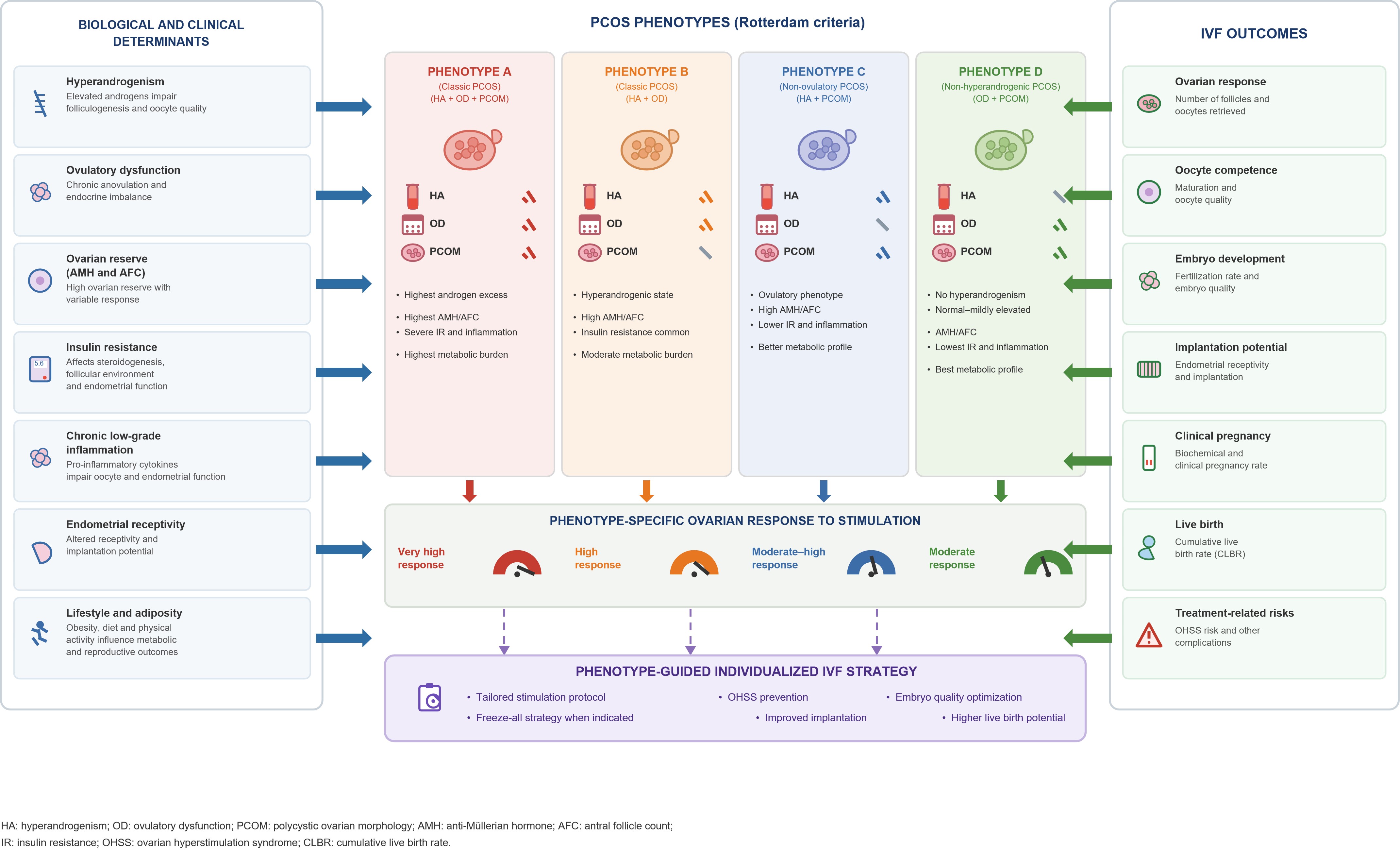
Current evidence demonstrates substantial variation in reproductive prognosis across PCOS phenotypes (Figure 4). Reproductive outcomes appear to follow a continuum extending from the high-risk hyperandrogenic Phenotype A to the non-hyperandrogenic Phenotype D, reflecting differences in ovarian response, embryo competence, implantation success, CLBR, and treatment-related complications [9,14] (Table 2).
|
Outcome |
A |
B |
C |
D |
|
Oocyte yield |
↑↑↑ |
↑ |
↑↑ |
↑ |
|
Mature oocytes |
↑↑ |
↑ |
↑↑ |
↑ |
|
Embryo quality |
↓ |
↓ |
↔ |
↑ |
|
OHSS risk |
↑↑↑ |
↓ |
↑↑ |
↓ |
|
Implantation |
↓ |
↓ |
↔ |
↑ |
|
Clinical pregnancy |
↓ |
↓ |
↑ |
↑↑ |
|
CLBR |
Lowest |
Intermediate |
High |
Highest |
|
Pregnancy complications |
High |
Moderate |
Moderate |
Low |
|
This table summarizes the available evidence regarding ovarian response, oocyte competence, embryo quality, implantation rates, Cumulative Live Birth Rates (CLBR), and pregnancy-related complications among the four Rotterdam phenotypes. Hyperandrogenic phenotypes generally demonstrate greater ovarian responsiveness and treatment-related risks, whereas non-hyperandrogenic phenotypes tend to exhibit more favorable reproductive outcomes. Abbreviations: IVF: In Vitro Fertilization; OHS: Ovarian Hyperstimulation Syndrome; CLBR: Cumulative Live Birth Rate. |
||||
Embryo morphokinetics and time-lapse imaging
Morphokinetic assessment provides a comprehensive framework for understanding and evaluating in vitro embryo development [36]. Embryo morphokinetics may be influenced not only by external factors, such as in vitro culture conditions, but also by patient-related comorbidities. PCOS has been reported to adversely affect embryo morphokinetics, particularly by reducing the synchrony of embryonic cleavage events [37,38]. Altered early embryonic development has been reported in women with hyperandrogenic PCOS, characterized by accelerated development up to the morula stage and a higher miscarriage rate. In contrast, no significant differences in morphokinetic parameters have been observed between women with normoandrogenic PCOS and those without PCOS. Taken together, these findings suggest that hyperandrogenism may play an important role in the adverse effects of specific PCOS phenotypic characteristics on embryo morphokinetics [39,40]. In addition to hyperandrogenism, metabolic disturbances associated with PCOS may also influence oocyte and embryo competence. Abnormal insulin levels associated with peripheral insulin resistance in PCOS may disrupt metabolic homeostasis and energy-related pathways in oocytes and developing embryos. These metabolic disturbances may consequently impair oocyte quality and reduce the likelihood of high-quality blastocyst formation [41].
Time-Lapse Imaging (TLI) technology enables continuous monitoring of preimplantation embryo development without disturbing culture conditions, thereby providing detailed and dynamic information on the temporal progression of embryonic development. TLI may also facilitate the assessment of oocyte and embryo quality and contribute to the prediction of implantation and pregnancy outcomes [37,42]. One of the developmental events assessed using TLI is Direct Unequal Cleavage at the first cleavage division (DUC-1), characterized by unequal blastomere division. The incidence of DUC-1 has been reported to be higher in patients with PCOS and a high ovarian response than in non-PCOS patients with a high ovarian response and those with a normal ovarian response. However, after excluding DUC-1 embryos, the developmental kinetics of embryos derived from high-responding patients with and without PCOS were reported to be similar for the majority of morphokinetic parameters [43]. These findings suggest that the morphokinetic alterations observed in PCOS may be driven, at least in part, by specific abnormal cleavage patterns rather than by a generalized acceleration or delay in embryo development.
Cumulative live birth rate as a clinically relevant outcome
In women with PCOS, the number of oocytes retrieved has been shown to be strongly associated with the Cumulative Live Birth Rate (CLBR) and the proportion of surplus embryos. However, retrieval of an excessively high number of oocytes does not necessarily translate into a higher CLBR and appears to provide no additional benefit in terms of achieving a live birth [44].
The total number of embryos suitable for transfer has been identified as one of the most important predictors of cumulative live birth in women with PCOS [45]. This is particularly relevant in PCOS, where a high ovarian response may generate multiple embryos suitable for subsequent transfer cycles. Given the increased risk of excessive ovarian response and OHSS in women with PCOS, IVF success should not be evaluated solely on the basis of fresh embryo transfer outcomes. A more comprehensive assessment of treatment success should incorporate the outcomes of both fresh and frozen–thawed embryo transfer cycles. Indeed, frozen embryo transfer has been associated with a higher live birth rate and a lower risk of OHSS than fresh embryo transfer in infertile women with PCOS. Therefore, the CLBR represents an important outcome measure for evaluating the overall prognosis of women with PCOS undergoing IVF [46].
Personalized IVF Management According to PCOS Phenotype
The recognition of phenotypic diversity in PCOS has important implications for assisted reproductive treatment. Although current IVF protocols are primarily guided by age, ovarian reserve markers, and previous treatment history, these parameters do not fully reflect the endocrine and metabolic differences observed among PCOS phenotypes. Consequently, incorporating phenotypic characteristics into treatment planning may facilitate more individualized and clinically relevant management strategies [12].
Rather than applying a uniform approach to all women with PCOS, phenotype-based assessment may assist clinicians in predicting ovarian response, identifying treatment-related risks, and selecting appropriate stimulation protocols. Such an approach may improve both treatment efficiency and patient safety.
Phenotype A: prevention of excessive ovarian response
Among all phenotypes, Phenotype A presents the greatest challenge during IVF treatment. Excessive ovarian responsiveness places these patients at increased risk of OHSS and related complications. Therefore, the primary objective should not be maximizing oocyte yield but achieving a safe and effective pathway to live birth [10,12,15].
Strategies such as GnRH antagonist protocols, GnRH agonist triggering, individualized gonadotropin dosing, elective cryopreservation ("freeze-all"), and single embryo transfer may substantially reduce treatment-related risks while maintaining reproductive success [13] (Table 3).
Phenotype B: optimizing endocrine and metabolic conditions
Phenotype B occupies an intermediate position within the PCOS spectrum. Although ovarian response is generally less pronounced than in Phenotype A, persistent hyperandrogenism and metabolic dysfunction may negatively influence reproductive outcomes [11,12].
Tailored stimulation protocols, combined with pre-treatment metabolic assessment and optimization of modifiable metabolic factors, may improve treatment efficiency and reproductive outcomes in this subgroup [47,48] (Table 3).
Phenotype C: individualized timing of reproductive intervention
The preservation of ovulatory function distinguishes Phenotype C from the other PCOS phenotypes. Because spontaneous conception remains possible, fertility counseling and appropriate timing of assisted reproductive intervention may be particularly important in this group [9,14].
When IVF is required, stimulation strategies should aim to achieve adequate ovarian response while avoiding unnecessary treatment intensity. Clinical decision-making should focus on balancing treatment effectiveness with the patient's remaining spontaneous reproductive potential [13] (Table 3).
Phenotype D: conventional IVF and enhanced reproductive potential
Phenotype D is generally associated with the most advantageous reproductive profile among the Rotterdam phenotypes. Reduced metabolic dysfunction and the absence of hyperandrogenism appear to contribute to enhanced reproductive potential and lower treatment-related risks [10].
Compared with hyperandrogenic phenotypes, women with Phenotype D generally have a lower risk of excessive ovarian response and OHSS. Consequently, intensive OHSS-prevention strategies may be required less frequently in this subgroup. Nevertheless, ovarian stimulation protocols should always be individualized according to age, ovarian reserve, BMI, and previous treatment response [9,10].
These observations reinforce the concept that different PCOS phenotypes may benefit from different management strategies during assisted reproduction [12,14] (Table 3).
|
Phenotype |
Major Risk |
IVF Strategy |
Expected Benefit |
|
A |
OHSS |
GnRH antagonist + agonist trigger + freeze-all |
Reduced OHSS |
|
B |
Suboptimal response |
Individualized gonadotropin dosing |
Improved oocyte yield |
|
C |
Delayed treatment |
Early fertility counselling |
Optimized timing |
|
D |
Mild phenotype |
Standard IVF protocols |
High cumulative live birth rate |
|
This table proposes phenotype-guided reproductive management strategies based on the endocrine and metabolic characteristics of each PCOS phenotype. Suggested approaches include individualized ovarian stimulation protocols, OHSS prevention strategies, metabolic optimization, and fertility counseling. The recommendations are intended to illustrate how phenotypic classification may contribute to personalized reproductive medicine and improved treatment outcomes. Abbreviations: IVF: In Vitro Fertilization; OHSS: Ovarian Hyperstimulation Syndrome; GnRH: Gonadotropin-Releasing Hormone. |
|||
Individualization of ovarian stimulation strategies
Ovarian stimulation protocols used during IVF treatment may influence oocyte and embryo development in women with PCOS and thereby affect treatment outcomes. The GnRH antagonist protocol is commonly preferred in women with PCOS because of its shorter duration of ovarian stimulation, lower gonadotropin requirements, and reduced incidence of OHSS. GnRH antagonist protocols have been reported to represent a safer and more cost-effective treatment option than the conventional long GnRH agonist protocol, without compromising IVF clinical outcomes [14,46,49].
In addition to conventional stimulation protocols, a study conducted in a specific subgroup of Chinese women with PCOS suggested that mild ovarian stimulation may be considered as an alternative approach, offering potential advantages such as better control of the number of oocytes retrieved and reduced overall treatment costs [44].
In women with PCOS, a lower initial Follicle-Stimulating Hormone (FSH) dose has been associated with a lower number of oocytes retrieved, whereas a higher starting FSH dose may significantly increase the risk of excessive ovarian response and OHSS. Patients receiving higher starting doses of gonadotropins have also been reported to have lower CLBRs than those receiving lower doses. Therefore, the initial FSH dose should be individualized according to patient-specific clinical characteristics and parameters such as Body Mass Index (BMI), basal FSH (bFSH), basal LH (bLH), AMH levels, and AFC. Such an individualized approach may contribute to improving the clinical outcomes of IVF treatment [25,46]. Therefore, individualized selection of the stimulation protocol and gonadotropin starting dose according to patient-specific clinical and ovarian reserve characteristics may contribute to optimizing ovarian response while improving treatment safety and cost-effectiveness in women with PCOS.
Clinical implications of phenotype-guided IVF management
Phenotypic classification may provide clinically relevant information beyond traditional ovarian reserve markers alone by capturing endocrine, metabolic, and reproductive heterogeneity among women with PCOS [50]. Integrating endocrine and metabolic characteristics into treatment planning may improve risk stratification, facilitate protocol selection, and support more individualized patient counseling [12,14].
Importantly, phenotype-guided management should be viewed as a complementary clinical framework rather than a replacement for established predictors such as ovarian reserve markers, age, and previous treatment response. Current clinical decision-making should continue to incorporate established prognostic factors while recognizing that phenotypic differences may provide additional information regarding ovarian responsiveness, metabolic risk, and treatment-related complications [9,50]. Nevertheless, current evidence suggests that consideration of phenotypic differences during IVF planning may contribute to improved risk assessment, more individualized treatment selection, and ultimately safer and more efficient reproductive care [9,12,14].
Future prospective studies are warranted to validate phenotype-guided treatment algorithms and determine whether integrating clinical phenotypes with molecular biomarkers and AI-assisted predictive models can further improve reproductive outcomes in women with PCOS [50,51]
Emerging Biomarkers, Evolving Concepts, and Future Directions
The growing recognition of phenotype-specific reproductive behavior in women with PCOS has highlighted the need for more refined approaches to patient stratification and treatment planning. Although ovarian reserve markers such as AMH, AFC, age, and BMI remain central components of fertility assessment, these parameters alone do not fully explain the substantial variability in ovarian response and reproductive outcomes observed among women with PCOS [12,13]. Consequently, increasing attention has focused on the identification of novel biomarkers capable of supporting a more individualized approach to reproductive management.
Among currently available biomarkers, AMH remains one of the most clinically valuable predictors of ovarian reserve and ovarian responsiveness during controlled ovarian stimulation. However, emerging evidence suggests that its predictive value may be influenced by the underlying PCOS phenotype and associated metabolic characteristics. Integrating AMH measurements with phenotypic classification may therefore improve assessment of ovarian response and treatment-related risk, particularly in women with hyperandrogenic phenotypes [13,18].
Beyond conventional ovarian reserve markers, increasing interest has focused on the follicular microenvironment. Alterations in follicular fluid composition, including inflammatory cytokines, oxidative stress markers, metabolites, growth factors, and lipid mediators, have been associated with differences in oocyte competence and embryo development in women with PCOS [5,7]. These emerging biomarkers may provide additional insight into reproductive potential and could eventually complement traditional predictors used in IVF practice.
Advances in molecular medicine have further expanded interest in genetic, epigenetic, transcriptomic, proteomic, and metabolomic profiling. Such approaches have the potential to identify biological signatures associated with ovarian responsiveness, embryo development, implantation success, and pregnancy outcomes. Future classification systems may therefore extend beyond clinical phenotypes and incorporate molecular characteristics capable of improving prediction of treatment response [51].
Artificial intelligence (AI) and machine-learning technologies are expected to play an increasingly important role in reproductive medicine. By integrating clinical, hormonal, ultrasonographic, metabolic, and molecular data, AI-driven models may facilitate more accurate prediction of ovarian response, treatment-related complications, embryo competence, and live birth probability. Such approaches have the potential to improve patient stratification, optimize treatment selection, and support individualized reproductive care [51]. In this context, current phenotype-based approaches may serve as an important foundation for future precision reproductive medicine strategies.
Beyond technological advances, evolving biological insights are also reshaping how PCOS is conceptualized and classified. The increasing recognition of endocrine, metabolic, and reproductive heterogeneity among women diagnosed with PCOS has challenged the traditional view of the syndrome as a single clinical entity. The 2023 International Evidence-Based Guideline emphasized that PCOS encompasses a broad spectrum of phenotypes characterized by substantial differences in metabolic risk, reproductive function, and long-term health outcomes, highlighting the importance of individualized assessment and management [50].
Children born to women with PCOS have been reported to be at an increased risk of adverse health outcomes. In particular, higher risks of preterm birth, Low Birth Weight (LBW), and Fetal Growth Restriction (FGR) have been observed among offspring of women with PCOS [52]. Furthermore, evidence suggests mildly impaired cardiometabolic health in the offspring of women with PCOS, with these alterations appearing to be more pronounced in female offspring [53]. The hyperandrogenic and hyperinsulinemic intrauterine environment associated with maternal PCOS may contribute to abnormal fetal growth, preterm birth, and an increased risk of diabetes, all of which may subsequently increase the risk of cardiovascular disease later in life [54]. The potential consequences of maternal PCOS in offspring may extend beyond metabolic disturbances, with evidence suggesting possible long-term effects on neurodevelopmental and psychiatric health. Children exposed to maternal PCOS have been reported to have an increased risk of Autism Spectrum Disorder (ASD), Attention-Deficit/Hyperactivity Disorder (ADHD), Chronic Tic Disorders (CTDs), anxiety disorders, other behavioral and emotional disorders, and neurological malformations [55].
As understanding of the disorder has expanded, concerns have emerged regarding whether the term polycystic ovary syndrome adequately reflects its biological complexity. The designation “polycystic” is increasingly considered misleading because the ovarian structures observed in affected women represent arrested follicles rather than true pathological cysts. Furthermore, the condition extends far beyond ovarian morphology, encompassing endocrine dysfunction, metabolic abnormalities, reproductive impairment, and long-term cardiometabolic consequences [50].
Reflecting these concerns, an international multidisciplinary initiative led by Teede and colleagues proposed replacing the term PCOS with Polyendocrine Metabolic Ovarian Syndrome (PMOS). This nomenclature was introduced to better capture the multisystem nature of the disorder and to acknowledge that the condition represents a complex endocrine–metabolic syndrome with heterogeneous reproductive manifestations rather than a disorder defined primarily by ovarian morphology [56].
Importantly, this evolving conceptual framework aligns closely with the growing emphasis on phenotype-guided reproductive medicine. Viewing PCOS/PMOS as a spectrum of biologically distinct reproductive and metabolic phenotypes reinforces the rationale for individualized IVF management. Future approaches are likely to integrate clinical phenotypes with endocrine markers, metabolic characteristics, molecular profiling, and AI-assisted predictive models, enabling more accurate patient stratification, improved treatment selection, and ultimately better reproductive outcomes [50,51] (Table 4).
|
Clinical Finding |
Suggested Evaluation |
Clinical Action |
|
AMH > X |
OHSS risk assessment |
Low-dose stimulation |
|
Hyperandrogenism |
Metabolic screening |
Pre-IVF optimization |
|
Obesity |
Weight management |
Lifestyle intervention |
|
Phenotype A |
Intensive monitoring |
Freeze-all strategy |
|
Phenotype D |
Standard protocol |
Conventional transfer |
|
This table summarizes current and emerging biomarkers with potential applications in phenotype-guided reproductive medicine. These markers include ovarian reserve indicators, follicular fluid biomarkers, inflammatory mediators, oxidative stress markers, and molecular signatures that may contribute to improved prediction of ovarian response, embryo competence, implantation potential, and reproductive outcomes in women with PCOS. Abbreviations: AMH: Anti-Müllerian Hormone: AFC: Antral Follicle Count; ROS: Reactive Oxygen Species; IVF: In Vitro Fertilization. |
||
Taken together, these developments suggest that the future of reproductive medicine in PCOS/PMOS will depend on integrating clinical phenotypes with endocrine, metabolic, molecular, and computational biomarkers. Such an approach may facilitate more accurate patient stratification, improve treatment selection, reduce treatment-related complications, and ultimately enhance reproductive outcomes. While many of these emerging tools remain investigational, they collectively support a transition from conventional IVF management toward increasingly individualized reproductive care.
Conclusion
PCOS is increasingly recognized as a spectrum of biologically distinct reproductive and metabolic phenotypes rather than a single homogeneous disorder. Accumulating evidence indicates that these phenotypes differ in endocrine characteristics, metabolic characteristics, ovarian responsiveness, and reproductive potential, resulting in meaningful variations in IVF outcomes.
The available literature suggests that phenotype-specific differences influence key determinants of reproductive success, including ovarian response to stimulation, oocyte competence, embryo development, implantation potential, CLBR, and treatment-related complications. Hyperandrogenic phenotypes generally demonstrate diminished reproductive potential and greater treatment-related risks, whereas non-hyperandrogenic phenotypes tend to exhibit improved reproductive outcomes.
Importantly, phenotypic classification may provide clinically relevant information that complements conventional predictors such as age, ovarian reserve markers, and previous treatment history. Incorporating phenotype-specific characteristics into reproductive planning may improve risk stratification, facilitate individualized treatment selection, and support more effective patient counselling.
Although phenotype-guided management has not yet become a universally accepted standard of care, current evidence supports its potential value as a practical framework for individualized IVF management. Future integration of clinical phenotypes with emerging biomarkers, molecular profiling, and advanced predictive technologies may further refine patient stratification and optimize reproductive outcomes.
In conclusion, PCOS phenotypes should be viewed not only as diagnostic classifications but also as clinically meaningful indicators of reproductive behavior. Recognizing and integrating phenotype-specific differences into IVF management may represent an important step toward more individualized, safer, and more effective reproductive care for women with PCOS.
Acknowledgements
The authors would like to thank their colleagues for their academic support and constructive discussions during the preparation of this manuscript. No professional writing assistance was received for this study.
Funding
This study received no specific financial support from any public, commercial, or not-for-profit funding agency.
Disclosure
The authors declare that they have no financial or non-financial competing interests related to the content of this manuscript. The authors have no industrial affiliations, consultancies, stock ownership, honoraria, paid expert testimony, patent applications, or other financial relationships that could be perceived as influencing the work reported in this paper.
Data Availability
No new data were generated or analyzed in this study. This manuscript is based exclusively on previously published literature.
References
2. Jakubowska-Kowal KM, Skrzynska KJ, Gawlik-Starzyk AM. Prevalence and diagnosis of polycystic ovary syndrome (PCOS) in adolescents - what's new in 2023? Systematic review. Ginekol Pol. 2024;95(8):643–9.
3. Escobar-Morreale HF. Polycystic ovary syndrome: definition, aetiology, diagnosis and treatment. Nat Rev Endocrinol. 2018 May;14(5):270–84.
4. Stańczak NA, Grywalska E, Dudzińska E. The latest reports and treatment methods on polycystic ovary syndrome. Ann Med. 2024 Dec;56(1):2357737.
5. Palomba S, Daolio J, La Sala GB. Oocyte Competence in Women with Polycystic Ovary Syndrome. Trends Endocrinol Metab. 2017 Mar;28(3):186–98
6. Sreerangaraja Urs DB, Wu WH, Komrskova K, Postlerova P, Lin YF, Tzeng CR, et al. Mitochondrial Function in Modulating Human Granulosa Cell Steroidogenesis and Female Fertility. Int J Mol Sci. 2020 May 19;21(10):3592.
7. Palomba S. Is fertility reduced in ovulatory women with polycystic ovary syndrome? An opinion paper. Hum Reprod. 2021 Aug 18;36(9):2421–8.
8. Tang K, Wu L, Luo Y, Gong B. In vitro fertilization outcomes in women with polycystic ovary syndrome: A meta-analysis. Eur J Obstet Gynecol Reprod Biol. 2021 Apr;259:146–52.
9. De Vos M, Pareyn S, Drakopoulos P, Raimundo JM, Anckaert E, Santos-Ribeiro S, P et al. Cumulative live birth rates after IVF in patients with polycystic ovaries: phenotype matters. Reprod Biomed Online. 2018 Aug;37(2):163–71.
10. Patel A, Saraf A, Rao VA, Rao KA. Comparing Assisted Reproductive Outcomes in Women with Different Polycystic Ovary Syndrome Phenotypes Undergoing Frozen Embryo Transfer. J Hum Reprod Sci. 2023 Oct-Dec;16(4):340–5.
11. Ramezanali F, Ashrafi M, Hemat M, Arabipoor A, Jalali S, Moini A. Assisted reproductive outcomes in women with different polycystic ovary syndrome phenotypes: the predictive value of anti-Müllerian hormone. Reprod Biomed Online. 2016 May;32(5):503–12.
12. Cela V, Obino MER, Alberga Y, Pinelli S, Sergiampietri C, Casarosa E, et al. Ovarian response to controlled ovarian stimulation in women with different polycystic ovary syndrome phenotypes. Gynecol Endocrinol. 2018 Jun;34(6):518–23.
13. Thakre N, Homburg R. A review of IVF in PCOS patients at risk of ovarian hyperstimulation syndrome. Expert Rev Endocrinol Metab. 2019 Sep;14(5):315–9.
14. Si M, Qi X, Zhen X, Yang C, Tian T, Long X, et al. Dose Nomogram of Individualization of the Initial Follicle-Stimulating Hormone Dosage for Patients with Polycystic Ovary Syndrome Undergoing IVF/ICSI with the GnRH-Ant Protocol: A Retrospective Cohort Study. Adv Ther. 2023 Sep;40(9):3971–85.
15. Sahmay S, Atakul N, Oncul M, Tuten A, Aydogan B, Seyisoglu H. Serum anti-Mullerian hormone levels in the main phenotypes of polycystic ovary syndrome. Eur J Obstet Gynecol Reprod Biol. 2013 Sep;170(1):157–61.
16. Lizneva D, Gavrilova-Jordan L, Walker W, Azziz R. Androgen excess: Investigations and management. Best Pract Res Clin Obstet Gynaecol. 2016 Nov;37:98–118.
17. Su P, Chen C, Sun Y. Physiopathology of polycystic ovary syndrome in endocrinology, metabolism and inflammation. J Ovarian Res. 2025 Feb 20;18(1):34.
18. Cedars MI. Evaluation of Female Fertility-AMH and Ovarian Reserve Testing. J Clin Endocrinol Metab. 2022 May 17;107(6):1510–9.
19. Schwenck-Carvalho PJ, Maffazioli GN, Simões RS, Macchione RF, de Medeiros SF, Soares Júnior JM et al. Anti-Müllerian Hormone Levels Across Phenotypes in Polycystic Ovary Syndrome: A Systematic Review and Meta-analysis. J Endocr Soc. 2025 Dec 16;10(3):bvaf199.
20. Akgül ÖK, Aksoy NK. Association of serum AMH with laboratory and phenotype in women with PCOS: A retrospective study. J Adv Res Health Sci.;6(3):289–94.
21. van der Ham K, Laven JSE, Tay CT, Mousa A, Teede H, et al. Anti-müllerian hormone as a diagnostic biomarker for polycystic ovary syndrome and polycystic ovarian morphology: a systematic review and meta-analysis. Fertil Steril. 2024 Oct;122(4):727–39.
22. Arvis P, Rongières C, Pirrello O, Lehert P. Predicting the ovarian response: towards a determinant model and implications for practice. J Assist Reprod Genet. 2024 Jan;41(1):213–22.
23. Pan Y, Pan C, Zhang C. Unraveling the complexity of follicular fluid: insights into its composition, function, and clinical implications. J Ovarian Res. 2024 Nov 26;17(1):237.
24. Dai M, Hong L, Yin T, Liu S. Disturbed Follicular Microenvironment in Polycystic Ovary Syndrome: Relationship to Oocyte Quality and Infertility. Endocrinology. 2024 Feb 20;165(4):bqae023.
25. Si M, Xu W, Qi X, Jiang H, Zhao Y, Li R, et al. Metabolic Syndrome Rather Than Other Phenotypes in PCOS as a Predictive Indicator for Clinical Outcomes in IVF: Comprehensive Phenotypic Assessment across All PCOS Classifications. J Clin Med. 2023 Aug 2;12(15):5073.
26. Wegrzynowicz AK, Banerjee S, Grimes E, Huddleston R, Leyva Jaimes F, Cooney LG, et al. Single-cell analysis of follicular fluid reveals dysregulation of ovulatory immune function in Polycystic Ovary Syndrome patients undergoing ovarian stimulation†. Biol Reprod. 2026 May 13;114(5):1736–46.
27. Yang F, Wang Z, Sørensen HT, Janszky I, Gissler M, Yuan W, et al. Maternal polycystic ovary syndrome and Offspring's Risk of Cardiovascular diseases in Childhood and Young Adulthood. Nat Commun. 2024 Nov 29;15(1):10414.
28. Onal M, Agar M. Follicular fluid redox balance and DNA damage according to PCOS phenotype. Eur Rev Med Pharmacol Sci. 2023 Jun;27(12):5583–9.
29. Zhang J, Mai Q, Zhou C. Mitochondrial DNA copy number variation and gene mutations mediate polycystic ovary syndrome: research progress. Am J Transl Res. 2024 Nov 15;16(11):6303–13.
30. Yurtdaş G, Akdevelioğlu Y. A New Approach to Polycystic Ovary Syndrome: The Gut Microbiota. J Am Coll Nutr. 2020 May-Jun;39(4):371–82.
31. Hanna A, Abbas H, Yassine F, AlBush A, Bilen M. Systematic review of gut microbiota composition, metabolic alterations, and the effects of treatments on PCOS and gut microbiota across human and animal studies. Front Microbiol. 2025 May 14;16:1549499.
32. Liu R, Zhang C, Shi Y, Zhang F, Li L, Wang X, et al. Dysbiosis of Gut Microbiota Associated with Clinical Parameters in Polycystic Ovary Syndrome. Front Microbiol. 2017 Feb 28;8:324
33. Wang L, Zhou J, Gober HJ, Leung WT, Huang Z, Pan X, et al. Alterations in the intestinal microbiome associated with PCOS affect the clinical phenotype. Biomed Pharmacother. 2021 Jan;133:110958.
34. Patel S. Polycystic ovary syndrome (PCOS), an inflammatory, systemic, lifestyle endocrinopathy. J Steroid Biochem Mol Biol. 2018 Sep;182:27–36.
35. Ibibama JO, Madaki PD, Aliche KA, Frazzoli C, Orisakwe OE. Environmental Toxicants and Polycystic Ovary Syndrome: Implications for a Metabolic-Ovarian Framework (PMOS) - A Systematic Review. Biol Trace Elem Res. 2026 Jul 9.
36. Working Group on the update of the ESHRE/ALPHA Istanbul Consensus; Coticchio G, Ahlström A, Arroyo G, Balaban B, Campbell A, De Los Santos MJ, et al. The Istanbul consensus update: a revised ESHRE/ALPHA consensus on oocyte and embryo static and dynamic morphological assessment†,‡. Hum Reprod. 2025 Jun 1;40(6):989–1035.
37. Garçon VM, Dietrich JE, Strowitzki T, Freis A. Polycystic ovary syndrome (PCOS) affects relative embryo morphokinetics observed by time-lapse imaging: an observational study. Arch Gynecol Obstet. 2026 Feb 8;313(1):83.
38. Wang J, Guo Y, Zhang N, Li T. Research progress of time-lapse imaging technology and embryonic development potential: A review. Medicine (Baltimore). 2023 Sep 22;102(38):e35203.
39. Chappell NR, Barsky M, Shah J, Peavey M, Yang L, Sangi-Haghpeykar H, et al. Embryos from polycystic ovary syndrome patients with hyperandrogenemia reach morula stage faster than controls. F S Rep. 2020 Sep 2;1(2):125–32.
40. Wissing ML, Bjerge MR, Olesen AI, Hoest T, Mikkelsen AL. Impact of PCOS on early embryo cleavage kinetics. Reprod Biomed Online. 2014 Apr;28(4):508–14.
41. Shibli Abu Raya Y, Aslih N, Atzmon Y, Sharqawi M, Shavit M, Bilgory A, et al. Non-Male Factor Only-ICSI Can Overcome Oocyte Factor in PCOS Patients. J Clin Med. 2025 Jan 3;14(1):244.
42. Tam Le M, Van Nguyen T, Thanh Nguyen T, Thanh Thi Nguyen T, An Thi Nguyen T, Huy Vu Nguyen Q, et al. Does polycystic ovary syndrome affect morphokinetics or abnormalities in early embryonic development? Eur J Obstet Gynecol Reprod Biol X. 2019 May 17;3:100045.
43. Schachter-Safrai N, Karavani G, Esh-Broder E, Levitas E, Wainstock T, Har-Vardi I, et al. High ovarian response to ovarian stimulation: effect on morphokinetic milestones and cycle outcomes. J Assist Reprod Genet. 2021 Dec;38(12):3083–90.
44. Chen YH, Wang Q, Zhang YN, Han X, Li DH, Zhang CL. Cumulative live birth and surplus embryo incidence after frozen-thaw cycles in PCOS: how many oocytes do we need? J Assist Reprod Genet. 2017 Sep;34(9):1153–9.
45. Li HW, Lee VC, Lau EY, Yeung WS, Ho PC, Ng EH. Cumulative live-birth rate in women with polycystic ovary syndrome or isolated polycystic ovaries undergoing in-vitro fertilisation treatment. J Assist Reprod Genet. 2014 Feb;31(2):205–11.
46. Li Y, Xia L, Li Z, Zhang Z, Jiang R. Factors affecting cumulative live birth rate after the 1st oocyte retrieved in polycystic ovary syndrome patients in women during IVF/ICSI-ET. J Ovarian Res. 2023 Oct 13;16(1):201.
47. Huang S, Wang Z, Yang R, Li R, Qiao J. Embryological and pregnancy outcomes of IVF-ET in overweight/obese and normal-weight women with PCOS. Front Endocrinol (Lausanne). 2025 Mar 27;16:1552561.
48. Vale-Fernandes E, Moreira MV, Bernardino RL, Sousa D, Brandão R, Leal C, et al. Polycystic ovary syndrome and excessive body weight impact independently and synergically on fertility treatment outcomes. Reprod Biol Endocrinol. 2025 Jul 7;23(1):97.
49. Kadoura S, Alhalabi M, Nattouf AH. Conventional GnRH antagonist protocols versus long GnRH agonist protocol in IVF/ICSI cycles of polycystic ovary syndrome women: a systematic review and meta-analysis. Sci Rep. 2022 Mar 15;12(1):4456.
50. Teede HJ, Tay CT, Laven JJE, Dokras A, Moran LJ, Piltonen TT, et al. International PCOS Network. Recommendations from the 2023 international evidence-based guideline for the assessment and management of polycystic ovary syndrome. Eur J Endocrinol. 2023 Aug 2;189(2):G43–64.
51. Liu K, Zhang Y, Martin C, Ma X, Shen B. Translational Bioinformatics for Human Reproductive Biology Research: Examples, Opportunities and Challenges for a Future Reproductive Medicine. Int J Mol Sci. 2022 Dec 20;24(1):4.
52. Bahri Khomami M, Hashemi S, Shorakae S, Harrison CL, Piltonen TT, Romualdi D, et al. Systematic review and meta-analysis of birth outcomes in women with polycystic ovary syndrome. Nat Commun. 2024 Jul 4;15(1):5592.
53. Gunning MN, Sir Petermann T, Crisosto N, van Rijn BB, de Wilde MA, Christ JP, et al. Cardiometabolic health in offspring of women with PCOS compared to healthy controls: a systematic review and individual participant data meta-analysis. Hum Reprod Update. 2020 Jan 1;26(1):103–17.
54. Yang J, Han M, Wang T, Yan B, Wei Y, Wang H, et al. Changes in inflammatory factor levels in follicular fluid of patients with PCOS and their impact on assisted reproductive outcomes: A systematic review and meta-analysis. J Reprod Immunol. 2026 Aug;176:104937.
55. Cao Q, Du S, Li Z, Liu X, Zhao J, Yao Z, et al. Neuropsychiatric disorders in children of mothers with polycystic ovary syndrome: a systematic review and meta-analysis. BMC Psychiatry. 2026 Apr 4;26(1):411.
56. Teede HJ, Khomami MB, Morman R, Laven JSE, Joham AE, Costello MF, et al; Global Name Change Consortium. Polyendocrine metabolic ovarian syndrome, the new name for polycystic ovary syndrome: a multistep global consensus process. Lancet. 2026 Jun 6;407(10545):2329–39.