

Abstract
Duchenne muscular dystrophy (DMD) is a serious genetic disease that affects muscles, caused by loss-of-function mutations in the DMD gene, which encodes the structural protein dystrophin. The absence of dystrophin leads to destabilization of the dystrophin-associated glycoprotein complex (DAPC), compromises sarcolemmal integrity and trigger progressive muscle fiber degeneration. Current therapeutic strategies focus on restoring dystrophin expression using gene therapy approaches. Adeno-associated virus (AAV) vectors have emerged as the leading delivery platform, thanks to their preferential tropism for muscle tissue, favorable immunogenicity profile, and capacity for sustained transgene expression. Nevertheless, the restricted packaging capacity of AAV (~4.7 kb) constrains transgene design to truncated micro-dystrophin (μDys) constructs. Major challenges include immune responses against the viral capsid and transgene products, as well as limited functional gain observed in patients, that might be due to the lack of key domains in the μDys protein. In this review, we examine the inherent limitations of μDys gene therapy, discuss the rationale for incorporating additional dystrophin domains into therapeutic constructs, and explore the functional and structural domain combinations being investigated to advance next-generation treatments for DMD.
Keywords
Duchenne muscular dystrophy, Adeno-associated virus, Dystrophin, DMD gene
Duchenne Muscular Dystrophy and Dystrophin
Duchenne muscular dystrophy (DMD) is a rare severe muscle-wasting disease that affects roughly 1 in 3,500-5,000 male births [1,2]. DMD is an X-linked recessive disease that is caused by deleterious mutations in the DMD gene, the largest gene found in the human genome. Due to its size, the DMD gene exhibits a relatively high mutation rate, with more than 7,000 distinct mutations identified. The majority consist of large deletions or duplications (79%), followed by smaller sequence variants (20%), whereas deep intronic mutations and complex rearrangements occur less frequently [3].
DMD patients show progressive muscle wasting, declining skeletal muscle and cardiac function, and, at late onset, severe cardiac and respiratory impairments, leading to patient death in the second decade of life [4]. Despite significant advancements in care and treatment with corticoids, individuals with DMD generally live between 20 to 40 years, with severely impaired quality of life. Dystrophin is a large sub-sarcolemmal protein that enables the assembly of several cytosolic and transmembrane proteins into the dystrophin-associated glycoprotein complex (also known as DAPC) which anchors muscle cells to the extracellular matrix [5,6]. Dystrophin protein is expressed at the sarcolemma and is enriched at the costameres (assembly of subsarcolemmal proteins that connects the Z-disk of myofibrils to the sarcolemma and the basement membrane) [7] and at the myotendinous junction [8] and neuromuscular junction (NMJ) [9]. Dystrophin’s main function is to protect myofibers from mechanical stress due to muscle contraction. In the absence of dystrophin, the sarcolemma is damaged by the shearing force generated during force transmission, leading to membrane tearing and myofiber necrosis [7]. Dystrophin protein can be divided into four structural regions: the actin-binding domain (ABD) at the N-terminus, the central rod domain, the cysteine-rich domain (CR), and the C-terminal domain (C-term). The central rod domain is formed by 24 spectrin-like regions (from now on referred to as R), each of them composed of a 110-aminoacid motif that forms three antiparallel helix bundles [10] (Figure 1). Within the myofiber, dystrophin interacts with structural and signaling proteins which confer dystrophin a central role in the regulation of cell homeostasis [11,12].
Structural role of dystrophin: DAGC assembly
In striated muscle, dystrophin serves as a key structural linker between the sarcolemma, the cytoskeleton, and the dystroglycan complex (Figure 1). Its association with the sarcolemma has been demonstrated by Zhao et al., who showed that sarcolemmal localization of dystrophin fragments was restored only when the R1–R3 and R10–R12 regions were present [13]. This interaction is essential for maintaining membrane stability and protecting myofibers from contraction-induced damage. Dystrophin also interacts with the cytoskeleton through multiple binding sites, including F-actin, microtubules, and intermediate filaments. Two actin-binding regions have been described: an N-terminal domain predicted by Hammond Jr et al. and subsequently confirmed by several studies [14–18], and an internal actin-binding site located within R11–R17 [14]. Together, these domains promote lateral association with actin and contribute to filament stabilization. In parallel, dystrophin binds microtubules through R4–15 and R20–23, helping preserve microtubule lattice organization and resistance to force loss during eccentric contractions [19–21]. Dystrophin also associates with intermediate filaments via the N-terminal ABD1, R11–R15, and the CR domain, thereby contributing to costamere organization and overall cytoskeletal integrity [22–24]. Finally, through its interaction with the dystroglycan complex, mediated by the C-terminal and CR domains, dystrophin links the intracellular cytoskeleton to the extracellular matrix and thereby plays a central role in preserving muscle structure during contraction [25–28].
Signaling role of dystrophin
One of the less explored but increasingly recognized roles of dystrophin is its contribution to myofiber signaling (Figure 1). This function is mediated by its interaction with several cytosolic signaling and scaffolding proteins, placing dystrophin at the center of key pathways regulating muscle homeostasis. Among these, the R8–R9 spectrin-like repeats of the central rod domain are required for regulation of cell polarity and division of satellite cells [29]. In parallel, the R16–R17 repeats recruit neuronal nitric oxide synthase (nNOS) to the sarcolemma, an interaction further stabilized by the association of nNOS with α-dystrobrevin/syntrophin at the dystrophin C-terminal domain [30,31]. This signaling axis is essential for perfusion control, satellite cells activation, muscle repair, and contractility, as well as for NO-dependent regulation of HDAC2 and metabolic pathways such as GLUT4 trafficking and phosphofructokinase (PFK) activity [32–35]. Finally, the C-terminal domain recruits syntrophins and dystrobrevin, which organize additional signaling components and contribute to neuromuscular junction maturation and maintenance [36,37].
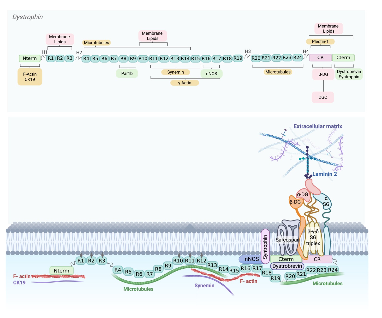
Figure 1. Dystrophin interacts with several structural and signaling proteins in the myofiber. Top panel: Schematic representation of dystrophin protein and its interactors. Sarcolemmal bindings are highlighted in pink, while cytoskeletal proteins (actin, microtubules and intermediate filaments) are indicated in yellow. Additionally, signaling proteins (Par1b, nNOS, dystrobrevin and syntrophin) are highlighted in green. Lower panel: Schematic representation of dystrophin in the skeletal muscle cell. Abbreviations used: Nterm: N-terminal; γ SG: γ Sarcoglycan; α SG: α Sarcoglycan; β-SG: β-Sarcoglycan; 𝝳 SG: 𝝳 Sarcoglycan; β-DG: β-Dystroglycan; α DG: α Dystroglycan; CR: Cystein Rich; Cterm: C terminal. Created in BioRender. Palmieri L. (2026) https://BioRender.com/ztj6ipq
Genetic Restoration Strategies for Dystrophin Expression
Therapeutic approaches for DMD fall into five main categories: (i) pharmacological, (ii) immunological (e.g. use of membrane-stabilizers antibodies, CART to target fibrosis) (iii) exon-skipping, (iv) stop-codon readthrough, (v) genetic intervention. Genetic interventions for DMD include CRISPR-Cas9–mediated genome editing and AAV-mediated gene delivery. CRISPR-Cas9 genome editing aims to correct the mutated DMD gene by restoring the open reading frame, thereby re-establishing proper dystrophin expression. While this approach holds considerable promises, its clinical translation remains challenging due to the limited progress made so far, the still preliminary nature of available clinical data [38,39], and its dependency on specific mutation types [40]. These factors currently place CRISPR-Cas9-based strategies at an earlier stage of clinical readiness compared to other genetic interventions such as AAV-mediated gene delivery.
The most developed and advanced genetic intervention is AAV-mediated gene delivery of a mini-gene encoding a shortened, partially functional dystrophin protein, a strategy that bypasses the need for mutation-specific correction and is therefore applicable to all DMD patients. Recombinant adeno-associated virus (rAAV) vectors are widely used experimentally and in clinical trials due to their ability to mediate long-term transgene expression in skeletal and cardiac muscle [41–45]. The primary limitation remains AAV's 4.7 kb packaging capacity versus dystrophin's 11.2 kb coding sequence, necessitating truncated micro- or midi-dystrophins (µ/midiDys) that retain essential functional domains [46]. This strategy draws from Becker muscular dystrophy (BMD), where ~46% gene deletions still yield milder phenotypes [47], inspiring diverse µ/midiDys constructs (Figure 2). Systemic delivery of single AAV vectors expressing µDys has shown broad muscle transduction, dystrophic phenotype improvement, and functional benefits in preclinical DMD models [46,48,49]. These preclinical advances have paved the way for clinical evaluation, and several ongoing clinical trials are now reporting initial outcomes (Table 1; Figure 2).
|
Company/Sponsor |
Clinical trial |
Clinical trial Phase/type |
Sample size |
Criteria inclusion |
Dose (vg/kg) |
SAE |
Status |
Investigational product name (AAV serotype) |
Notes |
Reference |
|
Sarepta |
NCT03769116 |
Phase 1/2 |
41 |
Children from 4 to 7 years old |
2,00E+14 |
Liver injury, hypertransalinasemia and rhabdomyolysis |
Completed |
SRP-9001(AAVrh74) |
|
[89] |
|
NCT04626674 (ENDEAVOR) |
Phase 1 |
55 |
Children from 4 to 8 years old |
1,33E+14 |
Immune-mediated myositis, myocarditis, increased transaminases |
Recruiting |
SRP-9001(AAVrh74) |
|
[90] |
|
|
NCT05096221 (EMBARK) |
Phase 3 |
126 |
Children from 4 to 7 years old |
1,33E+14 |
Myocarditis, left ventricular dysfunction, gamma-glutamyl transferase and transaminase increased, rhabdomyolysis |
Completed |
SRP-9001(AAVrh74) |
|
[91] |
|
|
NCT05881408 (ENVISION) |
Phase 3 |
148 |
Children from 4 to 7 years old |
1,33E+14 |
NA |
Active, not recruiting |
SRP-9001(AAVrh74) |
|
- |
|
|
NCT05967351 (EXPEDITION) |
Phase 3 |
400 |
Children from 4 to 7 years old |
NA |
NA |
Enrolling by invitation |
SRP-9001(AAVrh74) |
|
- |
|
|
NCT06128564 |
Phase 2 |
21 |
Children from 4 to 7 years old |
1,33E+14 |
NA |
Active, not recruiting |
SRP-9001(AAVrh74) |
|
[92] |
|
|
NCT06241950 |
Phase 1 |
6 |
Children from 4 to 9 years old |
1,33E+14 |
NA |
Completed |
SRP-9001(AAVrh74) |
|
- |
|
|
NCT06270719 (ENDURE) |
Phase 4 |
500 |
Children from 4 years old Mutations Not within Exons 8-9 |
NA |
NA |
Enrolling by invitation |
SRP-9001(AAVrh74) |
|
- |
|
|
NCT06597656 (HORIZON) |
Phase 1 |
16 |
Children from 4 to 8 years old Mutations Not within Exons 8-9 |
1,33E+14 |
NA |
Active, not recruiting |
SRP-9001(AAVrh74) |
|
- |
|
|
Solid Biosciences |
NCT03368742 (IGNITE-DMD) |
Phase 1/2 |
12 |
Children from 4 to 17 years old |
5E+13 - 2E+14 |
Complement activation within first weeks following dosing (3 patients) |
Active not recruiting |
SGT-001 (AAV9) |
Deprioritized |
- |
|
NCT06138639 (INSPIRE DUCHENNE) |
Phase 1/2 |
43 |
Children from 4 to 11 years old. Mutations Not within Exons 1-11, |
1,00E+14 |
NA |
Recruiting |
SGT-003 (AAV-SLB101) |
First clinical trial using myotropic AAV vectors (AAV-SLB101) |
- |
|
|
Pfizer |
NCT03362502 |
Phase 1B |
19 |
Children from 4 years old |
1E+14 - 3E+14 |
Dehydration, acute kidney injury, thrombocytopenia. One patient died of cardiogenic shock. |
Terminated |
PF-06939926 (AAV9) |
[93,94] |
|
|
NCT04281485 (CIFFREO) |
Phase 3 |
122 |
Children from 4 to 7 years old |
NA |
Myocarditis, liver injury |
Active not recruiting |
PF-06939926 (AAV9) |
[95] |
||
|
NCT05429372 (DAYLIGHT) |
Phase 2 |
10 |
Children from 2 to 3 years old |
2,00E+14 |
One patient died over one year after infusion (recurrent rhabdomyolysis) |
Terminated |
PF-06939926 (AAV9) |
|
- |
|
|
Genethon |
2023-505187-11-00 GNT-016 MDYF |
Phase 1/2/3 |
5 (estimated 50) |
Children from 6 to 10 years old Mutation not affecting exon 8 and/or 9 |
1E+13 - 3E+13 |
NA |
Recruiting |
GNT-0004 (AAV8) |
|
[54] |
|
Regenxbio |
NCT05693142 (AFFINITY DUCHENNE) |
Phase 1/2/3 |
65 |
Children from 1 to 11 years old Mutation in Exons 18 and above |
1E+14 - 2E+14 |
NA |
Recruiting |
RGX-202 (AAV8) |
|
[96] |
|
NCT06491927 |
Observational study |
19 |
Children from 1 to 11 years old Mutation in Exons 18 and above |
1E+14 - 2E+14 |
NA |
Enrolling by invitation |
RGX-202 (AAV8) |
|
- |
|
|
Insmed |
NCT06817382 (ASCEND) |
Phase 1 |
12 |
Children from 3 to 5 years old Mutation between Exons 18 and 58 |
NA |
NA |
Recruiting |
INS1201 (AAV9) |
First clinical trial with intratecal administration |
- |
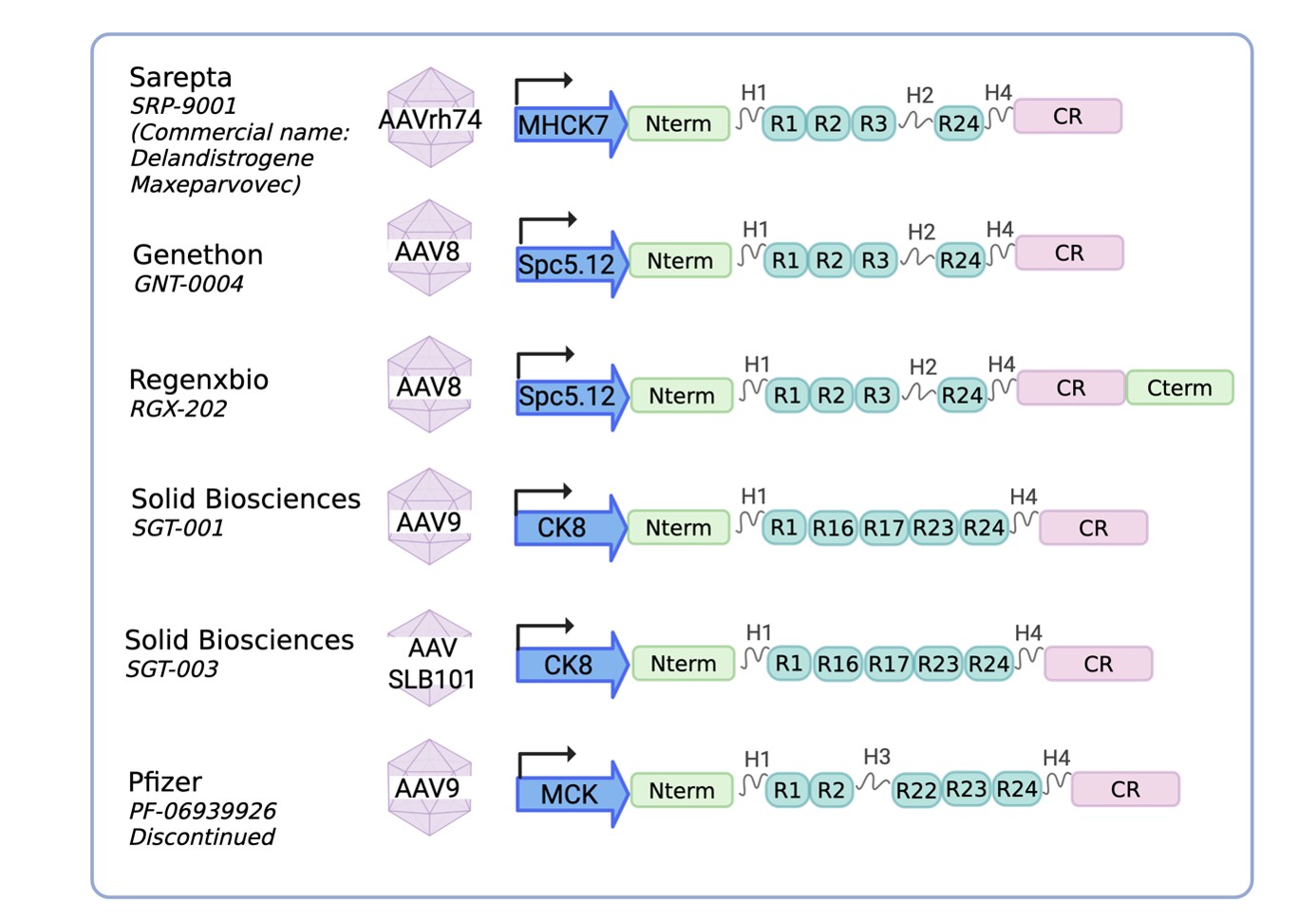
Figure 2. Micro-dystrophin (µDys) construct designs across clinical-stage gene therapy programs. All constructs are delivered via AAV vectors of different serotypes (AAVrh74, AAV8, AAV9, or AAV SLB101) and are driven by muscle-specific promoters (indicated in blue: MHCK7, Spc5.12, CK8, or MCK), selected to maximize muscle tropism and transgene expression. Despite sharing a common structural backbone (comprising an N-terminal domain (Nterm) and a cysteine-rich domain (CR), both essential for membrane anchoring and interaction with the dystrophin-associated protein complex (DAPC)) the constructs differ in their rod domain composition, particularly in the number and identity of spectrin-like repeats (R) and hinge regions (H) retained. These differences reflect distinct design strategies aimed at optimizing functional output while remaining within the AAV packaging size limit (~4.7 kb). The domain architecture of each µDys construct is shown for all gene therapy products currently in clinical development or approved by the FDA, including Delandistrogene Maxeparvovec (SRP-9001, Sarepta), GNT-0004 (Genethon), RGX-202 (Regenxbio), SGT-001 and SGT-003 (Solid Biosciences), and PF-06939926 (Pfizer, discontinued). Created in BioRender. Palmieri L. (2026) https://BioRender.com/ztj6ipq
Considerations on μDys gene therapy: safety issue and immunological response
Although preclinical models (including those developed by Sarepta, Pfizer, and Solid Biosciences) reported encouraging outcomes, the performance of mDys gene therapy across most clinical trials and publicly disclosed datasets has been more modest than anticipated, while also raising significant safety concerns in humans. Indeed, several serious adverse events (SAEs) have emerged in the context of AAV-mediated gene therapy for DMD. These events – including hepatotoxicity, myositis, and myocarditis – were observed in 6 of 18 clinical trials, notably the Sarepta trials (NCT03769116, NCT04626674, NCT05096221), the Solid Biosciences trial (NCT03368742), and the Pfizer trial (NCT03362502) (Table 1). Specifically, acute adverse effects, which arise within the first days to weeks following treatment and are driven by the innate immune response, may be triggered by CpG motifs present in the AAV genome [50] or by the capsid itself [51], which activates Toll-like receptor (TLR)–mediated immune signaling. Additionally, the AAV capsid can interact with the complement protein [52], leading to endothelial injury and thrombotic microangiopathy (TMA). TMA was observed in three independent clinical trials that shared no similarity in either the transgenes or capsid used; the only common denominator was the high AAV dose administered (> 1 x 10¹⁴ vg/kg) [53].
With respect to subacute and late SAEs, which manifest several months after gene therapy administration, these are thought to involve a T-cell response directed against the capsid and/or the μDys transgene. These subacute and late SAEs of μDys gene therapy clinical trials prompted a collaborative investigation among the various companies/sponsors developing μDys-based therapies to share and analyze safety data across clinical programs. This effort revealed that patients who developed subacute SAEs shared a common pathogenic variant involving exons 8 to 11 of the DMD gene, which encodes the highly immunogenic H1 region. Consequently, these patients were immunologically naïve to transgenes epitopes present in the gene therapy product. In consequence, clinical trial inclusion criteria changed to exclude patients carrying deletion in the region encompassing exons 8–11 [54].
Nevertheless, these adjustments in clinical trial criteria inclusion, four late-onset fatalities have been reported: three in the context of Elevidys clinical trials (Sarepta) and one in the PF-06939926 trial (Pfizer) [55,56]. The three children treated with Elevidys died 2–5 years post-dosing from causes that remain to be fully established. Nevertheless, elevated cardiac troponin-I concentrations in the blood of these patients may indicate a cardiac-related cause of death [57]. The patient who died 14 months after PF-06939926 administration exhibited a distinctive clinical course. This patient carried an in-frame deletion of exons 45–48, a mutation typically associated with the milder BMD phenotype yet presented a clinical course consistent with DMD [58], representing an apparent exception to this rule. The patient experienced recurrent rhabdomyolysis and ultimately died of hyperkalemia-induced cardiac arrest during the final episode. Notably, no immune response against the exogenous μDys transgene was detected, and the underlying mechanism remains uncertain.
These recent cardiac-related events have stimulated an active and timely area of research aimed at elucidating the molecular mechanisms underlying AAV-μDys-associated cardiac toxicity. According to Sweeney lab, μDys-associated cardiac toxicity arises from the displacement of utrophin (normally upregulated in the DMD heart and partially compensates for the absence of dystrophin). Because μDys is a truncated protein and less effective of utrophin in the heart, its replacement of utrophin is causing cardiac defects [59]. Nevertheless, the outcome of treating double knockout (dKO) mice (deficient in both utrophin and dystrophin) with AAV-mediated μDys gene transfer was likewise associated with cardiac toxicity [60]. Recent hypothesis identifies that accumulation of μDys at high concentrations may saturate the ubiquitin-proteosome system (UPS) and this proteostatic imbalance is causing cardiomyocytes toxicity [61,62]. In any case, further investigation is needed to elucidate the mechanisms underlying the cardiac abnormalities observed in the DMD context following μDys overexpression.
Considerations on μDys gene therapy: lack of functional primary endpoints
Although refined inclusion criteria (notably excluding patients with exons region 8–11) reduced SAEs incidence in children with DMD, the functional efficacy of μDys gene therapy products remain currently uncertain. Indeed, several clinical studies have failed to demonstrate statistically significant improvement over placebo in principle functional outcomes, such as North Star Ambulatory Assessment (NSAA). Among these, the Sarepta program is particularly noteworthy; in the phase III EMBARK trial, the primary endpoint changes from baseline in the NSAA score did not reach statistical significance between DMD patients after a single intravenous administration of delandistrogene moxeparvocec, the commercial μDys gene therapy product developed by Sarepta [63]. Nevertheless, in June 2024, the U.S. Food and Drug Administration granted full approval for Sarepta’s therapeutic product for ambulatory patients with DMD aged 4 years and older, based primarily on post-treatment expression of μDys. Among published clinical studies, the only statistically significant improvement in NSAA score has been reported in the EUDRACT study conducted by Genethon, which observed a 4.7-point increase in NSAA score (p<0.05). However, this finding was derived from a very small patient cohort (n = 5), compared with the substantially larger EMBARK study population (n = 126).
The discrepancy between efficacy in preclinical models and the limited clinical benefit has raised a critical concern regarding the insufficient level of transgene expression achieved in skeletal muscle tissue. In response, two principal strategies have been pursued: (i) substantial escalation of the administered dose in humans, reportedly up to tenfold higher than those used in preclinical studies [46,49,64–66] and (ii) the rational engineering of muscle-tropic rAAV capsids. The latter approach has particularly gained momentum, as illustrated by Solid Biosciences’ shift from its first-generation vector, SGT-001 (AAV9-based), to the more potent myotropic vector SGT-003, encapsidated with AAV-SLB101, a proprietary vector developed by Solid Biosciences. This next-generation platform has recently demonstrated encouraging early results. More broadly, advances in capsid bioengineering have led to the development of distinct classes of muscle-specific myotropic vectors known as AAVMYO, MyoAAV and LICA variants. These vectors share the common feature of displaying an integrin-binding RGD motif on the capsid surface, which enhances transduction efficiency in skeletal muscles of both mice and primates [67–69]. In addition, these engineered capsids exhibit liver-detargeting properties, thereby reducing hepatic exposure and the potential for liver-associated toxicities.
Novel Strategies for Large Dystrophin Delivery
The limited and inconsistent functional benefit observed in μDys human clinical trials has prompted the development of next generation approaches based on the delivery of larger dystrophin constructs. These strategies are grounded on the rationale that additional dystrophin domains may be required to achieve a more robust therapeutic effect. To overcome the packaging constraints of AAV vectors, this challenge has been addressed through multi-vector delivery approaches, in which the dystrophin coding sequence is split across two or three AAV vectors. Upon co-transduction of the same myofiber, these truncated sequences can recombine or reconstitute a longer, functional dystrophin protein. This reconstitution can occur through several mechanisms, including DNA trans-splicing, homologous recombination (HR), RNA/mRNA trans-splicing, or protein trans-splicing. The optimal vector design and choice of reassembly mechanism depend on the specific features of the transgene sequence as well as the biological properties of the target tissue. In DNA trans-splicing, AAV inverted terminal repeats (ITRs) drive tail-to-head genome concatenation: the 5′ vector delivers promoter, 5′ coding sequence (CDS), and splice donor (SD), while the 3′ vector provides splice acceptor (SA), 3′ CDS, and polyA signal. Subsequent ITR-mediated splicing generates intact full-length mRNA, though efficiency is limited by concatenation orientation. The overlapping HR approach offers a simpler alternative, relying on recombination between homologous CDS overlaps: the 5′ vector carries promoter and 5′ CDS, paired with a 3′ vector bearing 3′ CDS and polyA. This approach eliminates the need for exogenous splicing elements and can be extended to triple-vector full-length dystrophin delivery, although reconstitution efficiencies remain dependent on the transgene design and the target cell type [70]. An alternative strategy that relies on mRNA trans-splicing to reconstitute large transcripts. In this approach, two engineered DNA constructs encode the 5’ and 3’ segments of a larger pre-mRNA. These fragments incorporate a shared intronic hybridization domain (HD) which promotes trans-splicing and enables the precise joining of the two partial transcripts into a full-length mRNA [71]. Despite promising experimental results, this approach has not achieved a sufficient level of pre-mRNA trans-splicing, to support its implementation in dual-AAV strategies. Methods such as DNA trans-splicing AAV [65,72] and overlapping HR [73,74] have enabled the transfer of midiDys genes in dystrophin-null mice. However, these approaches remain limited by relatively low reconstitution efficiency, necessitating higher doses of AAVs, which in turn increase the risk of immune response and associated toxicities.
Another method for creating larger proteins from smaller transcription units involves the use of split-inteins to fuse two proteins. Split-inteins, protein orthologs of introns, are small polypeptides that undergo a distinct post-translational process called protein trans-splicing (PTS). During PTS, the N- and C-terminal residues (exteins) flanking the intein are naturally ligated, resulting in the formation of one functional protein while removing the reconstituted intein [75]. Work from Pinto and colleagues highlights GP41-1 as the most performant intein in vivo [76]. Intein-mediated PTS has been extensively investigated in the context of retinal and liver disease [77,78] as well as in DMD [79–81]. These studies have demonstrated significant amelioration of the dystrophic phenotype and physiological improvement in both young and old mdx mouse models. Remarkably, the inteins PTS technology has been successfully used for the delivery of full-length dystrophin transgene [81,82]. Tasfaout et al. demonstrated that full-length dystrophin improves cardiac function at later stage of disease in mdx, although evidence for long-term cardiac efficacy is lacking [82]. More notably, recent work from Tian et al. demonstrates that EV-mediated delivery of full-length DMD mRNA achieves higher therapeutic efficacy than AAV-μDys in mdx mice, although a fair comparison at equivalent dystrophin expression levels is lacking [83] Nevertheless, comparisons between full-length dystrophin and μDys have shown resulted broadly similar levels of functional improvement and histological rescue in skeletal muscle [81].
Rethinking Dystrophin Domain Inclusion: Is Larger Always Better?
Despite the remarkable progress achieved in delivering full-length dystrophin, several important considerations remain. First, the introduction of full-length dystrophin may increase immune reactivity due to the presence of novel epitopes not naturally expressed in patients. Second, the use of dual or triple AAV vector systems to reconstitute larger dystrophin constructs inherently requires higher vector doses, which can in turn increase immune responses against the viral capsids. Accordingly, the development of effective immune-modulating or tolerance-induction strategies will likely be essential to fully realize the therapeutic potential of full-length dystrophin in humans [50]. Third, the limited functional improvement observed in mdx mouse models compared with the current gold-standard μDys raises questions about the added therapeutic value of these approaches. However, this apparent equivalence may reflect the limitations of the mouse mdx model itself. The use of more representative preclinical models of DMD, such as rat models [84] or human organoids models [87], may better reveal potential differences and therapeutic benefits of full-length dystrophin constructs relative to μDys.
Another key consideration in addressing DMD is understanding the functional role of individual dystrophin domains and how different domains combinations influence therapeutic outcomes. The delivery of full-length dystrophin remains technically challenging, requiring multiple viral vectors, higher doses, and potentially introducing novel epitopes, raising the possibility that complete restoration of the protein may not be necessary. Instead, intermediate-length constructs may provide sufficient therapeutic benefit. This concept is supported by observations in patients carrying in-frame mutations in the DMD gene, who produce partially functional dystrophin variants associated with BMD. These cases highlight that certain domains are more critical than others, and their presence or absence in gene therapy constructs can markedly influence functional recovery. Notably, mutation hotspots in BMD patients are frequently observed within the ABD1 domain and in the spectrin-like repeat regions R17–R19 and R19–R23 [86]. One possible explanation is that some domains, particularly those involved in actin or microtubule binding, may exhibit a degree of functional redundancy. Alternatively, the relative contribution of dystrophin’s interactions with distinct cellular components may differ between domains. Given that dystrophin’s primary role is to link the contractile apparatus of myofibers to the sarcolemma and extracellular matrix, it is plausible that optimal combination of cytoskeletal-binding domains (for actin, microtubules, and intermediate filaments) and membrane-binding domains (via lipids or the DAPC) is sufficient to ensure functional integrity. For example, in the original design rationale of μDys constructs, the connection to intermediate filaments (mediated by R11–R15), an essential link to costameres and a key anchoring point for lateral force transmission in myofibers, was not originally incorporated [22]. Moreover, the relationship between specific dystrophin mutations and patient phenotypes provides key insights into the regions critical for dystrophin function. Notably, asymptomatic individuals often retain the DMD 3′ region (exons 56–79) as well as hinges 1 and 2, underscoring their importance in maintaining muscle function [86]. Although hinge regions have not been clearly associated with specific binding partner, they may nonetheless play a critical role in conferring elasticity and structural stability of the dystrophin protein [87]. Collectively, these observations suggest that preserving key structural domains may be sufficient to preserve dystrophin function, providing a strong rationale for the development of optimized, streamlined midi-dystrophin constructs.
Conclusion and Final Perspectives
Altogether, these findings underscore the importance of defining the minimal structural requirements for a functional dystrophin molecule. This body of evidence supports the development of optimized midi-dystrophin constructs that prioritize key functional domains, enabling the design of more efficient therapeutic transgenes. Looking ahead, the next phase in dystrophin gene therapy will likely rely on the rational design of constructs that balance functional sufficiency with efficient delivery. In this context, advances in structural prediction, including those enabled by AlphaFold-based approaches, offer a powerful framework to refine midi-dystrophin architecture in silico prior to experimental validation. Such strategies may help identify the minimal combinations of domains required to ensure mechanical stability, signaling capacity, and proper folding, while avoiding the complexity associated with multi-vector delivery systems. Beyond design considerations, the clinical translation of these constructs will depend on addressing several practical challenges. Among these is the precise titration required to co-deliver the dual- or triple-vector systems in the exact stoichiometry needed, so as to avoid expression of individual vectors in isolation. The removal of inteins derived from protein trans-splicing, which may trigger unwanted immune responses, should also be addressed, an approach already explored in the context of retinal genetic disorders [88]. Long-term safety must also be carefully evaluated, including the durability of transgene expression, the potential for immune responses against the vector or the transgene product, and the consequences of genomic persistence over time. Ultimately, integrating structural modeling with iterative functional testing should accelerate the development of more effective and clinically translatable dystrophin replacement therapies.
Authors Disclosure
F.R.’s lab is funded by the Association Française contre les Myopathies (AFM) via TRANSLAMUSCLE III program European MAGIC Project consortium (magic-horizon.eu, grant agreement No. 101080690). The authors declare no competing interests.
References
2. Mendell JR, Shilling C, Leslie ND, Flanigan KM, al-Dahhak R, Gastier-Foster J, et al. Evidence-based path to newborn screening for Duchenne muscular dystrophy. Ann Neurol. 2012 Mar;71(3):304–13.
3. Bladen CL, Salgado D, Monges S, Foncuberta ME, Kekou K, Kosma K, et al. The TREAT-NMD DMD Global Database: analysis of more than 7,000 Duchenne muscular dystrophy mutations. Hum Mutat. 2015 Apr;36(4):395–402.
4. Durbeej M, Campbell KP. Muscular dystrophies involving the dystrophin-glycoprotein complex: an overview of current mouse models. Curr Opin Genet Dev. 2002 Jun;12(3):349–61.
5. Campbell KP, Kahl SD. Association of dystrophin and an integral membrane glycoprotein. Nature. 1989 Mar 16;338(6212):259–62.
6. Rando TA. The dystrophin-glycoprotein complex, cellular signaling, and the regulation of cell survival in the muscular dystrophies. Muscle Nerve. 2001 Dec;24(12):1575–94.
7. Michalak M, Opas M. Functions of dystrophin and dystrophin associated proteins. Curr Opin Neurol. 1997 Oct;10(5):436–42.
8. Samitt CE, Bonilla E. Immunocytochemical study of dystrophin at the myotendinous junction. Muscle Nerve. 1990 Jun;13(6):493–500.
9. Bewick GS, Young C, Slater CR. Spatial relationships of utrophin, dystrophin, beta-dystroglycan and beta-spectrin to acetylcholine receptor clusters during postnatal maturation of the rat neuromuscular junction. J Neurocytol. 1996 Jul;25(7):367–79.
10. Chan Y, Kunkel LM. In vitro expressed dystrophin fragments do not associate with each other. FEBS Lett. 1997 Jun 30;410(2-3):153–9.
11. Wan L, Ge X, Xu Q, Huang G, Yang T, Campbell KP, Yan Z, Wu J. Structure and assembly of the dystrophin glycoprotein complex. Nature. 2025 Jan;637(8048):1252–60.
12. Wilson DGS, Tinker A, Iskratsch T. The role of the dystrophin glycoprotein complex in muscle cell mechanotransduction. Commun Biol. 2022 Sep 27;5(1):1022.
13. Zhao J, Kodippili K, Yue Y, Hakim CH, Wasala L, Pan X, et al. Dystrophin contains multiple independent membrane-binding domains. Hum Mol Genet. 2016 Sep 1;25(17):3647–53.
14. Amann KJ, Renley BA, Ervasti JM. A cluster of basic repeats in the dystrophin rod domain binds F-actin through an electrostatic interaction. J Biol Chem. 1998 Oct 23;273(43):28419–23.
15. Fealey ME, Horn B, Coffman C, Miller R, Lin AY, Thompson AR, et al. Dynamics of Dystrophin's Actin-Binding Domain. Biophys J. 2018 Aug 7;115(3):445–54.
16. Koenig M, Monaco AP, Kunkel LM. The complete sequence of dystrophin predicts a rod-shaped cytoskeletal protein. Cell. 1988 Apr 22;53(2):219–28.
17. Orlova A, Rybakova IN, Prochniewicz E, Thomas DD, Ervasti JM, Egelman EH. Binding of dystrophin's tandem calponin homology domain to F-actin is modulated by actin's structure. Biophys J. 2001 Apr;80(4):1926–31.
18. Renley BA, Rybakova IN, Amann KJ, Ervasti JM. Dystrophin binding to nonmuscle actin. Cell Motil Cytoskeleton. 1998;41(3):264–70.
19. Belanto JJ, Mader TL, Eckhoff MD, Strandjord DM, Banks GB, Gardner MK, et al. Microtubule binding distinguishes dystrophin from utrophin. Proc Natl Acad Sci U S A. 2014 Apr 15;111(15):5723–8.
20. Nelson DM, Fasbender EK, Jakubiak MC, Lindsay A, Lowe DA, Ervasti JM. Rapid, redox-mediated mechanical susceptibility of the cortical microtubule lattice in skeletal muscle. Redox Biol. 2020 Oct;37:101730.
21. Prins KW, Humston JL, Mehta A, Tate V, Ralston E, Ervasti JM. Dystrophin is a microtubule-associated protein. J Cell Biol. 2009 Aug 10;186(3):363–9.
22. Bhosle RC, Michele DE, Campbell KP, Li Z, Robson RM. Interactions of intermediate filament protein synemin with dystrophin and utrophin. Biochem Biophys Res Commun. 2006 Aug 4;346(3):768–77.
23. Rezniczek GA, Konieczny P, Nikolic B, Reipert S, Schneller D, Abrahamsberg C, et al. Plectin 1f scaffolding at the sarcolemma of dystrophic (mdx) muscle fibers through multiple interactions with beta-dystroglycan. J Cell Biol. 2007 Mar 26;176(7):965–77.
24. Stone MR, O'Neill A, Catino D, Bloch RJ. Specific interaction of the actin-binding domain of dystrophin with intermediate filaments containing keratin 19. Mol Biol Cell. 2005 Sep;16(9):4280–93.
25. Ervasti JM, Campbell KP. A role for the dystrophin-glycoprotein complex as a transmembrane linker between laminin and actin. J Cell Biol. 1993 Aug;122(4):809–23.
26. Huang X, Poy F, Zhang R, Joachimiak A, Sudol M, Eck MJ. Structure of a WW domain containing fragment of dystrophin in complex with beta-dystroglycan. Nat Struct Biol. 2000 Aug;7(8):634–8.
27. Jung D, Yang B, Meyer J, Chamberlain JS, Campbell KP. Identification and characterization of the dystrophin anchoring site on beta-dystroglycan. J Biol Chem. 1995 Nov 10;270(45):27305–10.
28. Rentschler S, Linn H, Deininger K, Bedford MT, Espanel X, Sudol M. The WW domain of dystrophin requires EF-hands region to interact with beta-dystroglycan. Biol Chem. 1999 Apr;380(4):431–42.
29. Dumont NA, Wang YX, von Maltzahn J, Pasut A, Bentzinger CF, Brun CE, et al Dystrophin expression in muscle stem cells regulates their polarity and asymmetric division. Nat Med. 2015 Dec;21(12):1455–63.
30. Brenman JE, Chao DS, Xia H, Aldape K, Bredt DS. Nitric oxide synthase complexed with dystrophin and absent from skeletal muscle sarcolemma in Duchenne muscular dystrophy. Cell. 1995 Sep 8;82(5):743–52.
31. Brenman JE, Chao DS, Gee SH, McGee AW, Craven SE, Santillano DR, et al. Interaction of nitric oxide synthase with the postsynaptic density protein PSD-95 and alpha1-syntrophin mediated by PDZ domains. Cell. 1996 Mar 8;84(5):757–67.
32. Anderson JE. A role for nitric oxide in muscle repair: nitric oxide-mediated activation of muscle satellite cells. Mol Biol Cell. 2000 May;11(5):1859–74.
33. Colussi C, Mozzetta C, Gurtner A, Illi B, Rosati J, Straino S, et al. HDAC2 blockade by nitric oxide and histone deacetylase inhibitors reveals a common target in Duchenne muscular dystrophy treatment. Proc Natl Acad Sci U S A. 2008 Dec 9;105(49):19183–7.
34. Percival JM, Anderson KN, Gregorevic P, Chamberlain JS, Froehner SC. Functional deficits in nNOSmu-deficient skeletal muscle: myopathy in nNOS knockout mice. PLoS One. 2008;3(10):e3387.
35. Zhao J, Yang HT, Wasala L, Zhang K, Yue Y, Duan D, et al. Dystrophin R16/17 protein therapy restores sarcolemmal nNOS in trans and improves muscle perfusion and function. Mol Med. 2019 Jul 2;25(1):31.
36. Rudolf R, Straka T. Nicotinic acetylcholine receptor at vertebrate motor endplates: Endocytosis, recycling, and degradation. Neurosci Lett. 2019 Oct 15;711:134434.
37. Schmidt N, Akaaboune M, Gajendran N, Martinez-Pena y Valenzuela I, Wakefield S, Thurnheer R, et al. Neuregulin/ErbB regulate neuromuscular junction development by phosphorylation of α-dystrobrevin. J Cell Biol. 2011 Dec 26;195(7):1171–84.
38. Amoasii L, Hildyard JCW, Li H, Sanchez-Ortiz E, Mireault A, Caballero D, et al. Gene editing restores dystrophin expression in a canine model of Duchenne muscular dystrophy. Science. 2018 Oct 5;362(6410):86–91.
39. Lek A, Keeler A, Flotte TR. Death after High-Dose rAAV9 Gene Therapy in a Patient with Duchenne's Muscular Dystrophy. Reply. N Engl J Med. 2023 Dec 7;389(23):2211.
40. Min YL, Li H, Rodriguez-Caycedo C, Mireault AA, Huang J, Shelton JM, et al. CRISPR-Cas9 corrects Duchenne muscular dystrophy exon 44 deletion mutations in mice and human cells. Sci Adv. 2019 Mar 6;5(3):eaav4324.
41. Asokan A, Conway JC, Phillips JL, Li C, Hegge J, Sinnott R, et al. Reengineering a receptor footprint of adeno-associated virus enables selective and systemic gene transfer to muscle. Nat Biotechnol. 2010 Jan;28(1):79–82.
42. Gregorevic P, Blankinship MJ, Allen JM, Crawford RW, Meuse L, Miller DG, et al. Systemic delivery of genes to striated muscles using adeno-associated viral vectors. Nat Med. 2004 Aug;10(8):828–34.
43. Inagaki K, Fuess S, Storm TA, Gibson GA, Mctiernan CF, Kay MA, et al. Robust systemic transduction with AAV9 vectors in mice: efficient global cardiac gene transfer superior to that of AAV8. Mol Ther. 2006 Jul;14(1):45–53.
44. Kotterman MA, Schaffer DV. Engineering adeno-associated viruses for clinical gene therapy. Nat Rev Genet. 2014 Jul;15(7):445–51.
45. Wang Z, Zhu T, Qiao C, Zhou L, Wang B, Zhang J, et al. Adeno-associated virus serotype 8 efficiently delivers genes to muscle and heart. Nat Biotechnol. 2005 Mar;23(3):321–8.
46. Birch SM, Lawlor MW, Conlon TJ, Guo LJ, Crudele JM, Hawkins EC, et al. Assessment of systemic AAV-microdystrophin gene therapy in the GRMD model of Duchenne muscular dystrophy. Sci Transl Med. 2023 Jan 4;15(677):eabo1815.
47. England SB, Nicholson LV, Johnson MA, Forrest SM, Love DR, Zubrzycka-Gaarn EE, et al. Very mild muscular dystrophy associated with the deletion of 46% of dystrophin. Nature. 1990 Jan 11;343(6254):180–2.
48. Bostick B, Shin JH, Yue Y, Wasala NB, Lai Y, Duan D. AAV micro-dystrophin gene therapy alleviates stress-induced cardiac death but not myocardial fibrosis in >21-m-old mdx mice, an end-stage model of Duchenne muscular dystrophy cardiomyopathy. J Mol Cell Cardiol. 2012 Aug;53(2):217–22.
49. Le Guiner C, Servais L, Montus M, Larcher T, Fraysse B, Moullec S, et al. Long-term microdystrophin gene therapy is effective in a canine model of Duchenne muscular dystrophy. Nat Commun. 2017 Jul 25;8:16105.
50. Kumar SRP, Duan D, Herzog RW. Immune Responses to Muscle-Directed Adeno-Associated Viral Gene Transfer in Clinical Studies. Hum Gene Ther. 2023 May;34(9-10):365–71.
51. Zhu J, Huang X, Yang Y. The TLR9-MyD88 pathway is critical for adaptive immune responses to adeno-associated virus gene therapy vectors in mice. J Clin Invest. 2009 Aug;119(8):2388–98.
52. Zaiss AK, Cotter MJ, White LR, Clark SA, Wong NC, Holers VM, et al. Complement is an essential component of the immune response to adeno-associated virus vectors. J Virol. 2008 Mar;82(6):2727–40.
53. Shieh PB, Proud C, Diamond T, Chapin CA, Ahmad J, Salama AD, et al. Learnings from Patient Mortality after Delandistrogene Moxeparvovec Administration: A Report of Two Cases and Expert Committee Considerations for Future Mitigation and Management. Hum Gene Ther. 2026 Mar;37(5-6):262–79.
54. Bönnemann CG, Belluscio BA, Braun S, Morris C, Singh T, Muntoni F. Dystrophin Immunity after Gene Therapy for Duchenne's Muscular Dystrophy. N Engl J Med. 2023 Jun 15;388(24):2294–6.
55. Byrne BJ, Flanigan KM, Matesanz SE, Finkel RS, Waldrop MA, D'Ambrosio ES, et al. Current clinical applications of AAV-mediated gene therapy. Mol Ther. 2025 Jun 4;33(6):2479–516.
56. Duan D, Herzog RW. Deaths in gene therapy of Duchenne muscular dystrophy and other diseases: Underlying mechanisms and mitigating strategies. Mol Ther. 2026 Apr 1;34(4):1893–908.
57. Keselman DJ, Small JC, Seneviratne T, McCague S, Kaschak TL, Yum SW, et al. Real-world outcomes of delandistrogene moxeparvovec gene therapy: Motor outcomes and emerging safety concerns. Mol Ther. 2026 Jan 7;34(1):123–30.
58. Anthony K, Arechavala-Gomeza V, Ricotti V, Torelli S, Feng L, Janghra N, et al. Biochemical characterization of patients with in-frame or out-of-frame DMD deletions pertinent to exon 44 or 45 skipping. JAMA Neurol. 2014 Jan;71(1):32–40.
59. Hart CC, Lee YI, Xie J, Gao G, Lin BL, Hammers DW, et al. Potential limitations of microdystrophin gene therapy for Duchenne muscular dystrophy. JCI Insight. 2024 May 7;9(11):e165869.
60. Forand A, Moog S, Mougenot N, Lemaitre M, Sevoz-Couche C, Guesmia Z, et al. Long-Term Dystrophin Replacement Therapy in Duchenne Muscular Dystrophy Causes Cardiac Inflammation. JACC Basic Transl Sci. 2025 Jun;10(6):759–82.
61. Le Guiner C, Toumaniantz G, Larcher T, Marchand S, Buscara L, Cedrone G, et al. Dose- and genotype-dependent cardiac arrhythmia and sudden death in rats following microdystrophin gene therapy. Mol Ther. 2026 Jan 7;34(1):140–60.
62. McLendon PM, Robbins J. Proteotoxicity and cardiac dysfunction. Circulation research. 2015 May 22;116(11):1863–82.
63. Mendell JR, Muntoni F, McDonald CM, Mercuri EM, Ciafaloni E, Komaki H, et al. AAV gene therapy for Duchenne muscular dystrophy: the EMBARK phase 3 randomized trial. Nat Med. 2025 Jan;31(1):332–41.
64. Gregorevic P, Blankinship MJ, Allen JM, Crawford RW, Meuse L, Miller DG, et al. Systemic delivery of genes to striated muscles using adeno-associated viral vectors. Nat Med. 2004 Aug;10(8):828–34.
65. Lai Y, Thomas GD, Yue Y, Yang HT, Li D, Long C, et al. Dystrophins carrying spectrin-like repeats 16 and 17 anchor nNOS to the sarcolemma and enhance exercise performance in a mouse model of muscular dystrophy. J Clin Invest. 2009 Mar;119(3):624–35.
66. Yue Y, Ghosh A, Long C, Bostick B, Smith BF, Kornegay JN, et al. A single intravenous injection of adeno-associated virus serotype-9 leads to whole body skeletal muscle transduction in dogs. Mol Ther. 2008 Dec;16(12):1944–52.
67. Tabebordbar M, Lagerborg KA, Stanton A, King EM, Ye S, Tellez L, et al. Directed evolution of a family of AAV capsid variants enabling potent muscle-directed gene delivery across species. Cell. 2021 Sep 16;184(19):4919–4938.e22.
68. El Andari J, Renaud-Gabardos E, Tulalamba W, Weinmann J, Mangin L, Pham QH, et al. Semirational bioengineering of AAV vectors with increased potency and specificity for systemic gene therapy of muscle disorders. Sci Adv. 2022 Sep 23;8(38):eabn4704.
69. Vu Hong A, Suel L, Petat E, Dubois A, Le Brun PR, Guerchet N, et al. An engineered AAV targeting integrin alpha V beta 6 presents improved myotropism across species. Nat Commun. 2024 Sep 11;15(1):7965.
70. Lostal W, Kodippili K, Yue Y, Duan D. Full-length dystrophin reconstitution with adeno-associated viral vectors. Hum Gene Ther. 2014 Jun;25(6):552–62.
71. Pergolizzi RG, Ropper AE, Dragos R, Reid AC, Nakayama K, Tan Y, et al. In vivo trans-splicing of 5' and 3' segments of pre-mRNA directed by corresponding DNA sequences delivered by gene transfer. Mol Ther. 2003 Dec;8(6):999–1008.
72. Zhang Y, Yue Y, Li L, Hakim CH, Zhang K, Thomas GD, et al. Dual AAV therapy ameliorates exercise-induced muscle injury and functional ischemia in murine models of Duchenne muscular dystrophy. Hum Mol Genet. 2013 Sep 15;22(18):3720–9.
73. Albini S, Palmieri L, Dubois A, Bourg N, Lostal W, Richard I. Assessment of Therapeutic Potential of a Dual AAV Approach for Duchenne Muscular Dystrophy. Int J Mol Sci. 2023 Jul 13;24(14):11421.
74. Odom GL, Gregorevic P, Allen JM, Chamberlain JS. Gene therapy of mdx mice with large truncated dystrophins generated by recombination using rAAV6. Mol Ther. 2011 Jan;19(1):36–45.
75. Saleh L, Perler FB. Protein splicing in cis and in trans. Chem Rec. 2006;6(4):183–93.
76. Pinto F, Thornton EL, Wang B. An expanded library of orthogonal split inteins enables modular multi-peptide assemblies. Nat Commun. 2020 Mar 23;11(1):1529.
77. Esposito F, Lyubenova H, Tornabene P, Auricchio S, Iuliano A, Nusco E, et al. Liver gene therapy with intein-mediated F8 trans-splicing corrects mouse haemophilia A. EMBO Mol Med. 2022 Jun 8;14(6):e15199.
78. Tornabene P, Trapani I, Minopoli R, Centrulo M, Lupo M, de Simone S, Tiberi P, Dell'Aquila F, Marrocco E, Iodice C, Iuliano A, Gesualdo C, Rossi S, Giaquinto L, Albert S, Hoyng CB, Polishchuk E, Cremers FPM, Surace EM, Simonelli F, De Matteis MA, Polishchuk R, Auricchio A. Intein-mediated protein trans-splicing expands adeno-associated virus transfer capacity in the retina. Sci Transl Med. 2019 May 15;11(492):eaav4523.
79. Palmieri L, Ferrand M, Vu Hong A, Richard I, Albini S. In Silico Structural Prediction for the Generation of Novel Performant Midi-Dystrophins Based on Intein-Mediated Dual AAV Approach. Int J Mol Sci. 2024 Sep 27;25(19):10444.
80. Tasfaout H, Halbert CL, McMillen TS, Allen JM, Reyes TR, Flint GV, et al. Split intein-mediated protein trans-splicing to express large dystrophins. Nature. 2024 Aug;632(8023):192–200.
81. Zhou Y, Zhang C, Xiao W, Herzog RW, Han R. Systemic delivery of full-length dystrophin in Duchenne muscular dystrophy mice. Nat Commun. 2024 Jul 21;15(1):6141.
82. Tasfaout H, McMillen TS, Reyes TR, Halbert CL, Tian R, Regnier M, et al. Expression of full-length dystrophin reverses muscular dystrophy defects in young and old mdx4cv mice. J Clin Invest. 2025 Jun 10;135(15):e189075.
83. Tian Y, Liu Y, Tong Y, Huntoon K, Yang L, Shi J, et al. Skeletal-muscle-targeted non-viral delivery of full-length DMD mRNA for Duchenne muscular dystrophy. Nat Biomed Eng. 2026 Jun 11.
84. Taglietti V, Kefi K, Bronisz-Budzyńska I, Mirciloglu B, Rodrigues M, Cardone N, et al. Duchenne muscular dystrophy trajectory in R-DMDdel52 preclinical rat model identifies COMP as biomarker of fibrosis. Acta Neuropathol Commun. 2022 Apr 25;10(1):60.
85. Palmieri L, Bimbi G, Ferrand M, Marcello M, Pili L, Hong AV, et al. Disease exacerbation in human DMD MYOrganoids enables gene therapy evaluation and unveils persistence of fibrotic activity. NPJ Regen Med. 2026 Jan 2;11(1):2.
86. Fortunato F, Tonelli L, Farnè M, Selvatici R, Ferlini A. DMD deletions underlining mild dystrophinopathies: literature review highlights phenotype-related mutation clusters and provides insights about genetic mechanisms and prognosis. Front Neurol. 2024 Jan 15;14:1288721.
87. Wasala LP, Watkins TB, Wasala NB, Burke MJ, Yue Y, Lai Y, et al. The Implication of Hinge 1 and Hinge 4 in Micro-Dystrophin Gene Therapy for Duchenne Muscular Dystrophy. Hum Gene Ther. 2023 May;34(9-10):459–70.
88. Tornabene P, Trapani I, Centrulo M, Marrocco E, Minopoli R, Lupo M, et al. Inclusion of a degron reduces levelsof undesired inteins after AAV-mediated proteintrans-splicing in the retina. Mol Ther Methods Clin Dev. 2021 Oct 19;23:448–59.
89. Mendell JR, Shieh PB, McDonald CM, Sahenk Z, Lehman KJ, Lowes LP, et al. Expression of SRP-9001 dystrophin and stabilization of motor function up to 2 years post-treatment with delandistrogene moxeparvovec gene therapy in individuals with Duchenne muscular dystrophy. Front Cell Dev Biol. 2023 Jul 11;11:1167762.
90. Zaidman CM, Proud CM, McDonald CM, Lehman KJ, Goedeker NL, Mason S, et al. Delandistrogene Moxeparvovec Gene Therapy in Ambulatory Patients (Aged ≥4 to <8 Years) with Duchenne Muscular Dystrophy: 1-Year Interim Results from Study SRP-9001-103 (ENDEAVOR). Ann Neurol. 2023 Nov;94(5):955–68.
91. Mendell JR, Muntoni F, McDonald CM, Mercuri EM, Ciafaloni E, Komaki H, et al. Two-Year Outcomes Following Delandistrogene Moxeparvovec Treatment in Ambulatory Patients with Duchenne Muscular Dystrophy: Phase 3 EMBARK Trial. Neurol Ther. 2026 Apr;15(2):545–59.
92. Mendell JR, Muntoni F, McDonald CM, Mercuri EM, Ciafaloni E, Komaki H, et al. LB: Delandistrogene moxeparvovec in Duchenne muscular dystrophy: Functional and safety outcomes up to 3 years post-infusion in the EMBARK study [Internet]. MDA Clinical & Scientific Conference; 2026. Available from: https://www.mdaconference.org/abstract-library/lb-delandistrogene-moxeparvovec-in-duchenne-muscular-dystrophy-functional-and-safety-outcomes-up-to-3-years-post-infusion-in-the-embark-study/
93. Sherlock SP, Levy DI, McIntosh A, Shieh PB, Smith EC, McDonnell TG, et al. Cardiac safety of fordadistrogene movaparvovec gene therapy in Duchenne muscular dystrophy: Initial observations from a phase 1b trial. Mol Ther. 2025 Sep 3;33(9):4216–25.
94. Byrne BJ, Butterfield RJ, Shieh PB, Smith EC, Licht C, Binks M, et al. Complement activation in a phase Ib study of fordadistrogene movaparvovec for Duchenne muscular dystrophy. Mol Ther. 2025 Sep 3;33(9):4226–38.
95. Muntoni F, Nascimento A, Shin J, Guglieri M, Stettner GM, Veerapandiyan A, et al. Safety and efficacy of fordadistrogene movaparvovec in ambulatory participants with Duchenne muscular dystrophy (CIFFREO): a phase 3, double-blind, randomised, placebo-controlled study. Lancet Neurol. 2026 Mar;25(3):245–55.
96. Rocha CT, Veerapandiyan A, Iannaccone ST, Kuntz N, Harper A, Boulos N, et al. RGX-202, an investigational gene therapy for the treatment of Duchenne muscular dystrophy: Interim Phase I/II clinical data [Internet]. MDA Clinical & Scientific Conference; 2026. Available from: https://www.mdaconference.org/abstract-library/rgx-202-an-investigational-gene-therapy-for-the-treatment-of-duchenne-muscular-dystrophy-interim-phase-i-ii-clinical-data/