Abstract
Introduction: Ptosis or the dropping of an eyelid can affect both the adult and pediatric populations and can be caused by various factors. Ptosis is not very common and in children, the common causes include orbital cellulitis, congenital ptosis, Cranial Nerve III palsy, and Horner’s syndrome. However, unilateral acute ptosis from an infectious disease process is rare and there are only a few documented cases of such occurrences. Acute ptosis is a medical emergency and prompt diagnosis, and management is critical for the resolution of symptoms and prevention of serious complications.
Case: We present an interesting case of a 14-year-old female with pansinusitis who experienced unilateral left-sided ptosis. Initially prescribed oral antibiotics for sinusitis, the patient developed ptosis, requiring inpatient hospital admission and intravenous antibiotics. On admission, the patient had leukocytosis, elevated CRP, and ESR levels. CT facial bones showed pansinusitis and ruled out orbital cellulitis, acute intracranial abnormalities, and venous sinus thrombosis.
After aggressive treatment with intravenous antibiotics and systemic steroids, the sinusitis resolved, as did her unilateral ptosis and facial swelling.
Discussion: This is a unique presentation of ptosis that was treated successfully allowing for prompt and complete resolution. We want to highlight the establishment of a varying diagnosis and treatment courses which led to the final resolution of the acute ptosis. The patient was treated with meropenem and dexamethasone. The adjunctive use of corticosteroids, in this case, was beneficial.
Introduction
Ptosis or drooping of the upper eyelid can affect both the adult and pediatric populations and can be caused by various factors, the most common being congenital ptosis [1]. Acquired ptosis is most commonly aponeurotic ptosis, and this is mostly seen in older adults [1]. Ptosis in the pediatric population is rare, with an occurrence of about 7.9 in 100,000 [2]. Two muscles are involved in the elevation of the eyelid: the levator palpebrae superioris, innervated by the superior branch of cranial nerve III, and the superior tarsal muscle (Müller's muscle), innervated by the cervical sympathetic system [3,4]. Acute ptosis can be secondary to a multitude of conditions such as Bell’s palsy, acute botulism, or third cranial nerve paralysis. Another cause of ptosis can be orbital cellulitis; however, this is exceedingly rare. The hallmark features of orbital cellulitis include fever, diffuse bulbar chemosis, diplopia due to ophthalmoplegia, and proptosis [5].
The symptoms of ptosis due to sinusitis may be accompanied by other symptoms of sinusitis such as headache, nasal congestion, and facial pain or pressure. Unilateral acute ptosis as a result of sinusitis is rare and there are only a few documented cases of such occurrences. There was a case of chronic, not acute, rhinosinusitis causing Horner syndrome in a 40-year-old male [6].
Horner syndrome is a triad of ptosis, miosis, and anhidrosis.
Acute ptosis is a medical emergency and prompt diagnosis, and management is critical for the resolution of symptoms and prevention of serious complications. Infections can extend posteriorly into the orbit, leading to significant visual and cerebral complications [7]. Frontal sinusitis is the most common predisposing pathology leading to brain abscess. Although these complications are rare due to antibiotics, they are still possible and prompt treatment of sinusitis is necessary to prevent such complications.
We would like to introduce an interesting case of a 14-year-old female with pansinusitis who presented with unilateral left-sided ptosis. After aggressive intravenous antibiotic and steroid treatment, the sinusitis resolved as did the unilateral ptosis. The progression of her ptosis is documented in the photos shown below.
The collection and evaluation of this patient’s health information were HIPAA-compliant.
Case Presentation
We are presenting a 14-year-old female with no past medical history who presented to the ER with new onset left eyelid ptosis, facial swelling, and bilateral retro-orbital pain. The patient had presented to the ER twice in the past week; the first time she presented with ear pain, was diagnosed with otitis media, and was discharged on amoxicillin. On the next visit, she presented with congestion, body aches, and headaches and was diagnosed with sinusitis and discharged on cefdinir. Both times, the mother stated that she did not give the patient the antibiotics she was discharged with. On her third visit she presented with more severe symptoms including dropping of the left eyelid, facial swelling, fevers, and bilateral eye pain but denied pain with EOM, vision changes, dizziness, dysphagia, neck pain, ear pain, hearing issues, history of ear infections, chest pain, SOB, diarrhea, or weakness. The patient was subsequently admitted to the pediatric unit. On admission, labs were notable for leukocytosis of 22.3, elevated C reactive protein of 7.7, and ESR of 25. CT facial bones were notable for pansinusitis with near-complete opacification of the paranasal sinuses with an air-fluid level in the right maxillary sinus and near- complete opacification of the left maxillary sinus. Air fluid levels were noted in the frontal sinuses bilaterally and complete opacification of the ethmoid sinuses. CT head was negative for acute intracranial abnormalities and venous sinus thrombosis. The respiratory viral panel was negative.
The patient was admitted and treated for pansinusitis with IV meropenem for seven days and three doses of dexamethasone with a satisfactory response. There was an immediate improvement in the facial swelling and ptosis on day one of treatment and along with the resolution of her fevers. Her labs showed improvement with a WBC count dropping down to 14.9K and CRP of 6.6. After three days of treatment, the facial swelling and ptosis were fully resolved. The patient was discharged home after completing seven days of inpatient treatment with meropenem and Augmentin. Instructions were given to continue Augmentin for an additional seven days with a follow up appointment with ENT outpatient.
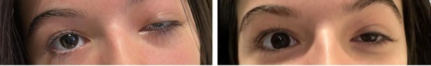
A. B.
C. D.
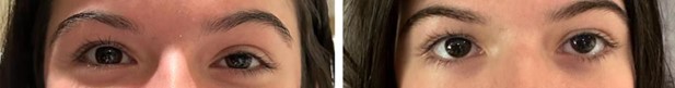
Figure 1. Progression of left eye ptosis. 1A. Day 1 of presentation. 1B. Day 2 with treatment. 1C. Day 3 with treatment. 1D. Day 4 with treatment and complete resolution of symptoms.
Discussion
Ptosis related to sinusitis is a relatively uncommon occurrence, but it can occur as a result of swelling and pressure from the sinuses impacting the function of the levator muscle, which is responsible for lifting the eyelid. In this case, the ptosis was temporary and resolved once the underlying sinusitis was treated. Due to the absence of proptosis, ophthalmoplegia, and impairment of visual acuity, orbital cellulitis was ruled out. This patient was initially suspected to have preseptal cellulitis, but CT findings showed pansinusitis with no evidence of preseptal cellulitis. Most documented cases of ptosis related to sinusitis are due to the infection causing orbital sinusitis. There is one reported case of pansinusitis that resulted in eyelid ptosis in a 9- year-old male, however, it was also associated with contralateral otitis media and was due to orbital cellulitis [8]. As such this remains to be a unique case of unilateral ptosis with no ophthalmoplegia or pupillary involvement as a result of pansinusitis.
The common treatment for acute pansinusitis is Augmentin, however, meropenem was used due the complexity of the case. Treatment with dexamethasone and meropenem allowed for prompt recovery of the patient’s symptoms including her ptosis. Furthermore, physical examinations including a thorough neurological exam and CT imaging was helpful in understanding the cause of her symptoms, effectively ruling out orbital cellulitis and preseptal cellulitis. The patient's pictures demonstrate the daily progress from admission, up to day four of treatment. The benefit of steroids as adjunctive therapy in acute sinusitis varies and five randomized controlled trials comparing systemic steroids to placebo were compared in 2014 in a paper titled “Systemic corticosteroids for acute sinusitis”[9] where it was concluded that oral corticosteroids as monotherapy were ineffective in adults, but the use of it as adjunctive therapy is modestly beneficial for short-term use as demonstrated in our patient who responded positively with the use of short-term corticosteroids for pansinusitis.
In summary, sinusitis should be suspected in patients who present with ptosis and prompt treatment should be administered to prevent any long-term complications. This patient had a successful resolution with the use of intravenous antibiotics and short-term corticosteroids. The images below showcase the clinical course of the ptosis as documented within this case report. Our limitations include only having a singular case and not finding many similar cases during our literature review. In the future we would like to conduct an analysis on all the pediatric ptosis cases that are admitted to our hospital and determine which are the most common etiologies and successful treatments.
Consents
The patient's mother consented to publication of the case by signing a consent form. Furthermore, this case report does not include any personal information that might lead to the identification of the patient.
Funding
No funding was provided for this case report.
Acknowledgements
None.
References
2. Griepentrog GJ, Diehl NN, Mohney BG: Incidence and demographics of childhood ptosis. Ophthalmology. 2011, 118:1180-3.
3. Patel R, Harper-Shankie M, Patel E, Sivaswamy L. Unilateral Ptosis. The Journal of Pediatrics. 167(5):1160- 1160.E1.
4. Pavone P, Cho SY, Praticò AD, Falsaperla R, Ruggieri M, Jin DK. Ptosis in childhood: A clinical sign of several disorders: Case series reports and literature review. Medicine (Baltimore). 2018, 97:12124-10.
5. Botting AM, McIntosh D, Mahadevan M. Paediatric pre- and post-septal peri-orbital infections are different diseases. A retrospective review of 262 cases. International Journal of Pediatric Otorhinolaryngology. 2008;72:377.
6. Rissardo JP, Caprara ALF, Silveira JOF, Jauris PGM. Isolated Horner's Syndrome Secondary To Rhinosinusitis: A Case Report And Literature Review. The Egyptian Journal of Neurology, Psychiatry and Neurosurgery. 2020;56:30.
7. Pradhan P, Samal DK, Preetam C, Parida PK. Intraorbital and Intracranial Complications of Acute Rhinosinusitis: A Rare Case Report. Iranian Journal of Otorhinolaryngology. 2018 Sep;30(100):301-304.
8. Wilbanks ND, Filutowski OR, Maldonado MD, Karcioglu ZA. Isolated left upper eyelid ptosis with pansinusitis and contralateral otitis media in a 9-year-old boy. American Journal of Ophthalmology. 2018;11:6-9.
9. Venekamp RP, Thompson MJ, Hayward G, Heneghan CJ, Del Mar CB, Perera R, et al. Systemic corticosteroids for acute sinusitis. Cochrane Database of Systematic Reviews. 2014 Mar 25;(3):CD008115.