

Abstract
Objective: To examine whether stereopsis may serve as a tool for screening for convergence insufficiency (CI) and monitoring improvement in binocular function following treatment with vergence and accommodation exercises.
Methods: In a prospective crossover study 27 children aged 9 to 13 years with CI were randomized to sequentially perform six weeks of vergence and six weeks of accommodation exercises in different order. Stereoacuity was assessed and compared with 79 children with normal binocular vision (NBV).
Results: Children with CI had poorer stereoacuity than those with NBV, but stereoacuity showed low sensitivity and specificity for detecting CI. Stereoacuity normalized after the first six weeks of treatment, regardless of exercise type, with no further improvement during the following six weeks.
Conclusion: Sensitivity and specificity were too low for stereoacuity to be used as a screening tool for anomalies such as CI. Furthermore, stereoacuity appears to normalize before the patients have fully recovered suggesting that it is not a sensitive indicator improvement in binocular function.
Keywords
Stereoacuity, Convergence insufficiency, Foveal suppression, Associated phoria, Accommodation exercises, Vergence exercises
Introduction
Human binocular depth perception requires the ability of fuse the two retinal images and sensitivity to detect binocular disparity to derive stereopsis [1] (p. 251), which is absent at birth but develops rapidly from the age of 3–4 months [2] and reach adult level before the age of nine years [3].
Stereopsis is quantified in seconds of arc and can be measured by various stereoacuity tests which use different methods for separation of input for right and left eye such as polarization and red/green separation [4] (pp. 167–172). The literature on level of stereoacuity is scarce but a cross-sectional study of 12-year-old children found that 3.7 percent had stereoacuity poorer than 120 seconds of arc which was primarily due to amblyopia, manifest strabismus, and anisometropia [5].
It is, however, uncertain whether latent strabismus also negatively impact stereoacuity, yet it is plausible that sensitivity to disparate visual inputs is reduced due to partially uncompensated latent strabismus which cause fixation disparity and/or foveal suppression. A retrospective study found that below normal stereoacuity was associated with reduced positive fusional vergence amplitude which indicate that vergence anomalies such as convergence insufficiency may elevate stereoacuity threshold. The authors suggested that stereoacuity can serve as a predictor for convergence insufficiency but due to the retrospective nature of the study this warrants further prospective investigation [6].
While it is well-established that vergence and accommodation exercises improve motor fusion [7] it is unknown whether sensory fusion improve concurrently.
Hence, the purpose of the present study was to examine stereoacuity as a predictor for convergence insufficiency and assess changes in fixation disparity, angular extend of foveal suppression, and stereoacuity during treatment with vergence and accommodation exercises in children 9–13-year-old children with convergence insufficiency.
Materials and Methods
Study design and recruitment
Prospective single site randomized crossover study of 27 children with convergence insufficiency randomized to vergence exercises followed by accommodation exercises or the reverse with a comparison to a control group consisting of 79 children with normal binocular vision.
Participants aged 9–13 were recruited from 3 public schools situated near Danish College of Optometry and Visual Science (DCOVS). The preliminary examination for convergence insufficiency consisting of a measurement of the near point of convergence, positive fusional vergence at 40 cm, and unilateral cover test which was performed in all children at a designated time during school hours. This revealed 36 children with signs of convergence insufficiency (near point of convergence > 10 cm and positive fusional vergence < 20 prism diopters [pd]) who were offered an extended examination at DCOVS to confirm the diagnosis. Among these, 32 children (88.9%) turned up for the examination and the diagnosis of convergence insufficiency was confirmed in 27 children.
At one of the public schools the entire examination protocol was carried out and children with normal binocular vision from this school served as control group for the baseline comparison.
Inclusion criteria for intervention group with convergence insufficiency
-
Near point of convergence ≥ 10 cm
-
Exophoria at least 4 prism diopters larger at 40 cm than at 4 m
-
Positive fusional vergence at 40 cm ≤ 20 pd or failed Sheard criterion, i.e., positive fusional vergence less than twice the magnitude of the near phoria
-
Convergence insufficiency Symptom Survey score (CISS) ≥ 16
Inclusion criteria for normal binocular vision (control group)
-
Near point of convergence < 8 cm
-
Heterophoria at near between 2 pd esophoria and 8 pd exophoria
-
Met Sheard’s criterion and positive fusional vergence > 10 pd and negative fusional vergence > 7 pd
-
Convergence insufficiency Symptom Survey score < 16
Ethical approval
The study adhered to the tenets of the Declaration of Helsinki [8] and was approved by the Regional Ethical Comity (Central Denmark Region; 1-10-72-274-15). All parents gave both oral and written informed consent.
Examination protocol
-
Monocular and binocular high-contrast Early Treatment Diabetic Retinopathy Study (ETDRS) visual acuity was measured at 6 m (Topcon CC-100XP, Tokyo, Japan) and at 40 cm (Precision Vision, Cat. No. 2107, Woodstock, IL, USA) while the child wore habitual correction, if any.
-
Cycloplegic (cyclopentolate, 1%) and noncycloplegic autorefraction was measured using a Topcon TRK-2P Kerato-refractometer.
-
Unilateral cover test was performed at 6 m and 40 cm to rule out manifest strabismus.
-
Alternating prism cover test was performed at 6 m and 40 cm to measure phoria direction and magnitude.
-
Ocular motility test was performed to rule out paralytic or restrictive deviations.
-
Near point of convergence was measured in centimeters with a Royal Air Force (RAF) ruler (Bernell Corp, Mishawaka, IN, USA) and a non-accommodative target (a vertical line), which was moved slowly toward the child’s nose until diplopia was reported, or loss of fixation was observed.
-
Negative and positive fusional vergence were measured at 40 cm with a prism bar and the demand on fusional vergence was gradually increased until either diplopia was reported or eye movement toward the base of the prism observed. If the child perceived the letters as blurred prior to loss of fusion, the blur value was recorded.
-
Monocular accommodation amplitude was measured twice using a RAF ruler by slowly moving the object toward the child until the first sustained blur was reported. To increase precision, the result was measured and recorded in cm and subsequently converted to diopters during initial data analysis.
-
Stereoacuity was examined with three different tests at 40 cm positioned so that it was parallel to the child´s face with room light on. The three tests used different object types and size and mechanisms for separating input to left and right eye.
-
The Randot circles test (Randot® Stereotest, Stereo Optical Co. Inc., Chicago, IL, USA) at 40 cm. The test consists of 10 rectangles with three contour circles of approximately 0.86 degrees in angular extend with random dot ground with decreasing image disparity from 400, 200, 140, 100, 70, 50, 40, 30, 25 to 20 seconds of arc. One circle in each rectangle is seen in crossed disparity and the child reported which of the circles seemed to float or appear different. Images for right and left eye were cross-polarized, and the child wore cross-polarized glasses during testing.
-
The Original Randot stereo test (Original Randot Stereotest, Optical Co. Inc., Chicago, IL, USA) consists of 8 quadrants rotated 45 degrees with two horizontally and two vertically aligned random dot circles with decreasing disparity from 400, 200, 100, 70, 50, 40, 30 to 20 seconds of arc. Three circles are seen in uncrossed disparity and one seen in crossed disparity. The child reported which of the circles seemed to float or appear different. Images for right and left eye were cross-polarized, and the child wore cross-polarized glasses during testing.
-
The TNO test for stereoscopic vision (Laméris OoTech, Utrecht, The Netherlands) present images that requires stereoacuity from 480, 240, 120, 60, 30 to 15 seconds of arc. For each level of required stereoacuity the test presents two circles in crossed disparity with an angular extend of approximately 8.5 degrees, each of them with a sector of 65 degrees with no disparity. The child pointed out directions of the sectors with no disparity which are perceived as missing from the floating circles. Stimulus to right and left eye was separated by use of anaglyph testing, i.e., superimposed stereograms in complementary colors (red and green) for each eye, and red-green glasses were worn during testing.
If stereo acuity response was absent with all three stereo tests the Stereo Fly test (Stereo Optical Co. Inc., Chicago, IL, USA) was used to test if gross stereopsis (3552 seconds of arc) was present or not.
-
The associated phoria at 40 cm (i.e., the minimum amount of prism diopters which neutralizes fixation disparity) was assessed by use of the near Mallett unit (Keeler, Windsor, UK). The child wore cross polarized glasses while viewing two vertically aligned cross polarized vertical nonius lines (0.6 ´ 0.2 degrees in angular extend) and if the child reported an offset between the lines, a prism bar was introduced before right eye gradually increasing prism power until the child reported alignment of the lines. The central fusion stimulus visible to both eyes consists of the text characters OXO perpendicular to the two nonius lines on a black background embedded in newspaper style text serving as peripheral fusional and accommodative lock.
-
Angular extend of foveal suppression was assessed by use of the near Mallett unit (Keeler, Windsor, UK). The child wore cross polarized glasses while viewing optotypes in decreasing letter size from 20, 15, 10, 7, and 5 minutes of arc (0.60 to 0.00 logMAR). While the central letters in each line were seen with both eyes, the remaining letters were only seen with either left or right eye. The size of the letters at the first line where not all letters were read aloud was recoded as the angular extend of the foveal suppression.
-
The frequency of symptoms related to reading and other types of close work was assessed with the CISS, which has been validated for children aged 9 to 18 [9, 10]. The sum of scores was calculated (from 0 to 60); a higher score representing more frequent symptoms.
Cover test, positive fusional vergence, and monocular accommodation amplitude were performed with habitual correction, if any, and using 6/9 and 0.4/0.6 letters (0.2 logMAR) for distance and near measures, respectively.
In 27 of the 32 (84.4 %) children, the examination revealed convergence insufficiency and the diagnosis was confirmed at a re-examination 1 to 2 weeks later.
Exclusion criteria for both intervention and control group were:
-
Manifest comitant or incomitant strabismus.
-
Distance visual acuity poorer than 6/7.5 (0.1 logMAR).
-
Hypermetropia, astigmatism, or anisometropia ≥ 0.75 diopters.
-
Myopia ≥ 0.50 diopters.
-
Parental reported systemic disease.
-
Parental reported adherence of less than 15 minutes of exercises 5 days per week.
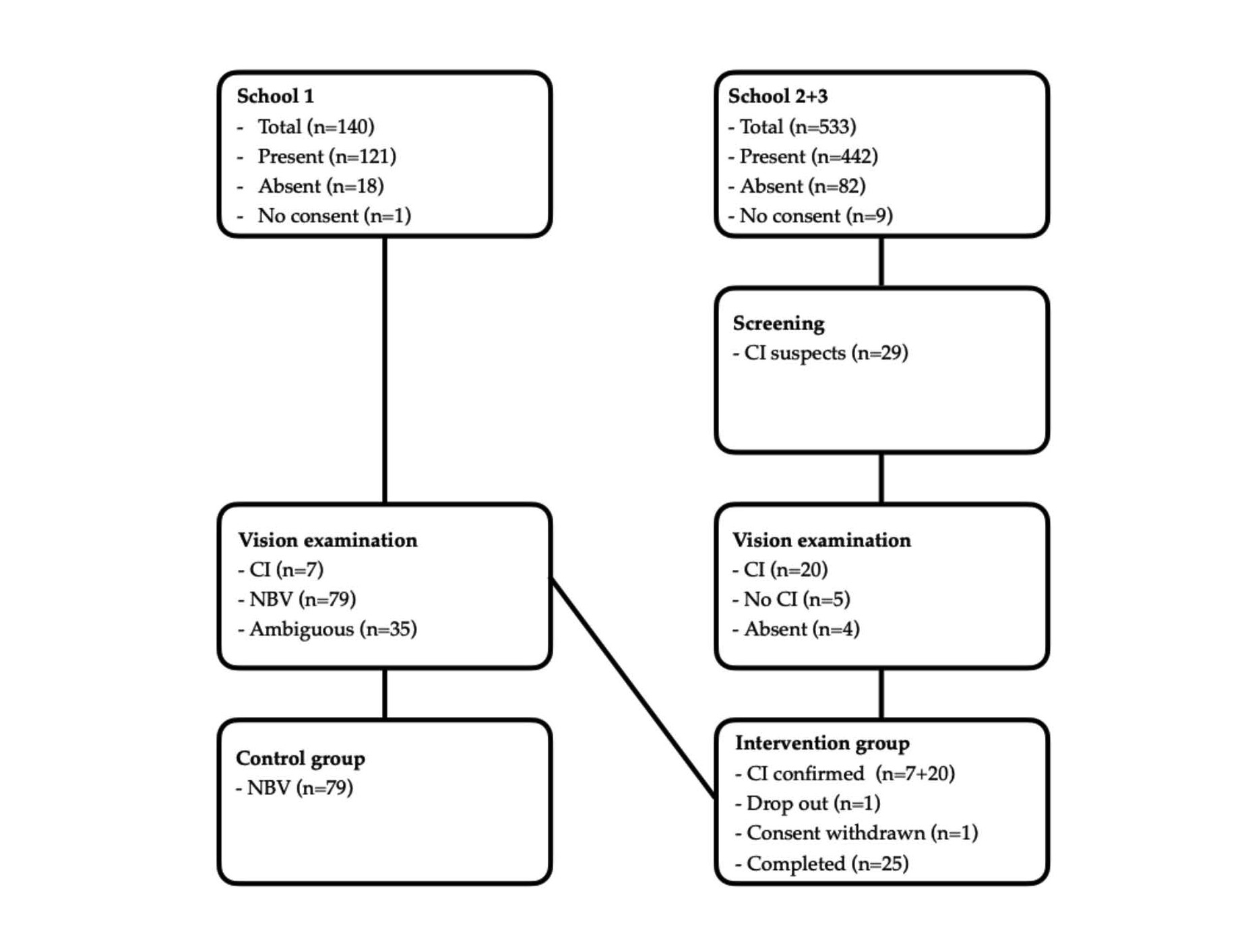
Figure 1. Flowchart of patient selection for convergence insufficiency (CI) and normal binocular vision (NBV).
|
|
Intervention groups |
Difference (p value) |
Control group |
Difference (p value) |
|||
|
|
A → V (n = 14) |
V → A (n = 11)
|
Between intervention groups |
(n = 79) |
Control |
||
|
Dist phoria** |
-0.1 ± 1.2 |
-0.1 ± 0.8 |
0.98 |
-0.3 ± 1.1 |
<0.001 |
||
|
Near phoria** |
-6.1 ± 3.0 |
-5.3 ± 2.8 |
0.36 |
-1.8 ± 3.1 |
<0.001 |
||
|
CISS** |
26.1 ± 9.9 |
23.5 ± 8.3 |
0.48 |
8.5 ± 6.8 |
<0.001 |
||
|
NPC** |
29.8 ± 11.4 |
23.6 ± 11.7 |
0.19 |
5.1 ± 1.2 |
<0.001 |
||
|
PFV** |
10.85 ± 5.1 |
9.5 ± 4.3 |
0.47 |
21.4 ± 8.2 |
<0.001 |
||
|
MAA** |
5.3 ± 2.8 |
5.5 ± 2.4 |
0.85 |
13.3 ± 4.0 |
<0.001 |
||
|
Foveal supp* |
420 (300; 420) |
180 (0; 900) |
0.44 |
0 (0; 0) |
<0.001 |
||
|
Ass. Phoria* |
-0 (-1; 0) |
-1 (-4; 0) |
0.10 |
0 (0; 0) |
<0.001 |
||
|
CISS: Convergence Insufficiency Symptom Survey; NPC: Near Point of Convergence (cm); PFV: Positive Fusional Vergence (pd); MAA: Monocular Amplitude of Accommodation (D); Foveal supp: Angular extend of foveal suppression (seconds of arc); Ass. Phoria: Associated Phoria (pd). A → V = Monocular accommodation followed by vergence exercises. |
|||||||
Sample size considerations
The calculations of sample size assumed a power of 80%, a p value of 5% and assumed that stereoacuity threshold was one octave lower in participants with normal binocular vision than in children with convergence insufficiency and that stereoacuity would reach normal level after vergence and accommodation exercises and resulted in a minimum of 18 children with CI and 54 in the control group after adjustment for unequal sample sizes.
Randomization
Children with convergence insufficiency were allocated to the two treatment groups using a computer-generated sequence to perform either 6 weeks of vergence exercises followed by 6 weeks of monocular accommodation exercises or the reverse order.
Each of the two 6-week treatments consisted of three 2-week phases after which the children received three new exercises for the next phase and examination protocol was repeated at all instruction sessions, except for CISS that was only assessed at week 6 and 12. The treatment program required that the child spent 20 minutes per day, 5 days per week performing exercises at home corresponding to a total of 20 hours during the 12-week treatment program.
Vergence exercises were performed binocularly while the children wore their habitual correction, if any. The basic principle of all vergence exercises was to increase or decrease convergence by inducing crossed and uncrossed diplopia while maintaining accommodation at a fixed distance by use of small optotypes or pictures. During all procedures, both vergence and accommodation loops were closed providing feedback for both accommodation and vergence systems, and stimuli to the two systems were separated in visual space.
Accommodation exercises were performed monocularly with an eye patch covering one eye and subsequently repeated with the patch covering the other eye while children wore their habitual correction, if any. The accommodative exercises were designed to induce hyperopic or myopic retinal defocus either by use of lenses or altering viewing and/or vertex distance. The eye patch ensured that the vergence loop was open by cancelling the sensory feedback to the vergence system.
All exercises were performed as suggested by the CITT study group and a detailed description was included in a previous study [11].
Data analysis
Outcome measures were stereoacuity measures with Randot circles test, Original Randot dot, and TNO test for stereoscopic vision.
Independent variables were oculomotor parameters (near point of convergence, positive fusional vergence, monocular amplitude of accommodation), sensory parameters (angular extend of foveal suppression), and sensory motor parameters (associated phoria). For comparison with stereoacuity the angular extend of foveal suppression was converted from minutes to seconds of arc by multiplying by 60.
Statistical analysis
Continuous data was tested for normal distribution using quantile-quantile plots and Shapiro-Wilks test and when assumption of normality was not met median and interquartile range was reported instead of mean and standard deviation. Differences in CISS, near point of convergence, positive fusional vergence, and monocular accommodation amplitude between control and intervention subgroups were analyzed with one-way ANOVA and post-test was performed using unpaired t-tests. Mann-Whitney U test was used to test for difference in Randot circles test between intervention and control group and sensitivity, specificity, and area under the Receiver Operating Characteristic curve were calculated.
Correlation between Randot circles test and associated phoria, angular extend of foveal suppression, near point of convergence, positive fusional vergence, monocular accommodation amplitude, and CISS were analyzed using Spearman rank correlation.
Changes from baseline to week 6 and 12 for Randot circles test, Original Randot stereo test, TNO test for stereoscopic vision, associated phoria, angular extend of foveal suppression, near point of convergence, positive fusional vergence, amplitude of accommodation and CISS were analyzed using repeated measures ANOVA and paired t-tests for post-tests for normally distributed data and Freidman test and Wilcoxon signed-rank test for post-test non-normal data. Since changes in stereoacuity did not differ between treatment groups data were not stratified by treatment group. Correlations between stereoacuity tests were analyzed with Spearman rank correlation while differences in level of stereoacuity were analyzed with Wilcoxon signed rank test.
Due to multiple comparisons all p values were adjusted by use of Bonferroni correction.
All statistical analyses by were performed in STATA (version 19.0, StataCorp, Texas, USA).
Results
Sensitivity
Figure 2 shows that normal stereoacuity was more common among children with normal binocular vision than children with convergence insufficiency and the threshold was significantly higher for the latter (p < 0.001). However, the area under the Receiver Operative Characteristics Curve was 0.74 and with a cutoff value of 40 seconds of arc sensitivity was 60.0% and specificity 70.9%. See Appendix 1 for ROC curve with sensitivity and specificity for all cutoff values.
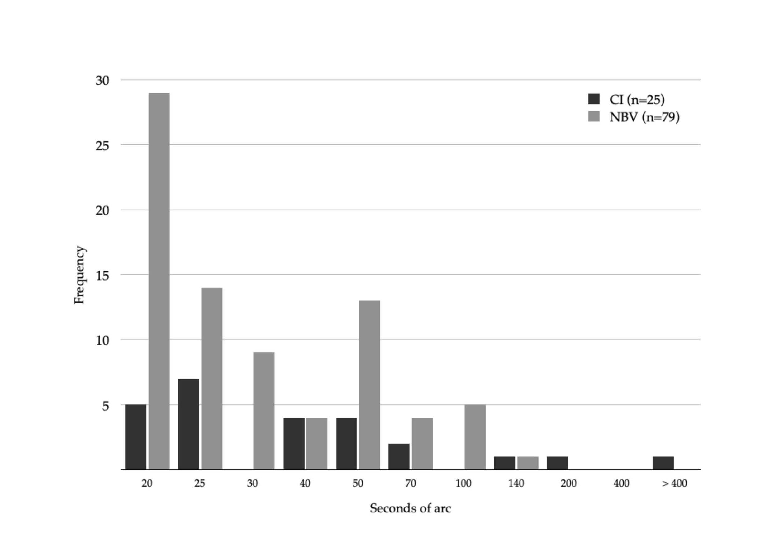
Figure 2. Baseline stereoacuity for children with convergence insufficiency (CI) and normal binocular vision (NBV).
Correlation
Table 2 shows that higher threshold with Randot circles test was correlated with larger angular extend of foveal suppression area, poorer near point of convergence, amplitude of accommodation, and positive fusional vergence at 40 cm but not magnitude of fixation disparity at 40 cm or CISS score.
|
|
rs |
p |
|
Foveal suppression (n = 103) |
0.50 |
< 0.001 |
|
Associated phoria (n = 102) |
-0.07 |
1.00 |
|
Near point of convergence (n = 104) |
0.29 |
0.016 |
|
Positive fusional vergence (n = 103) |
-0.27 |
0.041 |
|
Monocular amplitude of accommodation (n = 104) |
0.27 |
0.031 |
|
CISS (n = 103) |
0.24 |
0.084 |
Treatment
Table 3 shows that while all measures apart from associated phoria improved from baseline to week 6, only near point of convergence, positive fusional vergence, and CISS improved from week 6 to 12. Data was not stratified by intervention group since only TNO stereoacuity differed at week 6 (but not week 0, 2, 4, 8, 10 or 12) between intervention group. After Bonferroni correction no measures significantly differed between intervention groups. See Appendix 2 for stratified values.
|
|
Baseline |
Week 6 |
p week 0 to 6 |
Week 12 |
p week 6 to 12 |
|
Randot circles test * (seconds of arc) |
50 (30; 70) |
25 (20; 25) |
<0.001 |
20 (20; 25) |
1.0 |
|
Original Randot test * (seconds of arc) |
40 (20; 55) |
20 (20; 20) |
<0.001 |
20 (20; 20) |
1.0 |
|
TNO test * (seconds of arc) |
60 (60; 120) |
60 (45;60) |
<0.001 |
60 (30;60) |
0.18 |
|
Angular extend of foveal suppression * (seconds of arc) |
420 (0; 600) |
0 (0;0) |
<0.001 |
0 (0;0) |
1.0 |
|
Associated phoria * (pd) |
-0.5 (-3; 0) |
0 (-1; 0) |
1.0 |
0 (-0.5; 0) |
1.0 |
|
Near point of convergence ** (cm) |
27.0 ± 11.8 |
12.5 ± 10.0 |
<0.001 |
7.8 ± 5.1 |
0.018 |
|
Positive fusional vergence ** (pd) |
10.2 ± 4.7 |
25.5 ±12.1 |
<0.001 |
31.9 ± 11.5 |
0.045 |
|
Mono accommodation amplitude (D)** |
5.4 ± 2.6 |
9.3 ± 3.1 |
<0.001 |
11.6 ± 3.4 |
0.063 |
|
CISS ** |
25.0 ± 9.1 |
15.4 ± 11.1 |
<0.001 |
11.0 ± 10.8 |
0.021 |
|
* Median and interquartile range. ** Mean and standard deviation |
|||||
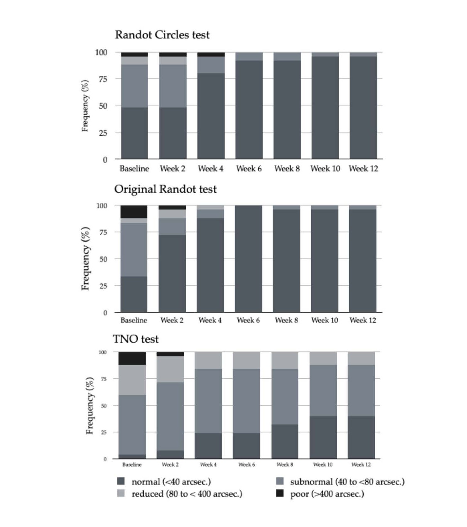
Figure 3. Changes in stereoacuity with Randot circles, Original Randot, and TNO stereoacuity test in two-week intervals for children with convergence insufficiency during treatment.
Difference between stereoacuity tests
For children with convergence insufficiency Randot circles test was positively correlated with the Original Randot test (rs = 0.83, p < 0.001) and TNO stereoacuity (rs = 0.51, p = 0.01) at baseline but not at week 6 or 12. TNO stereoacuity test threshold was significantly higher throughout the entire treatment program (p < 0.001). At baseline and week 12 stereoacuity threshold did not differ between Randot circles test and Original Randot test (both p > 0.33) while the latter was slightly better at week 6 (4 seconds of arc, p = 0.003).
Discussion
To the authors’ knowledge this is the first study to prospectively investigate improvement in stereoacuity as a result of vergence and monocular accommodation exercises in children with convergence insufficiency.
Within the first six weeks of treatment stereoacuity improved significantly and 96% (24/25) were within normal range (40” or better). All children with baseline stereoacuity poorer than normal range improved during treatment and improvement did not differ between children who received vergence and monocular accommodation exercise.
Reliability and validity
The diagnosis of convergence insufficiency complied with consensus criteria used in Cochrane reviews assessing treatment effect [7,12] The CISS questionnaire used to assess frequency of symptoms has also been shown to be suitable for separating convergence insufficiency from normal binocular vision in children aged 9 to 17 years [10]. The oculomotor measures used in the present study correlate with objective measures of vergence and neuroimaging which indicate that results reflect true measures of oculomotor disturbance [13].
Random dot stereoacuity measures have been shown to have good test-retest and for non-strabismic adults and children from the age of five years [14,15]. While no monocular cues are present in Random dot tests recent studies found binocular non-stereoscopic cues allow patients to deceive the test for coarse stereoacuity with the TNO and Randot circles test [16,17]. However, the stereoacuity measures were also found to be highly correlated with gold standard psychophysical tests [17] suggesting that the stereoacuity tests are valid measures of improvement in fine stereoacuity [18].
Sensitivity and specificity
Although stereoacuity was significantly poorer in children with convergence insufficiency than those with normal binocular vision, the area under the Receiver Operation Characteristics curve is only considered acceptable [19]. Forty percent of children with convergence insufficiency had normal stereoacuity (≤ 40”) at baseline indicating that stereopsis is not a potent screening tool for convergence insufficiency. A subgroup analysis where children with CI was divided into participants with normal (≤ 40“) and poor stereoacuity (> 40”) found no difference in distance or near phoria, near point of convergence, positive fusional vergence or accommodation amplitude (all p > 0.1) (data not shown). This further support the argument against stereoacuity as a screening tool for convergence insufficiency. This is contrary to manifest strabismus which is characterized by larger and constant deviations requiring a larger suppression area to cancel out diplopia and thereby deteriorating stereoacuity [20].
Correlation
In accordance with previous studies oculomotor parameter such as near point of convergence, positive fusional vergence, and amplitude of accommodation were correlated with stereoacuity [6,21] which suggests that the purpose of oculomotor function is to maintain vision single and clear. In the present study the strongest correlation was found between stereoacuity and angular extend of the foveal suppression suggesting that the impact of oculomotor parameters is mediated by foveal suppression reducing the sensitivity to fine disparity. The associated phoria was not correlated with stereoacuity which may be due to the high demand for precise fixation regardless of the magnitude of the associated phoria. Since the stereoacuity chart must be located within Panum´s fusional area to be perceived as single the retinal stimulation is within the foveola thereby maintaining high sensitivity of retinal disparity due to the small receptive fields [22,23]. Furthermore, it has been suggested that a minor misalignment of the ocular axis may be a purposeful adaptation which activates the fast fusional component more readily in patients with vergence anomalies [24].
Contrary to the oculomotor measures stereoacuity was not correlated with the frequency of symptoms (CISS) which may be due to the limited variance in stereoacuity threshold among participants corresponding to a very limited difference actual depth at close but not far distance. However, it is also possible that elevated stereoacuity threshold is of minor importance compared to symptoms such as blurred and double vision or that the CISS questionnaire is not sensitive to symptoms related to poor stereopsis. Regardless of the underlying cause, the lack of correlation suggests that stereoacuity should not be considered a key element in the diagnoses of convergence insufficiency.
Treatment
Data from the two intervention groups was combined since measures did not differ between treatment groups except TNO stereoacuity which only differed at week 6 but not two weeks before or after. Although it cannot be ruled out that this reflects a true difference in stereoacuity, it seems plausible that this single borderline significant result (p = 0.04) rather should be attributed to chance due to multiple comparisons.
During the first six weeks of treatment all measures improved significantly except associated phoria, while only near point of convergence, positive fusional vergence, and CISS improved from week 6 to 12. The lack of change in associated phoria during treatment may further support that a minor misalignment of the ocular axis may be a purposeful adaptation rather than a reflection of binocular dysfunction. However, it cannot be ruled out that the central fusional stimulus, the peripheral fusional, and accommodative lock combined reduce sensitivity to changes in associated phoria that might be measurable with other types of fixation disparity charts with less feedback to vergence and accommodation systems.
The improvement in all three stereoacuity measures from baseline to week 6 may be explained by the reduction in foveal suppression area which then allows high-resolution stereoacuity. However, more than a third of the children (9/25) did not have foveal suppression at baseline but still showed improved stereoacuity at week 6 possibly due to improvement of near point of convergence, positive fusional vergence, and AA which most likely provide a clearer and more stable retinal images and thus reinforcing perception [25].
The lack of improvement in stereoacuity from week 6 to 12 may be due to floor effect, and it cannot be ruled out that improvement still occurred since the tests used measure above absolute human threshold [26,27] (p. 25). It is however also possible that optimum stereoacuity is achieved prior to full normalization of near point of convergence and positive fusional vergence since the static nature of the test situation does not require optimum oculomotor control [28] and stereoacuity threshold decrease with increasing stimulus time presentation [29,30]. Regardless of the underlying cause the lack of measured improvement during the last six weeks of exercises suggests that stereoacuity is not suited to detect subtle binocular disturbance.
Difference between stereoacuity tests
While all three measures of stereoacuity were correlated at baseline suggesting that they measure the same sensory entity, tests were not correlated at subsequent examinations during treatment which most likely reflects loss of statistical power as variance decrease when stereoacuity improves.
In accordance with previous studies [31] TNO stereoacuity threshold was higher than Randot circles and original Randot at all examinations which may partly be due to the larger increment in levels of stereoacuity threshold in the TNO test which bias results towards higher threshold. However, it is also likely that the higher threshold of the TNO test is due to type of dissociation of image for right and left eye as a previous study has shown that the threshold for TNO test decreases if dissociation is without color [32] suggesting that chromatic aberration causing retinal blur negatively impacts stereoacuity. Similarly, stereoacuity threshold has been shown to be higher for other commercially available tests when dissociation is obtained by color rather than polarization [33]. A third difference between TNO and the polarized tests used is the object size, however, previous studies indicate that while image size affects stereoacuity in strabismic patients, it plays a negligible role in non-strabismic patients [34].
The study has several limitations including the single site study design, the lack of distance stereoacuity measures and objective tracking home-based exercises which may have reduced treatment effect if parents overestimated their children’s adherence to exercises. Furthermore, the narrow age range and strict refractive inclusion criteria limits generalizability to broader age ranges and children with high ametropia. Future studies should also address long-term effect of vergence and accommodation exercises on stereoacuity.
In conclusion, poor stereoacuity often accompany oculomotor dysfunction and may be due to foveal suppression and/or inability to maintain clear and single vision, respectively. However, both sensitivity and specificity were too poor for stereoacuity to be used as a screening tool for non-strabismic anomalies such as convergence insufficiency. Furthermore, stereoacuity appears to normalize before the patients have fully recovered suggesting that it is not a subtle indicator of non-strabismic binocular interference. Differences in threshold between stereoacuity tests emphasize that stereoacuity tests cannot be used interchangeably.
Conflicts of Interest
There are no conflicts of interest to declare. The funders had no role in the design of the study in the collection, analyses, or interpretation of data; in the writing of the manuscript; or in the decision to publish the results.
Funding
This research was funded by The Synoptik Foundation and Dania Academy of Higher Education.
Author Contribution Statement
Both authors contributed to conceptualization, methodology, validation, statistical analysis, writing original draft, project administration, and funding acquisition. Both authors have read and agreed to the published version of the manuscript.
No GenAI was used. The authors have reviewed and edited the output and take full responsibility for the content of this publication.
References
2. Birch EE, Shimojo S, Held R. Preferential-looking assessment of fusion and stereopsis in infants aged 1-6 months. Invest Ophthalmol Vis Sci. 1985 Mar;26(3):366–70.
3. Romano PE, Romano JA, Puklin JE. Stereoacuity development in children with normal binocular single vision. Am J Ophthalmol. 1975 Jun;79(6):966–71.
4. Elliott DB. Clinical procedures in primary eye care E-Book. 5th ed. Amsterdam: Elsevier Health Sciences; 2020.
5. Robaei D, Huynh SC, Kifley A, Gole GA, Mitchell P. Stereoacuity and ocular associations at age 12 years: findings from a population-based study. J AAPOS. 2007 Aug;11(4):356–61.
6. Leshno A, Stolovitch C, Zloto O, Blum Meirovitch S, Mezad-Koursh D. Reduced stereoacuity as a predictor for clinically significant convergence insufficiency. Br J Ophthalmol. 2021 Jan;105(1):37–41.
7. Scheiman M, Kulp MT, Cotter SA, Lawrenson JG, Wang L, Li T. Interventions for convergence insufficiency: a network meta-analysis. Cochrane Database Syst Rev. 2020 Dec 2;12(12):CD006768.
8. World Medical Association. World Medical Association Declaration of Helsinki: Ethical Principles for Medical Research Involving Human Participants. JAMA. 2025 Jan 7;333(1):71–4.
9. Borsting EJ, Rouse MW, Mitchell GL, Scheiman M, Cotter SA, Cooper J, et al. Validity and reliability of the revised convergence insufficiency symptom survey in children aged 9 to 18 years. Optom Vis Sci. 2003 Dec;80(12):832–8.
10. Rouse M, Borsting E, Mitchell GL, Cotter SA, Kulp M, Scheiman M, et al. Validity of the convergence insufficiency symptom survey: a confirmatory study. Optom Vis Sci. 2009 Apr;86(4):357–63.
11. Maagaard ML, Nisted I, Bek T. Vergence Exercises for Six Weeks Induce Faster Recovery of Convergence Insufficiency Than Accommodation Exercises in School Children. Invest Ophthalmol Vis Sci. 2021 May 3;62(6):23.
12. Scheiman M, Gwiazda J, Li T. Non-surgical interventions for convergence insufficiency. Cochrane Database Syst Rev. 2011 Mar 16;(3):CD006768.
13. Alvarez TL, Scheiman M, Morales C, Gohel S, Sangoi A, Santos EM, et al. Underlying neurological mechanisms associated with symptomatic convergence insufficiency. Sci Rep. 2021 Mar 22;11(1):6545.
14. Yildirim C, Altinsoy HI, Yakut E. Distance stereoacuity norms for the mentor B-VAT II-SG video acuity tester in young children and young adults. J AAPOS. 1998 Feb;2(1):26–32.
15. Mehta J, O'Connor A. Test retest variability in stereoacuity measurements. Strabismus. 2023 Sep;31(3):188–96.
16. Charman WN, Jennings JA. Recognition of TNO stereotest figures in the absence of true stereopsis. Optom Vis Sci. 1995 Jul;72(7):535–6.
17. Chopin A, Chan SW, Guellai B, Bavelier D, Levi DM. Binocular non-stereoscopic cues can deceive clinical tests of stereopsis. Sci Rep. 2019 Apr 8;9(1):5789.
18. Fawcett SL, Birch EE. Validity of the Titmus and Randot circles tasks in children with known binocular vision disorders. J AAPOS. 2003 Oct;7(5):333–8.
19. Mandrekar JN. Receiver operating characteristic curve in diagnostic test assessment. J Thorac Oncol. 2010 Sep;5(9):1315–6.
20. Ciner EB, Ying GS, Kulp MT, Maguire MG, Quinn GE, Orel-Bixler D, et al. Stereoacuity of preschool children with and without vision disorders. Optom Vis Sci. 2014 Mar;91(3):351–8.
21. Wajuihian SO. Correlations between clinical measures and symptoms: Report 1: Stereoacuity with accommodative, vergence measures, and symptoms. J Optom. 2020 Jul-Sep;13(3):171–84.
22. Hubel DH, Wiesel TN, Yeagle EM, Lafer-Sousa R, Conway BR. Binocular stereoscopy in visual areas V-2, V-3, and V-3A of the macaque monkey. Cereb Cortex. 2015 Apr;25(4):959–71.
23. Watson AB. A formula for human retinal ganglion cell receptive field density as a function of visual field location. J Vis. 2014 Jun 30;14(7):15.
24. Schor CM. The relationship between fusional vergence eye movements and fixation disparity. Vision Res. 1979;19(12):1359–67.
25. Odell NV, Hatt SR, Leske DA, Adams WE, Holmes JM. The effect of induced monocular blur on measures of stereoacuity. J AAPOS. 2009 Apr;13(2):136v41.
26. Stevenson SB, Cormack LK, Schor CM. Hyperacuity, superresolution and gap resolution in human stereopsis. Vision Res. 1989;29(11):1597-605.
27. Von Noorden GK. Binocular vision and ocular motility. Theory and management of strabismus. 6th ed. St. Louis, MO: Mosby; 2002.
28. Sachsenweger M, Sachsenweger U. Stereoscopic acuity in ocular pursuit of moving objects. Dynamic stereoscopy and movement parallax: relevance to road safety and occupational medicine. Doc Ophthalmol. 1991;78(1-2):1–133.
29. McKee SP, Levi DM, Bowne SF. The imprecision of stereopsis. Vision Res. 1990;30(11):1763–79.
30. Kumar T, Glaser DA. Some temporal aspects of stereoacuity. Vision Res. 1994 Apr;34(7):913–25.
31. Antona B, Barrio A, Sanchez I, Gonzalez E, Gonzalez G. Intraexaminer repeatability and agreement in stereoacuity measurements made in young adults. Int J Ophthalmol. 2015 Apr 18;8(2):374–81.
32. Zhang Y, Meng B, Wu H. Evaluating the Mechanism by Which the TNO Stereo Test Overestimates Stereo Thresholds. J Ophthalmol. 2021 Jan 18;2021:6665638.
33. Yamada T, Scheiman M, Mitchell GL. A comparison of stereopsis testing between red/green targets and polarized targets in children with normal binocular vision. Optometry. 2008 Mar;79(3):138–42.
34. Pageau M, de Guise D, Saint-Amour D. Random-dot stereopsis in microstrabismic children: stimulus size matters. Optom Vis Sci. 2015 Feb;92(2):208–16.