
Commentary
Prion diseases, or transmissible spongiform encephalopathies (TSEs), represent a group of incurable and invariably fatal neurodegenerative disorders driven by the pathological misfolding of the prion protein (PrP) [1–6]. These diseases are characterized by a progressive clinical course and, despite their significant impact on human [7–17] and animal [1,10,18–30] health, currently lack effective therapeutic interventions [31–34] and are limited in early-stage diagnostics [1,7,31–33]. Given the broad host range of prion diseases, together with the absence of effective treatments and limited antemortem diagnostic options, continued prion disease research remains essential. However, research and clinical investigations of prion diseases present substantial occupational biosafety risks [35,36] because infectious prion proteins are highly resistant to many conventional decontamination procedures. In fact, because prions are misfolded proteins rather than microorganisms, they exhibit exceptional resistance to conventional decontamination methods, exceeding that of most microbial pathogens [37,38]. As a result, specialized and rigorous prion inactivation protocols are required for minimizing potential exposure in laboratory and clinical settings [39,40]. Formic acid treatment has been demonstrated to reduce prion infectivity and is commonly incorporated into biosafety practices for the decontamination of prion-contaminated tissues [6,23,41] and materials, however, the morphologic effects of formic acid decontamination on tissues was not reported previously.
Our recently published study, Shabaan et al. [6], evaluated the morphologic effects of formic acid treatment on central nervous system (CNS) brain tissue using brain hemispheres from transgenic mouse models with variable degrees and patterns of neurodegeneration, see Table 1. The potential influence of age on tissue morphology following treatment was also assessed. Formic acid-treated tissues were compared with untreated control tissues in two experimental groups: young wild-type mice (4 months old; n = 3/group), aged wild-type mice (11 months old; n = 3/group), and 5xFAD mice exhibiting Alzheimer’s-like pathology (11 months old; n = 4/group). Tissues from mice with prion disease-associated spongiform degeneration were also evaluated; however, an untreated control group was not included because of biohazard considerations. In this cohort, Tg1536 mice expressing cervid PRNP at CWD endpoint (9.4 months old; n = 5) were compared with non-diseased control-inoculated mice that received normal brain homogenate (9.4 months old; n = 6).
|
Mouse Model |
Control Untreated |
Formic Acid Protocol |
Purpose |
|||
|
|
Young (-4 months) |
Old (~11 months) |
Young (-4 months) |
Old (~11 months) |
Prion Endpoint (-9.4 months) |
|
|
WT C57BI/6N |
3 |
3 |
3 |
3 |
|
1. Determine if formic (fa) treatment causes morphologic changes in brain tissues of wildtype mice |
|
5xFAD |
|
4 |
|
4 |
|
Determine if fa morphologic changes are exacerbated in neurodegenerative disease mouse models when comparing fa vs untreated control |
|
Tg1536 CWD
|
|
|
|
5
|
Determine if fa affects morphology differently between prion disease mouse brain tissue and non-diseased mouse tissues |
|
|
Tg1536 NBH |
|
|
|
6 |
||
Our study [6] analyzed CNS brain tissues treated with a standard formic acid prion decontamination protocol [41] to address the following:
- Whether formic acid impacts tissue morphology and if so, whether tissue structure is maintained or too damaged for histologic studies.
- Whether tissues with neurodegeneration and normal, non-diseased tissues are impacted similarly.
- Whether morphologic changes induced by formic acid are distributed across brain regions.
- Whether age of animals modulated formic acid impacts.
Corresponding brain sections (right formic acid, left control) were compared in the study, with one standardized section per condition used at the location of 1800 μm from the medial edge of the brain hemisphere. All samples were processed for histology simultaneously, with the right hemisphere subjected to the additional formic acid steps. The formic acid protocol consisted of a fixation step, a 95% formic acid immersion for 1 hour, a second fixation step as stated in Biosafety in Microbiological and Biomedical Laboratories (BMBL) [41] and then stored in PBS. For a detailed description of the formic acid and histologic methods stated, refer to the original manuscript [6]. Cortex width and hippocampal area measurements were performed with the experimental conditions unknown to the individual performing the analysis. Cortex thickness layers 1–6 were measured immediately dorsal to the CA1 of the hippocampus and hippocampal area measurements were performed by encircling the entire structure. Two separate measurements were performed in Fiji ImageJ©, were averaged for each structure as stated, and used for statistical analyses. The Student’s t-test determined statistical significance for pairwise comparisons unless otherwise noted in the original manuscript [6].
Results indicated that all brain hemispheres grossly appeared smaller with formic acid treatment with both the cortical width and hippocampal area measurements significantly reduced in the formic acid treated samples as compared to untreated controls. Additionally, tissue integrity appeared to be compromised as the appearance of tears and artifacts were observed more readily in the formic acid treated histologic specimens compared to non-formic acid treated samples. We determined that neurodegenerative pathology did not have a great effect on the amount of morphologic changes following formic acid treatment by comparing 5xFAD transgenic mice [42] with wildtype mice. These results demonstrated that similar reductions occurred in cortex width and hippocampal areas in both models after formic acid treatment. At the cellular level, formic acid treatment on the tissues did not appear to interfere with detection of neurons or astrocytes using immunofluorescence staining, however, the preservation of antigenicity, fluorescence intensity, or quantitative accuracy were not measured. When formic acid treated tissues exhibiting endpoint prion disease from Tg1536 [43,44] CWD infected samples were compared with uninfected Tg1536 tissues, the cortical width and hippocampal area measurements were almost identical, suggesting spongiform degeneration did not greatly alter or exacerbate cortical and hippocampal size reduction in the post-formic acid tissues. Formic acid treatment appeared to reduce tissue size differently based on brain region when cortex and hippocampus width ratios were compared in the combined wildtype and 5xFAD mice. Age did not appear to alter the effects of formic acid treatment on the samples, as there were no significant differences when comparing the cortex size reduction between the young and old mice. A summary of findings is provided in Figure 1.
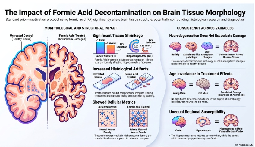
Figure 1.Overall results of study. Generated by AI platform, Notebook LM.
The authors acknowledge that the limited number of mice included in this exploratory study may restrict the generalizability of the observed effects. Given the study design and sample size, biological variability among animals is difficult to assess, and the findings may not be universal. The results of Shabaan et al [6]. demonstrate that standard prion decontamination protocols commonly used in research and clinical settings can significantly alter CNS tissue morphology. Although the mechanism for this was not analyzed in the study, these may include lipid alteration or extraction, collagen alteration, acid-induced tissue contraction, and protein denaturation. Because formic acid is used in prion diseased tissues to substantially alter prion proteins and promote decontamination of the pathogenic protein conformation, protein denaturation is a plausible mechanism. Formic acid has also been shown to solubilize hydrophobic proteins found in the CNS, such as myelin [45]. Additionally, the study found that the reduction in size was not uniform across brain regions, with the hippocampus showing a greater decrease than the cortex. This may be due to variations in cell density or protein and lipid content in the specific regions.
Changes in tissue size and architecture, together with treatment-associated artifacts such as tearing, may confound histologic interpretation and reduce the utility of tissue specimens for diagnostic and research applications. Because reproducible and accurate histologic findings are essential for reliable interpretation in both clinical and research contexts, understanding the morphologic effects of prion decontamination procedures is important for improving diagnostic confidence and minimizing confounding artifacts. These findings also reinforce the importance of using validated prion-inactivation methods when handling potentially infectious tissues. In the absence of data defining the tissue effects of formic acid treatment, personnel may be tempted to avoid prion-inactivating procedures to preserve tissue morphology; however, the severe consequences of potential prion transmission underscore the need to prioritize biosafety while continuing to develop improved decontamination approaches that better preserve histologic quality.
Decontamination of prion-contaminated instruments, equipment, and surfaces using alternatives to formic acid have been studied [24,37–40,46–53], although, to our knowledge, no approaches have been developed specifically for histology applications. Standard decontamination techniques include extremely harsh techniques such as sodium hypochlorite (NaOCl) or sodium hydroxide (NaOH) treatments, sometimes in combination with 134° autoclaving under long periods of time [38–41,48–53]. Other methods investigated for prion decontamination include enzymatic solutions, alkaline cleaners, peracetic acid, phenolic disinfectants, and vaporized gases such as hydrogen peroxide [37,46,47,50,54], often with greatest efficacy when used in combination. Thus, there remains a need to develop additional methods that are compatible with histologic applications; however, identifying approaches that are both sufficiently gentle to preserve tissue morphology and effective against highly resistant prions remains a substantial challenge. Promising future research approaches to preserve tissue morphology while decontaminating prions may include replacing formic acid with novel, milder chemical treatments; incorporating tissue-protective additives; or modifying the existing protocol by evaluating the efficacy of reduced formic acid exposure times.
Funding
This research was funded by the NIH COBRE award P20GM152335.
Acknowledgements
The commentary authors thank Dalia Shaaban, Lilia Crew, and Clairissa Kaylor, Sayre McElroy, Emma Guter, and June Pounder, the authors from “The impact of formic acid treatment on brain tissues for prion inactivation”, whose contributions drove curiosity, the reported experiments, and the manuscript. We would also like to thank the COBRE administrative core for discussions regarding this manuscript.
References
2. Prusiner SB. Prions. Proceedings of the National Academy of Sciences. 1998 Nov 10;95(23):13363–83.
3. Harris DA. Cellular biology of prion diseases. Clin Microbiol Rev. 1999 Jul;12(3):429–44.
4. Seerley AL, Rothfuss MT, Gray BM, Sebogo MA, Manakelew BA, Pounder JI, et al. Novel Prion Protein Gene (PRNP) Variants in Wild Montana Mule Deer. bioRxiv [Preprint]. 2026 Mar 19:2026.03.17.711390.
5. Das AS, Zou WQ. Prions: Beyond a Single Protein. Clin Microbiol Rev. 2016 Jul;29(3):633–58.
6. Shaaban D, Seerley A, Crew L, Kaylor C, McElroy S, Guter E, et al. The impact of formic acid treatment on brain tissues for prion inactivation. Acta Histochem. 2026 Jun;128(2):152324.
7. Nafe R, Arendt CT, Hattingen E. Human prion diseases and the prion protein - what is the current state of knowledge? Transl Neurosci. 2023 Oct 16;14(1):20220315.
8. Acevedo-Morantes CY, Wille H. The structure of human prions: from biology to structural models-considerations and pitfalls. Viruses. 2014 Oct 20;6(10):3875–92.
9. Bonda DJ, Manjila S, Mehndiratta P, Khan F, Miller BR, Onwuzulike K, et al. Human prion diseases: surgical lessons learned from iatrogenic prion transmission. Neurosurg Focus. 2016 Jul;41(1):E10.
10. Collinge J. Prion diseases of humans and animals: their causes and molecular basis. Annu Rev Neurosci. 2001;24:519–50.
11. Gao LP, Tian TT, Xiao K, Chen C, Zhou W, Liang DL, et al. Updated global epidemiology atlas of human prion diseases. Front Public Health. 2024 Jun 13;12:1411489.
12. Kobayashi A, Teruya K, Matsuura Y, Shirai T, Nakamura Y, Yamada M, et al. The influence of PRNP polymorphisms on human prion disease susceptibility: an update. Acta Neuropathol. 2015 Aug;130(2):159–70.
13. Piñar-Morales R, Barrero-Hernández F, Aliaga-Martínez L. Human prion diseases: An overview. Med Clin (Barc). 2023 Jun 23;160(12):554–60.
14. Pocchiari M, Puopolo M, Croes EA, Budka H, Gelpi E, Collins S, et al. Predictors of survival in sporadic Creutzfeldt-Jakob disease and other human transmissible spongiform encephalopathies. Brain. 2004 Oct;127(Pt 10):2348–59.
15. Prusiner SB. Human prion diseases and neurodegeneration. Curr Top Microbiol Immunol. 1996;207:1–17.
16. Prusiner SB, Hsiao KK. Human prion diseases. Annals of Neurology: Official Journal of the American Neurological Association and the Child Neurology Society. 1994 Apr;35(4):385–95.
17. Windl O, Dempster M, Estibeiro JP, Lathe R, de Silva R, Esmonde T, et al. Genetic basis of Creutzfeldt-Jakob disease in the United Kingdom: a systematic analysis of predisposing mutations and allelic variation in the PRNP gene. Hum Genet. 1996 Sep;98(3):259–64.
18. Prion Diseases in Animals: Poster Abstracts. Prion. 2013 Apr/May;7(sup1):18-53.
19. Costassa EV, Iulini B, Mazza M, Acutis P, Maurella C, Meloni D, et al. Pathogenesis and Transmission of Classical and Atypical BSE in Cattle. Food Saf (Tokyo). 2016 Dec 7;4(4):130–4.
20. Dudas S, Czub S. Atypical BSE: Current Knowledge and Knowledge Gaps. Food Saf (Tokyo). 2017 Feb 8;5(1):10–3.
21. Hamir AN, Gidlewski T, Spraker TR, Miller JM, Creekmore L, Crocheck M, et al. Preliminary observations of genetic susceptibility of elk (Cervus elaphus nelsoni) to chronic wasting disease by experimental oral inoculation. J Vet Diagn Invest. 2006 Jan;18(1):110–4.
22. Padilla D, Béringue V, Espinosa JC, Andreoletti O, Jaumain E, Reine F, et al. Sheep and goat BSE propagate more efficiently than cattle BSE in human PrP transgenic mice. PLoS Pathog. 2011 Mar;7(3):e1001319.
23. Taylor DM, Brown JM, Fernie K, McConnell I. The effect of formic acid on BSE and scrapie infectivity in fixed and unfixed brain-tissue. Vet Microbiol. 1997 Nov;58(2-4):167–74.
24. Giles K, Glidden DV, Beckwith R, Seoanes R, Peretz D, DeArmond SJ, et al. Resistance of bovine spongiform encephalopathy (BSE) prions to inactivation. PLoS Pathog. 2008 Nov;4(11):e1000206.
25. Baylis M, Goldmann W. The genetics of scrapie in sheep and goats. Curr Mol Med. 2004 Jun;4(4):385–96.
26. Goldmann W. Classic and atypical scrapie - a genetic perspective. Handb Clin Neurol. 2018;153:111–20.
27. Green KM, Browning SR, Seward TS, Jewell JE, Ross DL, Green MA, et al. The elk PRNP codon 132 polymorphism controls cervid and scrapie prion propagation. J Gen Virol. 2008 Feb;89(Pt 2):598–608.
28. Kurt TD, Sigurdson CJ. Cross-species transmission of CWD prions. Prion. 2016;10(1):83–91.
29. Arifin MI, Hannaoui S, Chang SC, Thapa S, Schatzl HM, Gilch S. Cervid Prion Protein Polymorphisms: Role in Chronic Wasting Disease Pathogenesis. Int J Mol Sci. 2021 Feb 25;22(5):2271.
30. Moazami-Goudarzi K, Andréoletti O, Vilotte JL, Béringue V. Review on PRNP genetics and susceptibility to chronic wasting disease of Cervidae. Vet Res. 2021 Oct 7;52(1):128.
31. Minikel EV, Zhao HT, Le J, O'Moore J, Pitstick R, Graffam S, et al. Prion protein lowering is a disease-modifying therapy across prion disease stages, strains and endpoints. Nucleic Acids Res. 2020 Nov 4;48(19):10615–31.
32. Corridon TL, O'Moore J, Seerley A, Sprague DA, Lian Y, Laversenne V, et al. PrP turnover in vivo and the time to effect of prion disease therapeutics. PLoS Pathog. 2026 May 26;22(5):e1014263.
33. Vallabh SM, Zou D, Pitstick R, O'Moore J, Peters J, Silvius D, et al. Therapeutic Trial of anle138b in Mouse Models of Genetic Prion Disease. J Virol. 2023 Feb 28;97(2):e0167222.
34. Liu F, Lü W, Liu L. New implications for prion diseases therapy and prophylaxis. Front Mol Neurosci. 2024 Mar 4;17:1324702.
35. Brandel JP, Vlaicu MB, Culeux A, Belondrade M, Bougard D, Grznarova K, et al. Variant Creutzfeldt-Jakob Disease Diagnosed 7.5 Years after Occupational Exposure. N Engl J Med. 2020 Jul 2;383(1):83–5.
36. Casassus B. France issues moratorium on prion research after fatal brain disease strikes two lab workers. Science. 2021 Jul 28;373:475–6.
37. Sakudo A, Yamashiro R, Onodera T. Recent Advances in Prion Inactivation by Plasma Sterilizer. Int J Mol Sci. 2022 Sep 6;23(18):10241.
38. Sakudo A, Ano Y, Onodera T, Nitta K, Shintani H, Ikuta K, et al. Fundamentals of prions and their inactivation (review). Int J Mol Med. 2011 Apr;27(4):483–9.
39. Belay ED, Schonberger LB, Brown P, Priola SA, Chesebro B, Will RG, et al. Disinfection and sterilization of prion-contaminated medical instruments. Infect Control Hosp Epidemiol. 2010 Dec;31(12):1304–6.
40. Rutala WA, Weber DJ; Society for Healthcare Epidemiology of America. Guideline for disinfection and sterilization of prion-contaminated medical instruments. Infect Control Hosp Epidemiol. 2010 Feb;31(2):107–17.
41. US Department of Health and Human Services. Biosafety in microbiological and biomedical laboratories (BMBL) 6th edition | CDC Laboratory Portal. Centers for Disease Control and Prevention; 2023. Available from: https://www.cdc.gov/labs/BMBL.html
42. Forner S, Kawauchi S, Balderrama-Gutierrez G, Kramár EA, Matheos DP, Phan J, et al. Systematic phenotyping and characterization of the 5xFAD mouse model of Alzheimer's disease. Sci Data. 2021 Oct 15;8(1):270.
43. Seelig DM, Mason GL, Telling GC, Hoover EA. Pathogenesis of chronic wasting disease in cervidized transgenic mice. Am J Pathol. 2010 Jun;176(6):2785–97.
44. Browning SR, Mason GL, Seward T, Green M, Eliason GA, Mathiason C, et al. Transmission of prions from mule deer and elk with chronic wasting disease to transgenic mice expressing cervid PrP. J Virol. 2004 Dec;78(23):13345–50.
45. McKenzie AT, Nnadi O, Slagell KD, Thorn EL, Farrell K, Crary JF. Fluid preservation in brain banking: a review. Free Neuropathol. 2024 Apr 23;5:10.
46. Herzog L, Reine F, Castille J, Passet B, Moudjou M, Bonnet R, et al. Optimization and evaluation of new decontamination procedures inactivating human prions. J Hosp Infect. 2025 Jun;160:109–17.
47. Baune C, Groveman BR, Hughson AG, Thomas T, Twardoski B, Priola S, et al. Efficacy of Wex-cide 128 disinfectant against multiple prion strains. PLoS One. 2023 Aug 24;18(8):e0290325.
48. Williams K, Hughson AG, Chesebro B, Race B. Inactivation of chronic wasting disease prions using sodium hypochlorite. PLoS One. 2019 Oct 4;14(10):e0223659.
49. Groveman BR, Race B, Hughson AG, Haigh CL. Sodium hypochlorite inactivation of human CJD prions. PLoS One. 2024 Nov 7;19(11):e0312837.
50. Fichet G, Comoy E, Duval C, Antloga K, Dehen C, Charbonnier A, et al. Novel methods for disinfection of prion-contaminated medical devices. Lancet. 2004 Aug 7-13;364(9433):521–6.
51. Schwenke KA, Wagenführ K, Thanheiser M, Beekes M. Kinetics of the reduction of Creutzfeldt-Jakob disease prion seeding activity by steam sterilization support the use of validated 134°C programmes. J Hosp Infect. 2023 Feb;132:125–32.
52. Sakudo A. Inactivation methods for prions. Curr Issues Mol Biol. 2020 Apr;36(1):23–32.
53. Hughson AG, Race B, Kraus A, Sangaré LR, Robins L, Groveman BR, et al. Inactivation of Prions and Amyloid Seeds with Hypochlorous Acid. PLoS Pathog. 2016 Sep 29;12(9):e1005914.
54. Sakudo A, Anraku D, Itarashiki T. Inactivation of Prions by Low-Temperature Sterilization Technology Using Vaporized Gas Derived from a Hydrogen Peroxide-Peracetic Acid Mixture. Pathogens. 2020 Dec 31;10(1):24.