

Abstract
Background: Intervertebral Differential Dynamics (IDD) Therapy is used as a targeted, non-surgical decompression intervention for selected patients with disc-related spinal pain. A previously published cohort reported significant short-term improvements in pain and disability, while paired magnetic resonance imaging (MRI) outcomes were reserved for subsequent analysis.
Objective: To evaluate paired MRI findings and clinical outcomes following IDD Therapy in adults with MRI-confirmed cervical or lumbar disc bulges and herniations.
Methods: This retrospective secondary analysis included 21 adults with 47 treated disc levels who completed a 30-session protocol. Previously published Visual Analog Scale (VAS) and Oswestry Disability Index (ODI) outcomes were linked to paired MRI measurements. Posterior disc contour extension, canal anteroposterior diameter, and disc height were analyzed separately for 14 lumbar and 7 cervical index lesions. Radiographic response and exploratory clinical-imaging correlations were also evaluated.
Results: Mean VAS improved from 5.8 to 1.5 and ODI from 18.5 to 6.5 in the parent cohort. Lumbar mean changes were -1.75 mm for disc contour extension, +1.31 mm for canal diameter, and +0.64 mm for disc height; cervical mean changes were -1.20 mm, +0.89 mm, and +0.53 mm, respectively. All region-specific changes were statistically significant. Global radiographic improvement was observed in 16 of 21 patients and 34 of 47 treated levels. Disc contour reduction was strongly associated with VAS improvement (r=0.95; 95% CI, 0.88-0.98; P<0.001).
Conclusions: Completion of IDD Therapy was associated with substantial clinical improvement and favorable paired-MRI changes in most patients in this selected cohort. Because the study was retrospective, lacked a comparator, used non-standardized clinical imaging, and included only treatment completers, the findings are hypothesis-generating rather than causal.
Keywords
Intervertebral Differential Dynamics Therapy, IDD Therapy, Non-surgical spinal decompression, Disc herniation, Disc bulge, Magnetic resonance imaging, Visual Analog Scale, Oswestry Disability Index, Conservative spine care
Introduction
Cervical and lumbar intervertebral disc disorders are common structural diagnoses associated with spine-related pain, disability, radicular symptoms, reduced quality of life, and work limitation. Lumbar disc herniation and disc bulging may contribute to low back pain, sciatica, sensory disturbance, motor weakness, and functional impairment. Cervical disc pathology may contribute to neck pain, radiculopathy, headache, upper-extremity symptoms, and activity limitation. Although many disc-related symptoms improve without surgery, a subset of patients experience persistent pain or disability despite medication, activity modification, home exercise, or general physical therapy [1,2]. The clinical cohort underlying the present analysis has been described previously [3].
Conservative care remains an important first-line approach for many patients with disc-related spinal pain when red flags, progressive neurologic deficit, cauda equina syndrome, myelopathy, fracture, infection, malignancy, or severe instability are absent [2]. Conservative care may include education, activity modification, exercise therapy, manual therapy, traction-based care, non-surgical spinal decompression, medication, injections, and multidisciplinary rehabilitation. Because these interventions vary in patient selection, treatment dosage, mechanism, and outcome measurement, the evidence base is heterogeneous. This is particularly true for traction and decompression-oriented interventions, where studies of non-specific low back pain may not apply to patients with MRI-confirmed, clinically concordant disc pathology [4–8].
Intervertebral Differential Dynamics (IDD) Therapy is a form of targeted, computer-controlled spinal decompression designed to unload selected cervical or lumbar disc levels through programmed distraction forces, patient positioning, and cyclical waveforms. Proposed mechanisms include modulation of mechanical loading, transient reduction of intradiscal pressure, improved nutrient exchange, reduction of neural element irritation, and increased tolerance to movement. These proposed mechanisms remain incompletely established in clinical populations; therefore, symptomatic improvement, structural imaging change, and causal inference must be distinguished carefully.
MRI is frequently used to confirm disc morphology, evaluate central canal and foraminal compromise, and identify neural element contact. However, imaging findings cannot be interpreted in isolation because disc degeneration, bulging, and protrusion are also observed in asymptomatic adults [9,10]. Longitudinal imaging is potentially valuable when it is paired with patient-reported outcomes and consistent anatomic measurements. A paired design allows each participant to serve as his or her own imaging comparator, but it does not eliminate confounding from natural history, co-interventions, measurement variability, or differences in scanners and positioning. For this reason, the present analysis emphasizes the direction and magnitude of interval change while avoiding the assumption that every radiographic change directly explains symptom improvement.
A previously published retrospective cohort study by Bartrom, Syed, and Graham evaluated clinical outcomes after IDD Therapy in adults with MRI-confirmed cervical and lumbar disc bulges or herniations [3]. That study included 21 patients with 47 treated disc levels who completed a 30-session protocol. Mean VAS pain scores improved from 5.8 to 1.5, and mean ODI scores improved from 18.5 to 6.5. No adverse events were reported. The parent article further noted that post-treatment MRI studies had been obtained to monitor objective findings and would be analyzed separately.
The present study extends that work by evaluating paired pre-treatment and post-treatment MRI findings in the same cohort and by exploring associations between radiographic changes and improvements in pain and disability. We hypothesized that patients completing the IDD Therapy protocol would demonstrate favorable changes in MRI markers of disc contour, canal dimensions, and disc height, and that improvement in selected MRI measures would be associated with clinical improvement.
The study was not designed to establish that IDD Therapy caused every observed imaging change. Disc morphology may change over time through natural history, and the relationship between MRI abnormalities and symptoms is complex [9–15]. The objective was therefore descriptive and exploratory: to characterize paired MRI and clinical outcomes in a selected, MRI-confirmed cohort that completed a standardized treatment protocol.
Materials and Methods
Study design
This was a retrospective secondary paired-imaging analysis of a previously published clinical outcomes cohort [3]. The parent study reported demographic characteristics, treatment details, level distribution, safety, and pre-treatment to post-treatment clinical outcomes. The present analysis evaluated paired MRI measurements, radiographic response classifications, and exploratory radiographic-clinical correlations that were not reported in the parent article.
The study is reported in accordance with principles of the Strengthening the Reporting of Observational Studies in Epidemiology (STROBE) statement [16]. The overlap with the parent publication is disclosed throughout the manuscript and in the accompanying cover letter.
Ethics approval and informed consent
The parent retrospective cohort was approved by the Indiana University School of Medicine Institutional Review Board (protocol No. 24292), and individual informed consent was waived for retrospective use of existing clinical data. The study was conducted in accordance with the Declaration of Helsinki. The paired-imaging analysis used de-identified clinical and imaging data from the same cohort.
Setting and participants
The parent cohort consisted of adults treated at Spine Restoration, a private spine clinic in Marion, Indiana, USA, between June 1, 2024, and July 1, 2025. Tyler Maitland Graham, DC, is currently affiliated with Balanced Chiropractic in Logansport, Indiana. Eligible patients were at least 18 years old, had MRI-confirmed cervical or lumbar disc bulge or herniation, completed 30 IDD Therapy sessions within a 3-month period, and had complete pre-treatment and post-treatment VAS and ODI data.
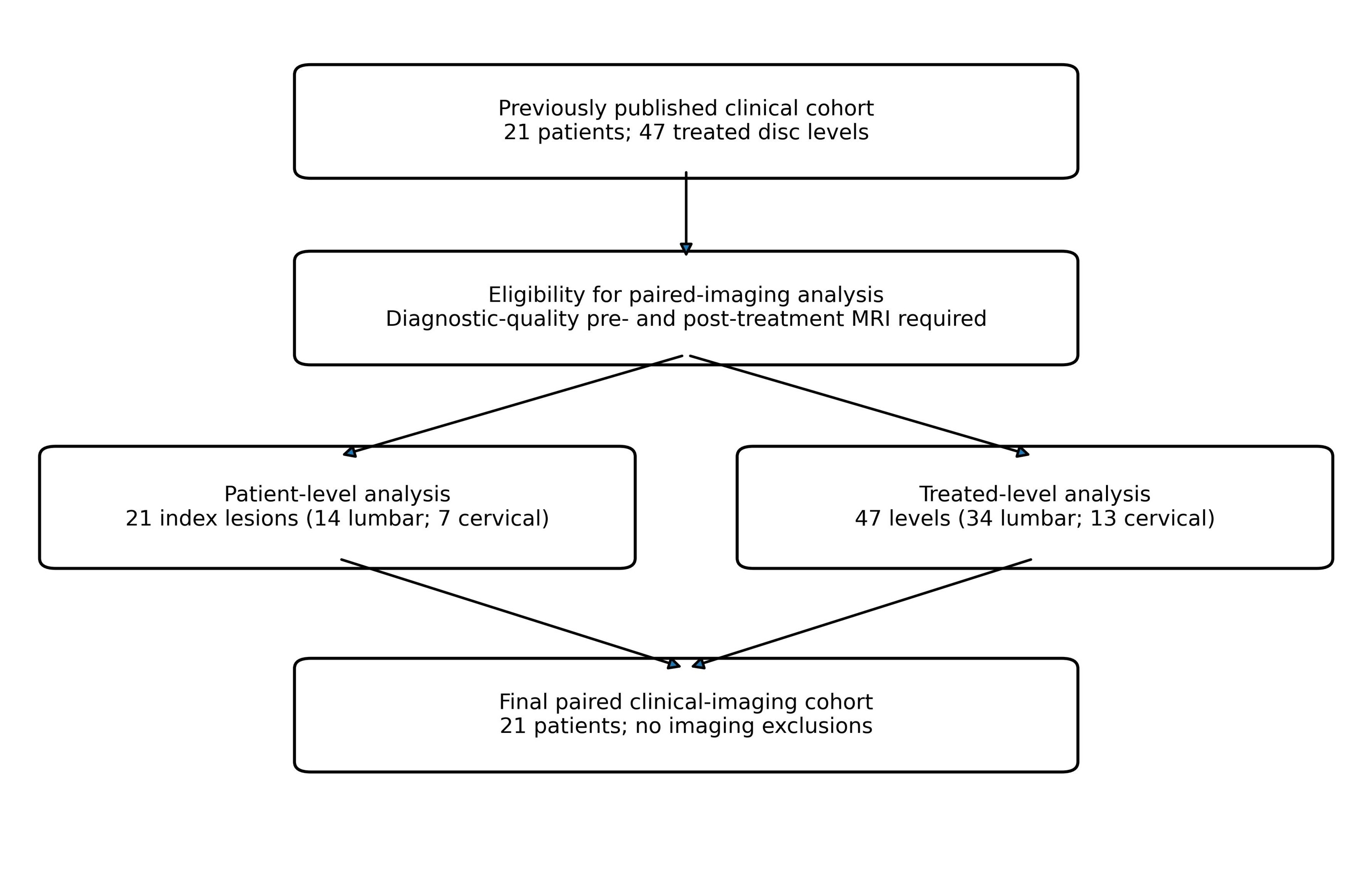
For the paired-imaging analysis, patients also required diagnostic-quality pre-treatment and post-treatment MRI studies of the same spinal region. All 21 patients in the parent cohort were included. The study flow is shown in Figure 1. The cohort accounted for 47 treated disc levels, including 34 lumbar and 13 cervical levels; two patients underwent treatment in both regions.
Inclusion criteria
- Age 18 years or older at the time of treatment.
- Primary diagnosis of cervical disc bulge, cervical disc herniation, lumbar disc bulge, or lumbar disc herniation.
- MRI confirmation of disc pathology before treatment.
- Completion of 30 IDD Therapy sessions within 3 months.
- Complete pre-treatment and post-treatment VAS and ODI data.
- Diagnostic-quality pre-treatment and post-treatment MRI available for comparison.
Exclusion criteria
Patients were excluded if they had active malignancy, pregnancy, prior surgery at the treatment region, multilevel fusion, grade II or higher spondylolisthesis, degenerative scoliosis, severe canal stenosis, discitis or osteomyelitis, adjacent segment disease, osteoporosis, spinal fracture, spondylolysis, abdominal aortic aneurysm, pacemaker, or a known genetic condition causing spinal instability. These criteria were intended to reduce clinical risk, improve cohort homogeneity, and exclude conditions for which decompression therapy may be inappropriate or substantially confounded by structural instability.
Data sources and quality assurance
Clinical data were abstracted from the electronic health record and from paper outcome forms completed by patients before clinician interaction. Data elements included age, sex, treated region and level, treatment completion, VAS, ODI, adverse events, and dates of MRI acquisition. Clinical values reported in the parent publication were retained without modification so that the imaging analysis remained linked to the original cohort [3].
Imaging data were matched to the clinical record using coded study identifiers. Pre-treatment and post-treatment examinations were reviewed for correct patient, region, level, and chronology before measurement. A standardized data dictionary was used to define each quantitative variable and response category. Range checks and internal consistency checks were applied to identify impossible values, duplicated records, and disagreement between the patient-level and treated-level datasets. Any discrepancy was resolved against the source record before analysis.
Treatment protocol
IDD Therapy was delivered with the Accu-SPINA system according to an established 30-session protocol. Treatment was individualized by region and target level. Initial distraction forces were calculated using protocol-based formulas. Distraction force was increased only when post-treatment and inter-treatment pain did not worsen and when radicular symptoms demonstrated centralization or clinical improvement.
For lumbar pathology, distraction force was increased in 5-pound increments as tolerated. For cervical pathology, force was increased in 2-pound increments. A Doctor of Chiropractic reassessed patients throughout care. Progression was based on symptom response and tolerance rather than achievement of a predetermined maximum force.
Concomitant care
Concomitant medications, home exercise, manual therapy, and other conservative interventions were not standardized by the retrospective protocol and were not captured with sufficient consistency for quantitative adjustment. Accordingly, the observed outcomes should be interpreted as results from a real-world treatment course rather than as isolated effects of IDD Therapy.
Clinical outcomes
Clinical outcomes were VAS pain score and ODI. VAS was recorded on a 0-to-10 scale, with higher scores indicating greater pain. ODI was administered using the standard, unmodified questionnaire and retained in the raw 0-to-50 units used in the parent study, with higher scores indicating greater disability [18]. Both outcomes were collected before initiation of IDD Therapy and after completion of the treatment protocol.
The primary clinical outcomes were mean changes in VAS and ODI. Exploratory responder analyses used a reduction of at least 2 points on VAS and at least 10 raw ODI points as clinically meaningful improvement thresholds, consistent with published work on interpretation of pain and disability change [19,20]. Because ODI is a lumbar-specific instrument, a post hoc lumbar-only sensitivity analysis was performed for the association between ODI improvement and disc contour reduction. Future prospective studies should use the Neck Disability Index for cervical participants [21].
MRI acquisition and timing
Baseline MRI was obtained before the first IDD Therapy session, and follow-up MRI was obtained after completion of the 30-session protocol. Diagnostic-quality sagittal and axial sequences were required. Images were included only when the same spinal region could be compared before and after treatment with sufficient quality to assess the index disc level.
The median interval from baseline MRI to follow-up MRI was 88 days (interquartile range, 74–104 days). The median interval from the final IDD Therapy session to follow-up MRI was 18 days (interquartile range, 10–29 days). Examinations were obtained as part of routine clinical care rather than under a single research imaging protocol. Consequently, scanner model, field strength, sequence parameters, slice thickness, patient positioning, and exact scan time of day were not standardized. Because MRI examinations were performed at outside outpatient imaging facilities during routine clinical scheduling, exact clock times were not consistently available in the study dataset. The absence of standardized MRI time of day is particularly relevant for interpretation of disc-height measurements, which may vary with diurnal hydration and loading. For this reason, disc-height findings were interpreted as secondary, supportive imaging measures rather than as standalone evidence of structural response.
Index-level selection
Because several patients had more than one treated level, one index symptomatic level was selected for patient-level analysis. The index level was defined as the level most concordant with the symptom distribution, neurologic findings, and baseline MRI severity. When multiple levels were clinically relevant, the level with the greatest baseline posterior disc contour abnormality and neural element involvement was selected.
A separate treated-level analysis was performed across all 47 treated disc levels. The patient-level index analysis was primary for radiographic-clinical correlation to avoid giving disproportionate statistical weight to patients with multiple treated levels.
MRI review and measurements
Paired MRI studies were reviewed independently by Tyler Maitland Graham, DC, and John Graham, DO, using a standardized measurement protocol. Images were de-identified, and reviewers were blinded to VAS and ODI change. Image chronology was available during paired comparison because interval change was the outcome of interest. An independent radiologist was not retained for the research measurements because the present study was an unfunded retrospective secondary analysis of existing clinical imaging, and no study-specific external radiology core or blinded radiologist review protocol had been established when the parent clinical cohort was collected. Routine diagnostic MRI reports were used clinically to confirm eligibility and diagnosis, whereas the research measurements reported here were performed using the prespecified paired-measurement protocol described below. To reduce measurement bias within the practical limits of the retrospective design, two clinician reviewers with spine-care experience assessed the images independently, used predefined anatomic landmarks, remained blinded to clinical outcome change, and resolved measurement discrepancies greater than 1.0 mm or disagreements in qualitative response category by consensus.
Reviewers used the same imaging plane and closest corresponding slice whenever possible. Measurements were recorded to the nearest 0.1 mm. When an exact slice match was unavailable, the slice showing the greatest abnormality at the index level was selected according to the prespecified protocol. Qualitative assessment considered disc contour, ventral thecal sac or cord contact, lateral recess narrowing, foraminal narrowing, and visible nerve root contact. Independent reader-level values were not retained after consensus adjudication; therefore, formal intraclass correlation coefficients could not be calculated.
Quantitative outcomes were maximal posterior disc contour extension, measured from the expected posterior vertebral body margin to the point of greatest posterior displacement; central canal anteroposterior diameter, measured at the point of greatest canal compromise; and mid-disc height, measured using consistent anterior-posterior landmarks. Foraminal narrowing and nerve root contact were graded qualitatively as absent, mild, moderate, or severe. Disc morphology was categorized as bulge, protrusion, or extrusion using accepted disc-nomenclature principles [17].
For descriptive quantitative response analyses, a change of at least 0.5 mm in the favorable direction was considered improved, a change of at least 0.5 mm in the adverse direction was considered worsened, and smaller changes were considered unchanged. The 0.5-mm threshold was prespecified as a pragmatic measurement-response threshold rather than a validated clinical threshold. Global radiographic response was categorized by reviewer consensus as improved, unchanged, or worsened after considering disc contour, canal and foraminal dimensions, neural element contact, and disc height. Global improvement required at least one favorable quantitative or qualitative change without clinically relevant worsening in another primary imaging domain.
Statistical analysis
Continuous variables were summarized as mean ± standard deviation or median with interquartile range. Categorical variables were summarized as counts and percentages. Previously published VAS and ODI summary statistics were retained from the parent article and were not independently re-estimated. Paired t tests were used for lumbar and cervical MRI measurements. Mean changes are reported with 95% confidence intervals, test statistics, two-sided P values, and absolute paired-sample Cohen's dz. Pearson correlation coefficients were reported with 95% confidence intervals calculated by Fisher z transformation. Analyses were performed in Python 3.13 using SciPy 1.17. P<0.05 was considered statistically significant.
Because the cohort was fixed by the available paired-imaging sample, no a priori sample-size calculation was performed. Correlation analyses were exploratory, and no adjustment for multiple comparisons was applied. Clinical-imaging correlations were conducted at the patient level; treated-level results were descriptive only, avoiding the assumption that multiple levels from the same patient were statistically independent. Distributional assumptions and influential observations were reviewed before interpreting parametric results. Source data, calculation files, and analysis outputs were retained as part of the study record.
Results
Cohort characteristics
The analysis included 21 patients with 47 treated disc levels. Mean age was 50.8 ± 15.5 years, and 11 patients (52.3%) were female. Thirty-four treated levels were lumbar and 13 were cervical; two patients had both cervical and lumbar treatment regions. Baseline characteristics are summarized in Table 1.
|
Characteristic |
Value |
|
Patients, n |
21 |
|
Age, mean ± SD, years |
50.8 ± 15.5 |
|
Female, n (%) |
11 (52.3%) |
|
Treated disc levels, n |
47 |
|
Treated levels per patient, mean ± SD |
2.1 ± 1.4 |
|
Lumbar treated levels, n (%) |
34 (72.3%) |
|
Cervical treated levels, n (%) |
13 (27.7%) |
|
Patients treated in both regions, n (%) |
2 (9.5%) |
|
Completed IDD Therapy sessions, n |
30 |
|
Baseline VAS, mean ± SD |
5.8 ± 1.9 |
|
Baseline ODI, mean ± SD (raw 0-50) |
18.5 ± 7.2 |
Treated-level distribution was C3-C4, 3 levels; C4-C5, 3; C5-C6, 3; C6-C7, 4; L2-L3, 2; L3-L4, 8; L4-L5, 14; and L5-S1, 10. Mean treated levels per patient was 2.1 ± 1.4 (Table 2). For the index-level imaging analysis, 14 patients had lumbar index lesions and 7 had cervical index lesions. Index morphology included 12 broad-based bulges, 6 focal protrusions, and 3 extrusions. Baseline neural element contact or compression was present in 18 of 21 patients. No patient was excluded from the imaging analysis because of missing or non-diagnostic follow-up imaging, and the patient-level and treated-level denominators therefore remained identical to the planned cohort shown in Figure 1.
|
Level |
Treated levels, n |
|
C3-C4 |
3 |
|
C4-C5 |
3 |
|
C5-C6 |
3 |
|
C6-C7 |
4 |
|
L2-L3 |
2 |
|
L3-L4 |
8 |
|
L4-L5 |
14 |
|
L5-S1 |
10 |
|
Total |
47 |
Clinical outcomes
Clinical outcomes were those reported in the parent cohort [3]. Mean VAS improved from 5.8 ± 1.9 before treatment to 1.5 ± 1.8 after treatment, a mean reduction of 4.3 points. Mean ODI improved from 18.5 ± 7.2 to 6.5 ± 6.1, a mean reduction of 12.0 points. Both changes were statistically significant and had large effect sizes (Table 3).
|
Outcome |
Pre-treatment |
Post-treatment |
Mean change |
P value |
Cohen's d |
|
VAS pain score |
5.8 ± 1.9 |
1.5 ± 1.8 |
-4.3 |
<0.001 |
2.16 |
|
ODI score (raw 0-50) |
18.5 ± 7.2 |
6.5 ± 6.1 |
-12.0 |
<0.001 |
1.69 |
|
a) Clinical values and effect sizes are reproduced from the parent publication and were not independently re-estimated in the present analysis. ODI: Oswestry Disability Index; VAS: Visual Analog Scale. |
|||||
The parent study subgroup analysis reported cervical VAS improvement from 7.0 ± 1.8 to 2.0 ± 1.4 and cervical ODI improvement from 23.3 ± 6.2 to 5.5 ± 3.3. The lumbar subgroup analysis reported VAS improvement from 5.6 ± 1.9 to 1.4 ± 1.9 and ODI improvement from 27.5 ± 7.1 to 6.7 ± 6.6, representing a mean lumbar ODI reduction of 20.8 points [3]. In the responder analysis, 18 of 21 patients met the VAS threshold and 15 of 21 met the ODI threshold.
MRI timing
Follow-up MRI was obtained a median of 88 days after baseline MRI (interquartile range, 74-104 days) and a median of 18 days after completion of the final IDD Therapy session (interquartile range, 10-29 days). Exact MRI appointment times were not consistently available across outside imaging facilities; therefore, time of day was not included as an analytic covariate. This limitation was considered most relevant to disc-height interpretation.
Patient-level MRI outcomes
MRI outcomes were analyzed separately by spinal region because cervical and lumbar dimensions are not directly comparable. Among 14 lumbar index lesions, posterior disc contour extension decreased from 6.05 ± 1.41 mm to 4.30 ± 0.45 mm (mean change, -1.75 mm; 95% CI, -2.46 to -1.04; t[13]=-5.36; P<0.001; |dz|=1.43). Canal AP diameter increased from 9.22 ± 1.26 mm to 10.53 ± 0.54 mm (mean change, +1.31 mm; 95% CI, +0.79 to +1.82; t[13]=5.51; P<0.001; |dz|=1.47), and disc height increased from 6.11 ± 0.41 mm to 6.75 ± 0.46 mm (mean change, +0.64 mm; 95% CI, +0.44 to +0.83; t[13]=6.97; P<0.001; |dz|=1.86). Among 7 cervical index lesions, disc contour extension decreased from 4.84 ± 0.69 mm to 3.64 ± 0.34 mm (mean change, -1.20 mm; 95% CI, -1.97 to -0.43; t[6]=-3.81; P=0.009; |dz|=1.44), canal AP diameter increased from 9.36 ± 0.75 mm to 10.24 ± 0.30 mm (mean change, +0.89 mm; 95% CI, +0.42 to +1.35; t[6]=4.64; P=0.004; |dz|=1.75), and disc height increased from 5.27 ± 0.33 mm to 5.80 ± 0.21 mm (mean change, +0.53 mm; 95% CI, +0.34 to +0.72; t[6]=6.79; P<0.001; |dz|=2.57) (Table 4 and Figure 2).
|
Region (n) |
Outcome |
Pre-treatment |
Post-treatment |
Mean change (95% CI) |
t (df) |
P value |
|dz| |
|
Lumbar (14) |
Posterior disc contour extension (mm) |
6.05 ± 1.41 |
4.30 ± 0.45 |
-1.75 (-2.46 to -1.04) |
-5.36 (13) |
<0.001 |
1.43 |
|
Lumbar (14) |
Canal AP diameter (mm) |
9.22 ± 1.26 |
10.53 ± 0.54 |
+1.31 (+0.79 to +1.82) |
5.51 (13) |
<0.001 |
1.47 |
|
Lumbar (14) |
Disc height (mm) |
6.11 ± 0.41 |
6.75 ± 0.46 |
+0.64 (+0.44 to +0.83) |
6.97 (13) |
<0.001 |
1.86 |
|
Cervical (7) |
Posterior disc contour extension (mm) |
4.84 ± 0.69 |
3.64 ± 0.34 |
-1.20 (-1.97 to -0.43) |
-3.81 (6) |
0.009 |
1.44 |
|
Cervical (7) |
Canal AP diameter (mm) |
9.36 ± 0.75 |
10.24 ± 0.30 |
+0.89 (+0.42 to +1.35) |
4.64 (6) |
0.004 |
1.75 |
|
Cervical (7) |
Disc height (mm) |
5.27 ± 0.33 |
5.80 ± 0.21 |
+0.53 (+0.34 to +0.72) |
6.79 (6) |
<0.001 |
2.57 |
|
a) Change is post-treatment minus pre-treatment. AP: Anteroposterior; CI: Confidence Interval; dz: Paired-sample Cohen effect size based on the standard deviation of change scores; MRI: Magnetic Resonance Imaging. |
|||||||
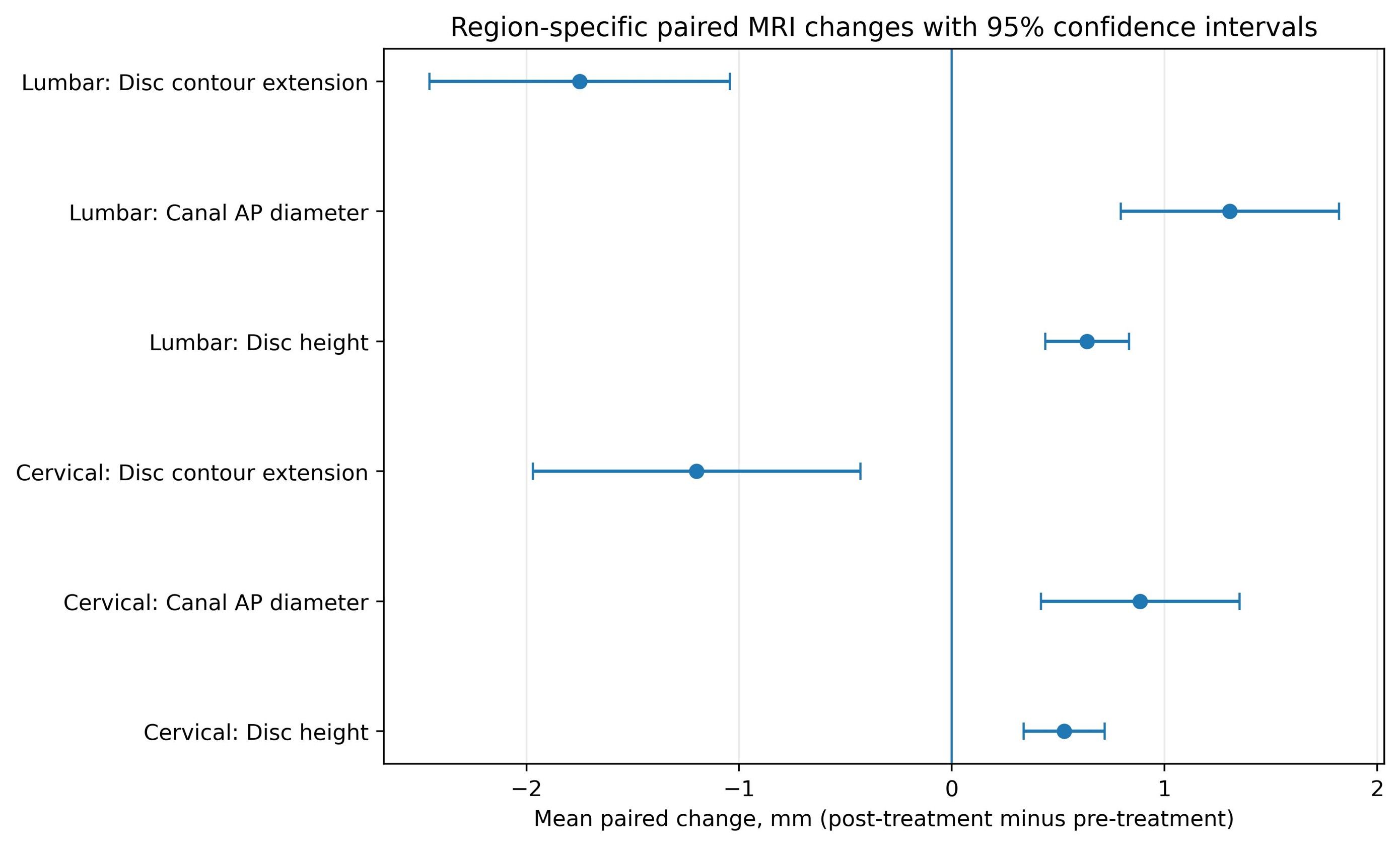
Figure 2. Region-specific paired MRI changes. Mean paired changes and 95% confidence intervals for posterior disc contour extension, canal anteroposterior diameter, and disc height, analyzed separately for lumbar and cervical index lesions. Negative values for disc contour extension and positive values for canal diameter and disc height indicate favorable change.
Using the prespecified 0.5-mm quantitative response threshold, posterior disc contour improved in 16 of 21 patients, canal AP diameter improved in 17, and disc height improved in 15 (Table 5). Global patient-level radiographic improvement was observed in 16 of 21 patients; four were classified as unchanged and one as worsened. The complete patient-level dataset is provided in Supplementary Table S1.
|
Quantitative domain |
Improved |
Unchanged |
Worsened |
|
Posterior disc contour extension |
16/21 (76.2%) |
5/21 (23.8%) |
0/21 (0.0%) |
|
Canal AP diameter |
17/21 (81.0%) |
4/21 (19.0%) |
0/21 (0.0%) |
|
Disc height |
15/21 (71.4%) |
6/21 (28.6%) |
0/21 (0.0%) |
|
a) Improved or worsened required a change of at least 0.5 mm in the corresponding direction; smaller changes were classified as unchanged. This was a pragmatic measurement-response threshold, not a validated clinical threshold. |
|||
Treated-level MRI outcomes
Across all 47 treated disc levels, 34 levels were classified as improved, 12 as unchanged, and 1 as worsened. Improvement was observed in 25 of 34 lumbar levels and 9 of 13 cervical levels (Table 6). Focal protrusions and extrusions appeared more likely to show visible interval change than broad-based degenerative bulges, although the study was not powered for formal morphology subgroup comparison.
|
Analysis |
Improved |
Unchanged |
Worsened |
|
Patient-level index lesions |
16/21 (76.2%) |
4/21 (19.0%) |
1/21 (4.8%) |
|
All treated levels |
34/47 (72.3%) |
12/47 (25.5%) |
1/47 (2.1%) |
|
Lumbar treated levels |
25/34 (73.5%) |
8/34 (23.5%) |
1/34 (2.9%) |
|
Cervical treated levels |
9/13 (69.2%) |
4/13 (30.8%) |
0/13 (0.0%) |
Radiographic-clinical correlation
Reduction in maximal posterior disc contour extension was strongly associated with VAS improvement (r=0.95; 95% CI, 0.88-0.98; P<0.001) and ODI improvement (r=0.91; 95% CI, 0.78-0.96; P<0.001). Because ODI is designed for low-back-related disability, a post hoc analysis restricted to the 14 lumbar index lesions was also performed and showed a similarly strong association between disc contour reduction and ODI improvement (r=0.96; 95% CI, 0.89-0.99; P<0.001). The association with VAS improvement is illustrated in Figure 3. These estimates are exploratory and may be unstable because of the small, selected all-completer cohort.
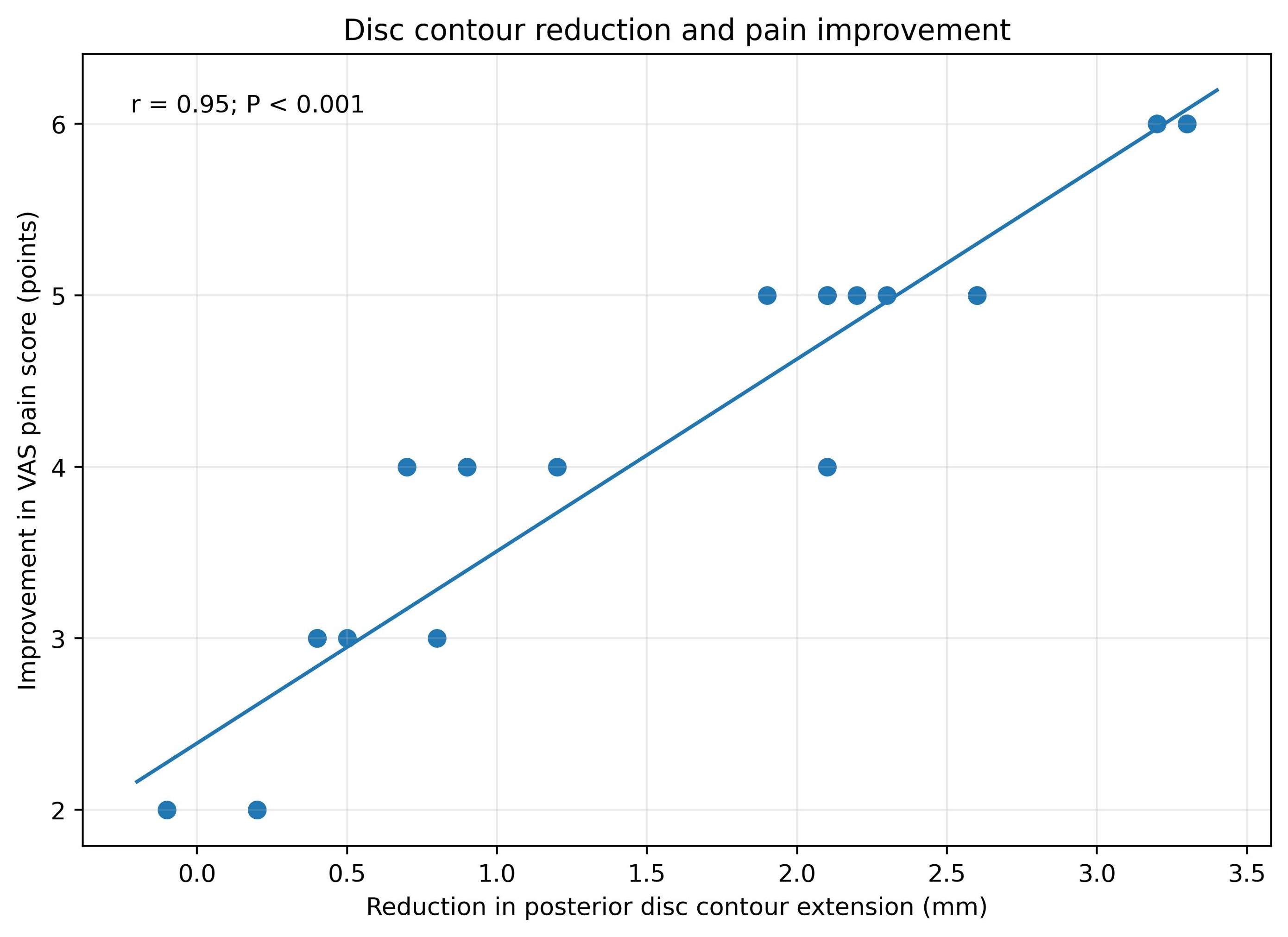
Figure 3. Association between reduction in posterior disc contour extension and improvement in VAS pain score. Each point represents one patient. The fitted line is shown for visualization (r=0.95; 95% CI, 0.88-0.98; P<0.001).
Safety
No serious adverse events were documented during the treatment period. No patient required emergency surgical referral during the IDD Therapy protocol, and no patient discontinued because of treatment-related worsening.
Discussion
This secondary paired-imaging analysis extends a previously published clinical outcomes cohort by evaluating structural MRI findings in the same patients. The principal observations were substantial reductions in pain and disability, favorable region-specific changes in posterior disc contour extension, canal diameter, and disc height, and a favorable global radiographic response in most patients. At the patient level, 76.2% of index lesions were classified as improved; at the treated-level analysis, 72.3% of levels were classified as improved.
The magnitude of the clinical change was large relative to commonly used thresholds for meaningful improvement in pain and disability [19,20]. Nevertheless, the clinical results were previously published and should not be interpreted as an independent replication. Their role in the present manuscript is to provide the clinical context needed to evaluate whether interval MRI changes tracked with patient improvement. The principal new contribution is the paired imaging analysis and its relationship to the previously reported outcomes.
Posterior disc contour extension was selected as the primary imaging measure because it offers a direct estimate of posterior displacement on sagittal or axial MRI. Cervical and lumbar raw dimensions were analyzed separately to avoid pooling anatomically dissimilar regions. Canal diameter and disc height were complementary measures, but each is sensitive to slice selection, patient positioning, hydration, and diurnal variation. The prespecified 0.5-mm response threshold improved transparency, although it was a pragmatic measurement threshold rather than a validated marker of clinically meaningful structural change.
The combination of clinical and imaging outcomes is more informative than either endpoint alone. Improvement in pain or disability can reflect reduced inflammation, improved movement tolerance, changes in pain processing, activity modification, regression to the mean, placebo effects, or natural history. Conversely, interval MRI improvement does not guarantee symptomatic recovery. The strong correlations observed between disc contour reduction and clinical improvement in this cohort should therefore be interpreted as exploratory and hypothesis-generating rather than as proof that the measured structural changes caused the clinical response. This caution is consistent with evidence that degenerative imaging findings may occur in both symptomatic and asymptomatic adults [9,10].
Spontaneous regression is a particularly important competing explanation. Lumbar disc herniations, especially extrusions and sequestrations, may decrease in size through dehydration, retraction, neovascularization, inflammatory resorption, and macrophage-mediated remodeling [11–14]. Serial imaging studies have documented regression during nonoperative care [13–15]. Without an untreated or active-comparator group, the present analysis cannot determine how much of the observed imaging change was attributable to IDD Therapy rather than natural history or co-interventions.
The findings should also be interpreted within the mixed literature on traction and non-surgical decompression. A randomized trial of IDD Therapy found no benefit over sham treatment in a broader low-back-pain population [4], whereas retrospective studies, a randomized trial of non-surgical decompression added to physical therapy, and a recent case series have reported improvements in selected patients [5–8]. Differences in diagnosis, treatment protocol, comparison intervention, adherence, and requirement for MRI-confirmed pathology may partly explain the inconsistent results. The present cohort was highly selected and required completion of all 30 sessions, which may increase internal consistency but also limit generalizability.
Prior imaging reports also differ substantially in design. Some studies have measured disc height or herniation dimensions after conservative care, while others have presented selected cases without uniform quantitative criteria [5,8,13–15]. The present analysis attempts to improve interpretability by specifying an index lesion, reporting both patient-level and treated-level results, and pairing imaging with clinical outcomes. Even so, comparison across studies remains difficult because definitions of bulge, protrusion, extrusion, improvement, and clinically meaningful structural change are not uniform. Standardized nomenclature and prespecified measurement thresholds would strengthen future research and make meta-analysis more feasible.
An additional consideration is the all-completer design. Requiring 30 sessions ensured that participants received the intended treatment exposure, but it excluded individuals who stopped early because of inconvenience, cost, lack of benefit, worsening symptoms, or unrelated circumstances. The cohort may therefore over-represent patients who tolerated care and perceived enough benefit to continue. A prospective effectiveness study should enroll patients at treatment initiation, report attrition and reasons for discontinuation, and analyze outcomes according to intention-to-treat principles in addition to per-protocol completion.
The pattern of imaging response may also depend on morphology. Focal protrusions and extrusions have a discrete contour that is easier to measure and may be more likely to demonstrate interval reduction. Broad-based degenerative bulges may represent chronic annular remodeling and disc height loss that are less likely to reverse over a short interval. The apparent difference in response by morphology in this cohort is descriptive and should not be interpreted as proof of differential treatment efficacy.
The observed correlations between disc contour reduction and clinical change were strong. Although internally consistent with the patient-level dataset, estimates of this magnitude should be interpreted cautiously. The sample was small, restricted to treatment completers, and drawn from a narrow range of clinically selected patients; each factor can increase correlation instability and limit external validity. The findings should therefore be treated as exploratory signals requiring independent replication rather than as precise estimates of a causal structure-outcome relationship.
The cervical subgroup deserves particular caution. The cohort included 13 cervical treated levels and 7 cervical index lesions. ODI was used in the parent study for the overall cohort, but it was developed for low-back-related disability [18]. Cervical outcomes should ideally include the Neck Disability Index and region-specific measures of radicular symptoms [21]. The use of ODI in cervical cases reduces interpretability of the disability results and should be addressed prospectively.
Strengths of this study include direct linkage to a previously reported cohort, MRI confirmation of disc pathology, completion of a standardized treatment course, paired imaging, and separation of patient-level from treated-level analyses. The patient-level approach reduces the risk of over-weighting participants with multiple treated levels, while the level-level analysis describes the distribution of radiographic response across the treated anatomy.
Clinically, the results support a cautious interpretation of follow-up MRI. Repeat imaging should not be used routinely as a surrogate for recovery, but paired MRI may provide useful objective information in selected patients when symptoms persist, neurologic findings change, or the clinical course is discordant with expectations. For research, the findings suggest that disc contour extension may be a more responsive short-term measure than disc height, although this hypothesis requires confirmation with standardized imaging and formal measurement-error analysis.
The study has several limitations. It was retrospective, single-center, small, and limited to treatment completers. The absence of a control group prevents causal inference, and spontaneous regression, regression to the mean, and concomitant care remain competing explanations. Imaging was obtained during routine care rather than under a standardized research protocol; scanner characteristics, sequence parameters, slice thickness, positioning, hydration, and exact time of day may therefore have varied. Because disc height may fluctuate over the course of a day, the lack of standardized MRI time of day is an important limitation, and disc-height changes should be interpreted as supportive rather than primary evidence. An independent radiologist did not perform the research measurements because this was an unfunded retrospective secondary analysis of existing clinical images rather than a prospectively designed imaging trial. Although routine clinical radiology reports supported the original diagnoses and eligibility determination, the paired research measurements were performed by two clinician reviewers using a standardized protocol and consensus adjudication. Formal inter-reader reliability could not be calculated because independent reader-level measurements were not retained after consensus adjudication. The 0.5-mm response threshold was pragmatic and has not been validated as a clinically meaningful MRI threshold. The cohort combined cervical and lumbar pathology, the parent clinical dataset used ODI in cervical cases, and co-interventions were not consistently captured. Clinical response influenced treatment progression, so treating clinicians could not be blinded. Finally, this was a secondary analysis of a previously published cohort, making transparent disclosure of overlap essential. Future prospective studies should use standardized MRI scheduling, preferably at similar times of day, and should incorporate independent radiologist or radiology-core review with retained reader-level measurements for formal reliability analysis.
Despite these limitations, paired clinical-imaging data from carefully characterized conservative-care cohorts are uncommon. The present findings provide estimates that may inform prospective study design, including expected effect sizes, imaging intervals, outcome selection, and morphology-based stratification. A future trial should compare IDD Therapy plus structured rehabilitation with structured rehabilitation alone, use prespecified MRI acquisition and blinded review, collect VAS or numerical pain ratings, ODI for lumbar cases, NDI for cervical cases, patient global impression of change, medication use, work status, and longer-term outcomes.
Conclusion
In this selected retrospective cohort, completion of a 30-session IDD Therapy protocol was associated with substantial clinical improvement and favorable region-specific paired-MRI changes in most patients with MRI-confirmed cervical or lumbar disc bulges and herniations. Posterior disc contour reduction was strongly associated with improvement in pain and disability, but the magnitude of these exploratory correlations requires independent replication. The study lacked a control group, used routine-care imaging rather than a standardized research protocol, and included only treatment completers; therefore, the findings do not establish causation. Prospective controlled studies with standardized MRI acquisition, retained independent reader measurements, formal reliability analysis, region-specific clinical outcomes, documented co-interventions, and longer follow-up are needed.
Data Availability Statement
De-identified data supporting the findings are available from the corresponding author upon reasonable request, subject to institutional, ethical, privacy, and regulatory requirements.
Conflicts of Interest
Tyler Maitland Graham, DC, provides chiropractic and non-surgical spinal decompression care in clinical practice. John Graham, DO, reports no conflicts of interest. The authors report no commercial funding, equipment sponsorship, consulting payments, royalties, or financial ownership interests related to IDD Therapy or the Accu-SPINA system.
Funding Statement
No external funding was received for this study or preparation of the manuscript.
Author Contributions
Tyler Maitland Graham, DC: conceptualization, methodology, investigation, clinical data curation, imaging review, project administration, formal analysis, visualization, writing - original draft, and writing - review and editing. John Graham, DO: independent imaging review, clinical interpretation, validation, and writing - review and editing. Both authors approved the final manuscript and accept accountability for the work.
Acknowledgments
None.
References
2. Kreiner DS, Hwang SW, Easa JE, Resnick DK, Baisden JL, Bess S, et al. An evidence-based clinical guideline for the diagnosis and treatment of lumbar disc herniation with radiculopathy. Spine J. 2014 Jan;14(1):180–91.
3. Bartrom S, Syed E, Graham T. Quantifying clinical outcomes of IDD Therapy in patients with cervical and lumbar disc bulges and herniations. J Phys Med Rehabil. 2026;8(1):10–16.
4. Schimmel JJ, de Kleuver M, Horsting PP, Spruit M, Jacobs WC, van Limbeek J. No effect of traction in patients with low back pain: a single centre, single blind, randomized controlled trial of Intervertebral Differential Dynamics Therapy. Eur Spine J. 2009 Dec;18(12):1843–50.
5. Apfel CC, Cakmakkaya OS, Martin W, Richmond C, Macario A, George E, et al. Restoration of disk height through non-surgical spinal decompression is associated with decreased discogenic low back pain: a retrospective cohort study. BMC Musculoskelet Disord. 2010 Jul 8;11:155.
6. Macario A, Richmond C, Auster M, Pergolizzi JV. Treatment of 94 outpatients with chronic discogenic low back pain with the DRX9000: a retrospective chart review. Pain Pract. 2008 Jan-Feb;8(1):11–7.
7. Amjad F, Mohseni-Bandpei MA, Gilani SA, Ahmad A, Hanif A. Effects of non-surgical decompression therapy in addition to routine physical therapy on pain, range of motion, endurance, functional disability and quality of life versus routine physical therapy alone in patients with lumbar radiculopathy; a randomized controlled trial. BMC Musculoskelet Disord. 2022 Mar 16;23(1):255.
8. Connolly J, Luginsland L, Welsh S, Bexley JA, Pirone J, Martin WR, et al. Clinical and imaging outcomes of non-surgical spinal decompression for lumbar intervertebral disc lesions: a case series. J Contemp Chiropr. 2025;8(1):81–87.
9. Brinjikji W, Luetmer PH, Comstock B, Bresnahan BW, Chen LE, Deyo RA, et al. Systematic literature review of imaging features of spinal degeneration in asymptomatic populations. AJNR Am J Neuroradiol. 2015;36(4):811–16.
10. Brinjikji W, Diehn FE, Jarvik JG, Carr CM, Kallmes DF, Murad MH, et al. MRI Findings of Disc Degeneration are More Prevalent in Adults with Low Back Pain than in Asymptomatic Controls: A Systematic Review and Meta-Analysis. AJNR Am J Neuroradiol. 2015 Dec;36(12):2394–99.
11. Chiu CC, Chuang TY, Chang KH, Wu CH, Lin PW, Hsu WY. The probability of spontaneous regression of lumbar herniated disc: a systematic review. Clin Rehabil. 2015 Feb;29(2):184–95.
12. Zhong M, Liu JT, Jiang H, Mo W, Yu PF, Li XC, et al. Incidence of Spontaneous Resorption of Lumbar Disc Herniation: A Meta-Analysis. Pain Physician. 2017 Jan-Feb;20(1):E45–E52.
13. Maigne JY, Rime B, Deligne B. Computed tomographic follow-up study of forty-eight cases of nonoperatively treated lumbar intervertebral disc herniation. Spine (Phila Pa 1976). 1992 Sep;17(9):1071–74.
14. Yukawa Y, Kato F, Matsubara Y, Kajino G, Nakamura S, Nitta H. Serial magnetic resonance imaging follow-up study of lumbar disc herniation conservatively treated for average 30 months: relation between reduction of herniation and degeneration of disc. J Spinal Disord. 1996 Jun;9(3):251–56.
15. BenEliyahu DJ. Magnetic resonance imaging and clinical follow-up: study of 27 patients receiving chiropractic care for cervical and lumbar disc herniations. J Manipulative Physiol Ther. 1996 Nov-Dec;19(9):597–606.
16. von Elm E, Altman DG, Egger M, Pocock SJ, Gøtzsche PC, Vandenbroucke JP. The Strengthening the Reporting of Observational Studies in Epidemiology (STROBE) statement: guidelines for reporting observational studies. Ann Intern Med. 2007;147(8):573–77.
17. Fardon DF, Williams AL, Dohring EJ, Murtagh FR, Gabriel Rothman SL, Sze GK. Lumbar disc nomenclature: version 2.0: recommendations of the combined task forces of the North American Spine Society, the American Society of Spine Radiology, and the American Society of Neuroradiology. Spine (Phila Pa 1976). 2014 Nov 15;39(24):E1448–65.
18. Fairbank JC, Pynsent PB. The Oswestry disability index. Spine. 2000 Nov 15;25(22):2940–53.
19. Bombardier C, Hayden J, Beaton DE. Minimal clinically important difference. Low back pain: outcome measures. J Rheumatol. 2001 Feb;28(2):431–38.
20. Emshoff R, Bertram S, Emshoff I. Clinically important difference thresholds of the visual analog scale: a conceptual model for identifying meaningful intraindividual changes for pain intensity. Pain. 2011 Oct;152(10):2277–82.
21. Vernon H, Mior S. The Neck Disability Index: a study of reliability and validity. J Manipulative Physiol Ther. 1991 Sep;14(7):409–15.