

Abstract
Background: The presence of lymphatic vessels in the human endometrium is sketchy and is unsettled. Using D2-40 as a marker of lymphatic vessels in the formalin-fixed and paraffin-embedded human endometrium, a reduced density of lymphatic vessels was reported in human functionalis. Normally cycling human endometrium is relatively difficult to study for ethical reasons.
Objectives: Uteri from menstrual, proliferative, and secretary cycle were collected to study lymphatic vessels compared to blood vessels by immunohistochemical staining for lymphatic vessels in the artificially cycling monkey endometrium. The full thickness of uterus was vertically sectioned and was collected from menstrual, proliferative to secretary cycle in the cycling endometrium. With frozen sections, we studied lymphatic and blood vessels by immunostaining with D2-40 and LYVE-1 for lymphatic vessels and von Willebrand’s factor for blood vessels, respectively.
Results: There was the consistent presence of lymphatic vessels in myometrium and basalis. Blood vessels supply the growing functionals while lymphatic vessels grew longitudinally from basalis to functionalis with D2-40 and LYVE-1 immunostaining in proliferative phase from lower to middle secretary functionalis in late secretary phase. There were dilated and bursting lymphatic vessels in upper functionalis at menstruation.
Conclusion: Ample blood vessels supply the entire endometrium while lymphatic vessels do not catch up with the fast-growing blood vessels in the functionalis. The presence of lymphatic vessels in the functionalis during the menstrual period implies the presence of sliver of lymphatic fluid in the menstrual fluid. The less lymphatic vessels in the late secretary functionalis may contribute to extreme interstitial endometrial edema during menstrual shedding.
Keywords
Blood vessels, D2-40, Immunocytochemistry, Frozen sections, Lymphatic vessels, LYVE-1
Introduction
Information on the endometrial lymphatic vessels is sketchy and unsettled: some authors claimed that lymphatic vessels were absent in the human endometrium [1–3] while more investigators increasingly agree that lymphatic vessels are present in the human endometrium: one group reported that lymphatic vessels were present in 62% of functionalis [4] and the other group reported that lymphatic vessels were present in the basalis alone using monoclonal D2-40 as a lymphatic vessel marker [4,5]. With formalin-fixed and paraffin-embedded (FFPE) human endometrium, Rogers et al reported lymphatic vessels in functionalis at a reduced density compared to basalis and myometrium using D2-40 as a lymphatic endothelial marker [6–9]. More specific markers for lymphatic vessels have become available. One is lymphatic vessel endothelial receptor-1 (LYVE-1), a transmembrane receptor for hyaluronan that is highly expressed by the by the lymphatic vessel endothelium: LYVE-1 is also expressed by liver sinusoids, various macrophages and some other cells [10]. Monoclonal and polyclonal antibodies against LYVE-1 have been used as immunocytochemical markers to study lymphatic vessels in various tissues [10]. Lymphatic vessels were clearly observed using D2-40 antibody [6–9] but LYVE-1 has not been extensively used to immunostain lymphatic vessels lymphatic vessels in the endometrium [5–9]. Other markers of lymphatic vessels include podoplanin, a membrane glycoprotein recognized by the monoclonal antibody D2-40, and VEGFR-3, which binds VEGF-C/D [6]. D2-40 antibody has been used to quantify lymphatic vessels density in the human endometrium throughout the menstrual cycle [8,9], and one study used LYVE-1 staining on the endometrium [11]. Polyclonal LYVE-1 antibody immunostained lymphatic vessel endothelium more than monoclonal D2-40 [10]. Lymphatic vessels were consistently more numerous in the basalis than in the functionalis during the menstrual cycle, but the status on lymphatic vessels during the menstrual cycle had not been reported to date [5–9]. In this study, LYVE-1 and D2-40 were used for lymphatic vessel markfers and von Willebrand’s factor (vWF) was used for blood vessel marker, respectively. The non-human primate model simulates the human menstrual cycle with an artificially hormone control cycle since it is not feasible to undergo a similar study in humans because of ethical issues and a variation of menstrual cycle among individual women.
Materials and Methods
Female artificially cycling rhesus monkeys
Animal care for the rhesus monkeys (Macaca mulatta) was provided by the veterinary staff of the Division of Animal Resources of the Oregon National Primate Research Center, in accordance with NIH policy for the care and use of laboratory animals. Female rhesus monkeys were oophorectomized and treated sequentially with estradiol (E2) and progesterone (P) to induce menstrual cycles described before [12–16]. To induce these cycles, the oophorectomized animals were first implanted sc with 3 cm Silastic capsules (Dow Corning Corp, Midland, MI) packed with crystalline E2 (Sigma) to stimulate development of an induced proliferative phase. After 14 days, a 6 cm Silastic capsule packed with crystalline P (Sigma) was implanted sc, and both implants remained in place for 14 days to induce the secretary phase. To induce menstruation, the P implant was removed while the E2 was left in place. Menstrual bleeding normally occurs 2–4 days after the P implant was removed. The human endometrium according to the exact menstrual cycle is hard to study from the ethical points of view and this study was carried out in rhesus monkeys. The numbers of the animals studied were as follows: the induced menstrual phase, 3 days after removing P, (E-3p): 5 cases, early proliferative phase, 7 days after removing P, (E-7p): 4 cases, late proliferative phase, 14 days after removing P, (E-14p):3 cases, early secretary phase, 7 days after implanting P, (E+7p): 3 cases, mid secretary phase, 10 days after implanting P, (E + 10P): one case and late secretary phase, 14 days after implanting P, (E+14p): 4 cases to total 19 animals.
Preparation of frozen sections
For each uterus, the thickness of endometrium including that of basalis, functionalis, and total endometrial thickness were measured with H- and -E-stained tissue slides in the FFPE embedded tissues. Fresh uterine tissues, about 1.0 x 1.0 x 0.5 cm, were tangentially excised including surface of endometrium to full thickness of myometrium and were microwave-irradiated for 7 sec in a microwave oven, embedded in OCT, frozen in liquid propane and sectioned at 5–7 microns [12–16]. Frozen sections (5–7 µm) were mounted on Super Frost Plus slides (Fisher Scientific, Pittsburgh, PA), microwave-irradiated again on ice for 3 sec, fixed in 2% paraformaldehyde in phosphate buffer at pH 7.4 for 10–15 min at room temperature and immersed twice for 2 min each time in 85% ethanol. To inhibit endogenous peroxidase activity, sections were incubated in a solution containing glucose oxidase (1 U/ml), sodium azide (1 mmol/L) and glucose (10 mmol/L) in PBS for 45 min at room temperature.
|
|
|
Frozen sections |
Paraffin-sections |
|
Goat anti-human LYVE-1 |
R and D System, Minneapolis, MN |
1:1200 |
1:100 |
|
Mouse monoclonal D2-40 |
Signet Laboratories, Dedham, Mass |
1:100 |
1:100 |
|
Rabbit human vWF |
Dako System, Carpenteria, CA |
1:800 |
1:100 |
Immunohistochemical staining for lymphatic and blood vessels with frozen sections
Frozen sections were incubated with goat anti-human LYVE-1 antibody at 1:1,000 dilutions, mouse D2-40 antibody at 1: 100 dilutions and rabbit anti-human von Willebrand’s factor (vWF) at 1:800 dilutions, respectively [17]. The frozen sections of spleen were immunostained for LYVE-1 and vWF to illustrate lymphatic and blood vessels as control tissue. For comparing between the frozen sections and the FFPE embedded section for immunostaining for lymphatic vessels and blood vessels, the frozen sections of spleen and the routinely FFPE embedded spleen were processed for immunostaining for LYVE-1 and vWF, respectively. FFPE embedded sections of spleen were treated for antigen retrieval procedure, then were incubated with 1:100 dilutions of both LYVE-1 and von Willebrand factor (vWF) antibodies, respectively (Table 2). Both the frozen and deparaffinized sections were incubated with the primary antibody overnight at 4°C. After rinsing and immersion in blocking serum, sections were incubated with the second antibody (1:200 dilution) for 30 min at room temperature. Final visualization was achieved with the ABC kit (Vector Laboratories, Burlingame, CA) and 0.025% diaminobenzidine tetrahydrochloride (Dojindo Molecular Technology, Rockville, MD) in Tris-buffer pH 7.6, 0.03% H2O2 (Fisher Scientific) to induce brown color. Tissue sections were then lightly counterstained with hematoxylin to facilitate identification of cellular elements. The measurement of thickness of functionalis and basalis was made in FFPE embedded sections stained for H. and E. [17].
|
Animals |
Basalis |
Functionalis |
Total Endometrium |
|
Menstrual phase, E - 3 (5) |
|||
|
1 |
1.0 mm |
0 |
1.0 mm |
|
2 |
0.8 |
0 |
0.8 |
|
3 |
0.8 |
0 |
0.8 |
|
4 |
0.8 |
0 |
0.8 |
|
5 |
0.8 |
0 |
0.8 |
|
Mean ± SE |
0.8 ± 0 |
0 ± 0 |
0.8 ± 0 |
|
Early proliferative phase, E - 7p (4) |
|||
|
1 |
0.4 mm |
1.7 mm |
2.1 mm |
|
2 |
0.4 |
1.8 |
2.2 |
|
3 |
0.4 |
1.9 |
2.3 |
|
4 |
0.2 |
1.7 |
1.9 |
|
Mean ± SE |
0.4 ± 0 |
1.8 ± 0 |
2.1 ± 0.2 |
|
Late proliferative phase, E - 14p (3) |
|||
|
1 |
0.6 mm |
2.8 mm |
3.4 mm |
|
2 |
0.4 |
2.1 |
2.5 |
|
3 |
0.4 |
2.1 |
2.5 |
|
Mean ± SE |
0.4 ± 0 |
2.3 ± 0.2 |
2.8 ± 0.3 |
|
Early secretary phase, E + 7p (3) |
|||
|
1 |
0.4 mm |
3.3 mm |
3.7 mm |
|
2 |
0.4 |
3.2 |
3.6 |
|
3 |
0.4 |
2.9 |
3.2 |
|
Mean ± SE |
0.4 ± 0 |
3.1 ± 0.1 |
3.5 ± 0.1 |
|
Mid secretary phase, E + 10p (1) |
|||
|
1 |
0.4 |
4.0 |
4.4 mm |
|
Late secretary phase, E + 14p (4) |
|||
|
1 |
0.4 mm |
2.8 mm |
3.2 mm |
|
2 |
0.4 |
2.8 |
3.2 |
|
3 |
0.6 |
2.9 |
3.5 |
|
4 |
0.4 |
2.8 |
3.2 |
Double immunostaining was performed with FFPE embedded spleen, which was immunostained initially for vWF in brown and then blue (Vecstatin and Vector SG, Burlingame, CA) for LYVE-1.
Results
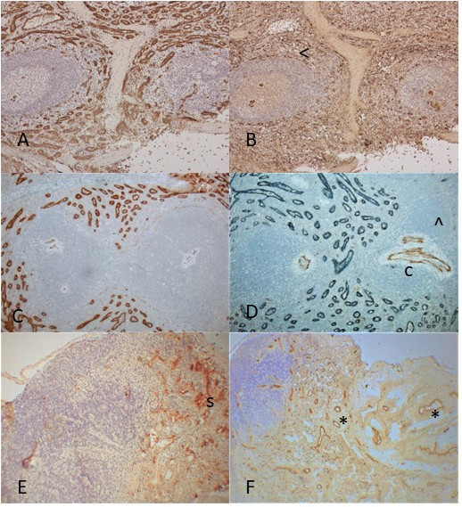
The frozen sections of spleen showed diffuse positive immunostaining for LYVE-1 in the lymphatic sinusoidal epithelia with no staining in the central arteries in the germinal center (Figure 1A). vWF immunostaining was positive in the central arteries as well as in the numerous thinner venous sinusoidal epithelia in the frozen section (Figure 1B), of which a few parts were immunostained in the FFPE embedded tissue sections (Figure 1D). The broader LYVE-1 positive sinusoidal space was immunostained a bit wider in the frozen sections than in the FFPE sections (Figures 1A and 1C). With routinely FFPE embedded splenic sections, double immunostaining for LYVE-1 and vWF was performed, which showed strong LYVE-1 immunostaining in the broader splenic sinusoids and positive vWF staining for the large central arteries in the germinal center with a part of blood sinusoid immunostained for vWF, the latter were mostly positively stained for vWF only in the frozen sections (Figures 1B and 1D). Thus, smaller vWF-positive venous sinusoids were mostly immunostained with the frozen sections with a small part of venous sinusoid immunotained in the FFPE embedded sections (Figure 1D). The frozen sections of lymph node showed subcapsular sinus (s) (Figure1E) where there were many dilated veins and some arteries (^), both were immunostained by vWF (Figure 1F).
In E-3p, 3 days after removing P capsule, which represented the menstrual phase, all showing sloughed off residual basalis (Figures 2A –2D), measuring 0.8 mm in thickness (Table 2). There were several thin lymphatic and venous vessels in the basalis as compared to many lymphatic and venous vessels in myometrium (Figures 2A and 2C), some of which extended vertically into the endometrial-myometrial interphase (Figures 2A–2D). Another case showed dilated lymphatic vessels by D2-40 (Figure 2C) and a few dilated venous vessels by vWF in the basalis (Figure 2D). All five cases revealed post-menstrual endometrium after menstrual bleeding.
In E-7p, 7 days after removing P capsule, there were functionalis, measuring 1.8 mm, larger than the basalis (Table 2). Lymphatic vessels in the basalis for LYVE-1 immunostaining showed a few vertical lymphatic vessels toward the endometrial surface in the lower functionalis but not by D2-40 immuostaining (Figures 2E and 2F). The myometrium contained abundant venous vessels and was mildly cross immunostained for D2-40 as seen in all stages of menstrual cycle (Figures 2C and 2F). The basalis consisted of round to oval short glands with slightly basophilic granular cytoplasm while the functionalis consisted of elongated, vertically tubular and clear cytoplasm with weak iridescent to eosinophilic staining (Figures 2E and 2F). Vertical blood vessels toward the endometrial surface were distributed in the entire endometrium with more blood vessels in the functionalis than in the basalis (Figure 2G).
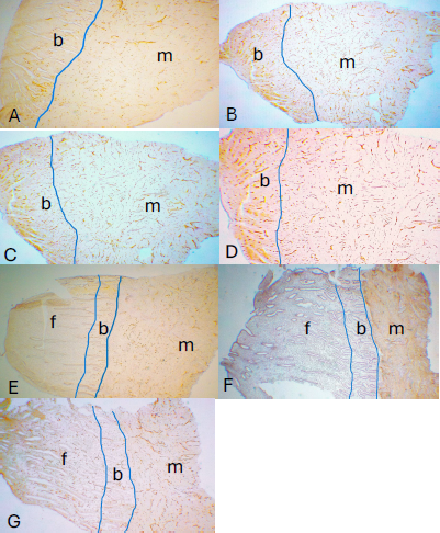
Figure 2. Endometrium 3 days after removing P capsule (E-3p) (A to D) and Endometrium 7 days after removing P capsule (E-7p) (E to G) Cases 1 to 5 from E-3p all showed the sloughed off residual basalis, measuring 0.8. Case 1 showed small linear lymphatic vessels in the basalis by LYVE-1 (A) and many venous vessels in the basalis (B). Case 2 showed dilated lymphatic vessels by D2-40 (C) and a few dilated veins in the basalis (D) while there were many vertical blood vessels in basalis and numerous linear blood vessels in the myometrium by vWF, some of which were circumferential in the myometrium (B and D). Cases from E-7p showed thin basalis and thin functionalis, containing a few small linear lymphatic vessels by LYVE-1 (E) and no obvious lymphatic vessels by D2-40 in the basalis (F). Numerous vertical venous vessels were more densely covered in the functionalis than basalis (G). There was mild cross immunostaining of D2-40 in the myometrium (F). A and E: LYVE-1, C and F: D2-40, B, D and G: vWF immunostained. b: basalis, f: functionalis, m: myometrium.
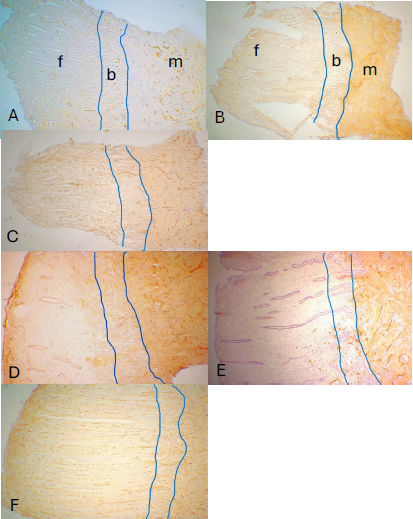
In E-14p, 14 days after removing P capsule, functionalis was a bit more enlarged, measuring 2.3 ± 0.2 mm, where vertical lymphatic and venous vessels toward the endometrial surface were immunostained with some of lymphatic vessels spreading into the lower functionalis from basalis (Figures 3A and 3B). Myometrium was richly supplied by venous vessels (Figure 3B) with mild cross immunostaining to D2-40 (3-C).
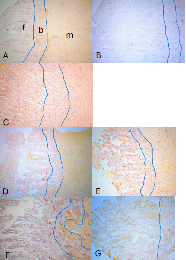
In E + 7p, 7 days after placing P capsule, there were larger functionalis, measuring 3.1 ± 0.1 mm with scattered small lymphatic vessels by LYVE-1 (Figure 4D) and a bit larger lymphatic vessels by D2-40 (Figure 4E) were present in the deep functionalis with numerous venous vessels in the entire functionalis (Figure 4F).
In E + 10p, 10 days after inserting P capsule, functionalis measured the largest at 4 mm in thickness where scattered lymphatic vessels in the basalis spread into the deep functionalis by LYVE-1 and D2-40 (Figures 4A and 4B) with covering venous vessels in the entire functionalis (Figure 4C).
In E+14p, 14 days after inserting P capsule, functionalis measured 3.3 ± 0.1 mm, where there were abundant lymphatic vessels in the basalis by both LYVE-1 and D2-40 immunostaining with LYVE-1 smaller lymphatic capillaries immunostained than D2-40 in the lower functionalis, some of which extended into the middle functionalis with LYVE-1 immunostaining (Figure 4D). D2-40 immunostained less but broader lymphatic vessels while LYVE-1 immunostained more but smaller lymphatic vessels (Figure 4D and 4F). Cases 1 and 2 showed elongated tubular functionalis containing elongated straight arteries accompanied by numerous small periarterial lymphatic vessels, corresponding to the late secretary phase (Figures 4D and 4E). Cases 3 and 4 presented short functionalis, where there was positive interstitial immunostaining for vWF in the subepithelial upper functionalis, which appeared to have vWF-positive oozing interstitial leaking blood into the upper functionals at the menstruation (Figures 4F and 4G). The similar interstitial positive immunostaining for vWF was also noted at the tip of E-3p Case 2 as shown in Figure 2D, which represented the residual phase of menstrual phase. All four cases did not show degenerative phase of menstrual endometrium.
Discussion
There was a difference in immunostaining patterns between frozen sections and FFPE-embedded sections of spleen: LYVE-1 positive splenic sinusoids were broader in the frozen sections than in the FFPE embedded sections (Figures 1A and 1B). There were two types of sinusoids in the splenic frozen sections: the broader, strongly LYVE-1 positive lymphatic sinusoids shown in both frozen sections and FFPE embedded sections and the thinner vWF positive venous sinusoids, which were depicted mostly in the frozen sections with only partly depicted FFPE embedded sections (Figures 1A and 1B). Thus, the thinner vWF positive venous sisunoids in the frozen section were not mostly immunostained in the FFPE sections, and this may prove the validity of using frozen section immunostaining for lymphatic and blood vessels. The broader LYVE-1 positive vessels appeared to be broader lymphatic sinusoids while the vWF positive thin capillaries in the frozen sections corresponded to the venous sinusoids (Figures 1C and 1D).
The endometrium consists of basalis and functionalis while basalis generally makes up about one third of endometrium while functionalis makes up more than two thirds of endometrium, the latter is the site of proliferation, secretion, and degeneration and is believed to regrow and regenerate from basalis, which is primarily driven by estrogen [18,19]. The basalis provides the regeneration capacity of the endometrium when functionalis has been desquamated [18,19]. Functionalis sheds at menstruation and subsequently regenerates from the remaining basalis, suggesting the putative endometrial stem cells residing in basalis [20].
On pathogenesis of endometriosis, lymphatic dissemination theory has been proposed for explaining the presence of endometrial and/or endometriotic tissue in lymphatic vessels. To support this theory, lymphatic vessel density is increased in the eutopic endometrium of women with endometriosis, which promotes for entering endometrial tissue into the lymphatic circulation [21]. Histopathologically, typical endometriosis consists of a few to more round or oval glands lined by simple layers of endometrial glands, which are surrounded by non-neoplastic endometrial stromal cells. Glands are variable from simple to dilated cysts [19]. Thus, glands forming endometriosis appear to be closer to that of basalis glands than functionalis glands.
Menstruation, marked by the bleeding and sloughing of the functionalis, is followed by repair and regenerative processes in the surviving tissue (primarily basalis) both in human and non-human primates [22–24]. In macaques, postmenstrual repair begins around day 3 after P withdrawal and includes the healing of the ragged surface by epithelial cells that migrate from the gland openings to form a new luminal epithelium, and the production of glandular fibronectin, which may facilitate surface healing [22–24]. Post menstrual repair is also characterized by the reappearance of estrogen receptors in the stroma and glands of the functionalis [22–24] and by the onset of mitosis in the newly constituted functionalis glands around day 5 after P withdrawal [13–16].
Arterial distribution in the uterus is well established: arcuate arteries are often circumferential in the myometrium and radial arteries are present close to the myometrium-endometrium junction. Basal arteries are in the basalis while spiral arteries, which are amplified post-ovulatory phase, are in the functionalis, which end up becoming surface arteries and eventually form surface capillary network [19,20,23,24]. Spinal arteries are characteristic of the human endometrium and have central roles in the menstruation process and in placenta formation [22–25]. Spiral arteries develop during the secretary phase under the influence of progesterone, coiling through the basalis into functionalis to supply a discrete region of the sub-epithelial capillary plexus and the plexus subsequently drain into venous sinuses [18,19]. Vertical surface arteries were present in the functionalis from the secretary phase monkey endometrium (Figures 4D and 4F). Bleeding during a normal menstrual cycle is a consequence of rhythmic vasospasm and relaxation of spiral arteries, resulting in a complete, yet self-limited sloughing of functionalis [18–20]. Endometrium is one of the highly vascularized organs with functionalis more vascularized than basalis. Endometrium’s rich vascularity is crucial for its function in menstruation and pregnancy [25]. From the current study by immunostaining blood vessels with the frozen sections, endometrium is immensely vascularized on par with the other most vascularized organs of liver, kidney and lungs. Most blood vessels immunostained with vWF were veins and arteries had not been reportedly immunostained except damaged ones with FFPE embedded sections [6]. With frozen sections, normal non-damaged arterial endothelium was also immunostained for vWF albeit weaker immunostained than veins. Vertical venous vessels toward the endometrial surface are diffusely distributed more in the functionalis than in the basalis as shown in this study. In contrast, the distribution of lymphatic vessels in the uterus is quite different: there were many small, linear circumferential lymphatic vessels in the myometrium, while basalis had preexisting small lymphatic vessels. Functionalis had no lymphatic vessels in the early proliferative phase and had a few lymphatic vessels in the lower functionalis in the proliferative and early secretary phase, which partly extended into the middle functionalis during late secretory phase by both D2-40 and LYVE-1 immunostaining. Blood vessels grow concomitantly to supply blood to the full thickness of functionalis with enough nutrients through the blood circulation, but lymphatic vessels have not grown to catch up with fast growing blood vessels in the secretary phase.
Rogers et al. extensively studied angiogenesis and lymphangiogenesis in the FFPE embedded human endometrium by measuring lymphatic vessels density (LVD) using monoclonal D2-40: no significant difference between proliferative and secretory phase endometrium LVD within the functionalis (proliferative phase: 16.7 ± 2.6 mm2 vs secretary phase: 16.2 ± 2.6 mm2), basalis (proliferative phase: 73.1 ± 3.7 mm2 vs secretary phase: 79.1 ± 7.5 mm2) and myometrium (proliferative phase: 63.4 ± 2.7 mm2 vs secretary phase: 60.3 ± 2.6 mm2) [7]. These data are quite different from ours using frozen sections for immunohistochemically staining using both D2-40 and LYVE-1 antibodies [7] as presented here: most abundant, residing lymphatic vessels were consistently present in the myometrium and in the basalis throughout the menstrual cycle while lymphatic vessels started growing longitudinally from the basalis to the deep functionalis in the early proliferative phase, which further grew during in the mid secretary phase and reached at the middle functionalis in the late secretary phase. With routinely FFPE embedded human endometrium, Donoghue et al. further studied the relative percentage of lymphatic vessels compared to the blood vessels and estimated that 13%, 43%, and 28% were lymphatic vessels in the functionalis, basalis, and myometrium, respectively [7]. As shown in Figures 2–4, there were many more blood vessels, especially vertically growing venous vessels immunostained in the frozen sections using vWF as a blood vessel marker.
By immunostaining with frozen sections, D2-40 immunostained less but thicker lymphatic vessels while LYVE-1 immunostained more but thinner lymphatic vessels (Figures 4C and 4D). We estimate the percentage of lymphatic vessels compared to that of blood vessels as less than 1% in the functionalis and less than 10% in basalis and less than 25% in the myometrium since most endometrial blood vessels are venous capillaries, which are immunostained with frozen sections but were not reportedly and properly immunostained with routinely FFPE embedded sections. In our experience, immunohistochemical staining for lymphatic and blood vessels did not provide longitudinally growing lymphatic and blood vessels in the FFPE embedded tissues. Using currently available antibodies to lymphatic and blood vessels, the previous authors using FFPE embedded tissue counted the limited positive staining in the high-power photomicrographs, not presenting vertically growing lymphatic and blood vessels in the low power photomicrographic perspective as we presented in this study [7–10]. Thus, it appears that those newly formed lymphatic vessels in the middle functionalis, which probably did not contain enough D2-40 protein, might not be positively immunostained with the FFPE embedded tissue sections.
Regarding immunocytochemical staining blood vessels with vWF antibody, vWF circulates in the blood stream and sticks to the surface endothelium of blood vessels, which is immunostained by vWF antibody in the venous vessels in the endometrium. The positive interstitial oozing immunostaining for vWF may represent the leaked vWF since the molecular weights of vWF varies between 500 and 20,000 Da [27,28] and the vWF with a smaller molecular weight may leak into the stroma in the late stage of the secretary phase as it was ready for menstrual bleeding [23,24]. Newly formed venous vessels in the functionalis are diffusely and clearly immunostained by vWF immunostaining in the frozen sections while arterial endothelium is less immunostained probably due to the less vWF antigen attached to the endothelial surface by the faster blood stream in the artery than in the vein (Figure 4F). Undamaged arteries were not reportedly immunostained with vWF antibody in the FFPE embedded tissue sections [6–9] in contrast to the consistently positive immunostaining in the non-damaged arteries and veins with frozen sections.
The lymphatic vessel system plays a major role in both fluid tissues and immune surveillance in the endometrium [29,30], but the functional significance of sparce lymphatic vessels in the functionalis remains speculative today [29,30]. Edema in the functionalis during the late phase of menstrual cycle is due to hormonal fluctuation, especially progesterone [23, 24]. The sparce lymphatic vessels in the functionalis causes an insufficient interstitial drainage, which results in interstitial edema in the functionalis, and this edema has been recognized as histological feature of the functionalis at the secretary phase of the menstrual cycle [23,24]. Maintaining fluid homeostasis is a key function of lymphatic vessels. Excess protein-rich fluid is removed from the tissue stroma via lymphatic system for return to the blood circulation [7,31–34]. Endometrium is one of the most vascularized tissues and more venous vessels grow concomitantly with less lymphatic vessels in normal and neoplastic tissues [35,36]. Endometrium is unique in that venous vessels are diffusely supplied in all stages of menstrual cycle compared to less lymphatic vessels in functionalis, especially in the upper part of functionalis during the late secretary phase (Figures 4D–4G).
To summarize the pathophysiology of lymphatic vessels in the menstrual bleeding, diffuse blood vessels supply the entire endometrium in both proliferative and secretary phase while lymphatic vessels reside in the basalis throughout the menstrual cycle. Lymphatic and diffuse blood vessels grow vertically toward the endometrial surface from the basalis to the lower functionalis in the early secretary phase, then, grow continuously to the mid to upper functionalis in the late secretary phase and burst during the menstrual shedding. Thus, lymph angiogenesis in the functionalis is not followed by the fast-growing angiogenesis, which is a cause of endometrial edema in the menstrual shedding through decreased absorption of interstitial fluid. The presence of lymphatic vessels in the functionalis during menstrual period implies the presence of sliver of lymphatic fluid in the menstrual fluid.
We conclude that immunostaining with frozen sections is superior to the FFPE embedded tissue for revealing lymphatic and blood vessels as shown in this study. There was a big difference in how much antibody needed for immunostaining between frozen sections and FFPE embedded sections: polyclonal LYVE-1 antibody needed 1: 100 dilutions for the FFPE embedded sections while frozen sections needed 1: 1,200 dilutions. The polyclonal vWF antibody needed 1: 100 dilutions for the FFPE embedded sections compared to 1: 800 dilutions needed for frozen sections. The monoclonal D2-40 needed the same 1: 100 dilutions for both frozen sections and FFPE embedded sections. The antibodies are expensive, so immunostaining with frozen sections is much more economic than with the FFPE embedded sections.
The hindside of using frozen sections at 5–7 µm in thickness is labor-intensive and cumbersome, and only small animal tissues (1 x 1 x 0.5 cm) are properly handled at our hand with monkey tissues [12–16]. With human tissues, slightly larger tissues (1.5 x 1 x 0.5 cm) are feasible to handle as human tissues are easier to freeze sectioning than animal tissues from rats, mice and monkeys. Despite these size limitations in handling the freezing sections compared to FFPE embedded tissues, immunostaining for lymphatic and blood vessels with the frozen sections is superior to the FFPE embedded tissues. The new findings from immunostaining with frozen sections will certainly shed light on involvement of lymphatic and blood vessels in pathophysiology and prognosis of human cervical and endometrial carcinoma and endometriosis.
Acknowledgement
We want to express our sincere thanks to Drs Robert M Brenner and Ov D. Slayden for kindly allowing us to use normal tissues of rhesus monkey at their research laboratory of the Oregon National Primate Research Center, Beaverton, OR, USA.
References
2. Blackwell PM, Fraser IS. Superficial lymphatics in the functional zone of normal human endometrium. Microvasc Res. 1981 Mar;21(2):142–52.
3. Uchino S, Ichikawa S, Okubo M, Nakamura Y, Iimura A. Methods of detection of lymphatics and their changes with oestrous cycle. Int Angiol. 1987 Jul-Sep;6(3):271–8.
4. Koukoulakis M, Giatromanolaki A, Sivridis E, Simpoulos C, Gatter KC, Harris AL et al. LYVE-1 immunohistochemical assessment of lymphangiogenesis in endometrial and lung cancer. J Clin Pathol. 2005 Feb;58(2):202–6.
5. Red-Horse K, Rivera J, Shatz A, Zhou Y, Winn V, Kipidzic, M. et al. Cytoptrophoblast induction of arterial apotptosis and lymphpangiogeneis in an in vitro model of human placentation. J Clin Invest. 2006 Oct;116(10):26543–52.
6. Scarvelli C, Weber E, Agliano M, Cirulli T, Nico, B et al. Lymphatics at crossroad of angiogenesis and lymphangiogenesis. J Anat. 2004 Jun;204(6):433–49.
7. Garling JE, Rogers, PAW. The endometrial lymphatic vasculature: function and dysfunction. Rev Endocr Metab Disord. 2012 Dec;13(4):265–75.
8. Rogers OPAW, Donoghue JF, Girling JE. Endometrial lymphangiogenesis. Placenta. 2008 Mar;29(Suppl A):S48–54.
9. Donoghue JF, Lederman FL, Susil BJ, Rogers PA. Lymphangiogenesis of normal endometrium and endometrial adenocarcinoma. Hum Reprod. 2007 Jun;22(6):1705–13.
10. Jackson DG. The lymphatics revisited: new perspective from the hyaluronan receptor LYVE-1. Trends Cardiovas Med 2003 Jan;13(1):1–7.
11. Salamosen LA, Shuster S, Stern R. Distribution of hyaluronan in human endometrium and across menstrual cycle: Implications for implantation and menstruation. Cell Tissue Res. 2001 Nov;306(2):335–40.
12. Slayden OD, Brenner RM. RU 486 action after estrogen priming in the endometrium and oviducts of rhesus monkeys (Macaca mulatta). J Clin Endocrinol Metab. 1994 Feb;78(2):440–8
13. Slayden OD, Brenner RM. Microwave stabilization enhances immunocytochemical detection of estrogen receptor in frozen sections of macaque oviduct. Endocrinol. 1995 Sep;136(9):4012–21.
14. Nayak NR, Critchley HO, Slayden OD, Menrad A, Chwalisz K, Baird DT, et al. Progesterone withdrawal up-regulates vascular endothelial growth factor receptor type 2 in the superficial zone stroma of the human and macaque endometrium: potential relevance to menstruation. J Clin Endocrinol Metab. 2000 Sep;85(9):3442–52.
15. Keator CS, Mah K, Ohm L, Slayden OD. Estrogen and progesterone regulate expression of the endothelins in the rhesus monkey endometrium. Human Reprod. 2011 Jul;26(7):1715–28.
16. Cao W, Mah K, Carroll RS, Slayden OD, Brenner RM. Progesterone withdrawal up-regulates fibronectin and integrins during menstruation and repair in the rhesus macaque endometrium. Hum Reprod. 2007 Dec;22(12):3223–31.
17. Tomita T. Immunohistochemical Staining for Lymphatic and Blood Vessels in Normal Tissues: Comparison between Routinely Paraffin-Embedded Tissues and Frozen Sections. Acta Med Acad. 2021 Apr;50(1):13–28.
18. Slayden OD, Keator CS. Role of progesterone in nonhuman primate implantation. Semin Reprod Med. 2007 Nov;25(6):418–430.
19. Maruyama T, Masuda H, Ono M, Kajitani T, Yoshimura Y. Human uterine stem/progenitor cells: their possible role in uterine physiology and pathology. Reproduction. 2010 Jul;140(1):11 –22.
20. Padykula HA. Regeneration in the primate uterus: the role of stem cells. Ann N Y Acad Sci. 1991;622:47–56.
21. Jerman LF, Hey-Cunningham AJ. The role of the lymphatic system in endometriosis: a comprehensive review of the literature. Biol Reprod. 2015 Mar;92(3):64.
22. Baltmez GW. Studies on the menstruating mucous membrane of human uterus. Contrb Embryl. 1933;24(142):143–86.
23. Mutter GL, Frenczy A. Anatomy and histology of the uterine corpus. In: Kurman RJ, Ed. Blaunstein’s Pathology of the Female Genital Tract, 5th ed. New York: Springer Verlag; 2002. pp. 383–419.
24. Buga GAB. The normal menstrual cycle. In: Kruger TF, Batha MH, Eds. Clinical Gynecology, 3rd ed. Cape Town: Juta & Co; 2007. pp. 73–87.
25. Rogers PA. Structure and function of endometrial blood vessels. Hum Reprod Update. 1996 Jan-Feb;2(1):57–62.
26. Pijnenborg R, Vercruysse L, Hanssens M. The uterine spiral arteries in human pregnancy: facts and controversies. Placenta. 2006 Sep-Oct;27(9-10):939–58.
27. Mannucci PM. von Willebrand factor: a marker of endothelial damage? Arterioscler Thromb Vasc Biol. 1998 Sep;18(9):1359–62.
28. Pusztaszeri MP, Seelentag W, Bosman FT. Immunohistochemical expression of endothelial markers CD31, CD34, von Willebrand factor, and Fli-1 in normal human tissues. J Histochem Cytochem. 2006 Apr;54(4):385–95.
29. Donoghue JF, McGavigan CJ, Lederman FL, Cann LM, Fu L, Dimitriadis E, et al. Dilated thin-walled blood and lymphatic vessels in human endometrium: a potential role for VEGF-D in progestin-induced break-through bleeding. PLoS One. 2012;7(2):e30916.
30. Red-Horse K. Lymphatic vessel dynamics in the uterine wall. Placenta. 2008 Mar;29 Suppl A(Suppl A):S55–9.
31. Schulte-Merker S, Sabine A, Petrova TV. Lymphatic vascular morphogenesis in development, physiology, and disease. J Cell Biol. 2011 May 16;193(4):607–18.
32. Shields JD. Lymphatics: at the interface of immunity, tolerance, and tumor metastasis. Microcirculation. 2011 Oct;18(7):517–31.
33. Al-Rawi MA, Mansel RE, Jiang WG. Molecular and cellular mechanisms of lymphangiogenesis. Eur J Surg Oncol. 2005 Mar;31(2):117–21.
34. Stacker SA, Williams SP, Karnezis T, Shayan R, Fox SB, Achen MG. Lymphangiogenesis and lymphatic vessel remodelling in cancer. Nat Rev Cancer. 2014 Mar;14(3):159–72.
35. Garnier L, Gkountidi AO, Hugues S. Tumor-Associated Lymphatic Vessel Features and Immunomodulatory Functions. Front Immunol. 2019 Apr 9;10:720.
36. Schledzewski K, Falkowski M, Moldenhauer G, Metharom P, Kzhyshkowska J, Ganss R, et al. Lymphatic endothelium-specific hyaluronan receptor LYVE-1 is expressed by stabilin-1+, F4/80+, CD11b+ macrophages in malignant tumours and wound healing tissue in vivo and in bone marrow cultures in vitro: implications for the assessment of lymphangiogenesis. J Pathol. 2006 May;209(1):67–77.