

Abstract
Oncolytic virotherapy (OVT) has emerged as a promising cancer immunotherapy strategy that combines selective tumor cell infection and lysis with immune activation. However, clinical responses remain variable, and innate antiviral immunity is a key determinant of this variability. Type I interferon (IFN) signaling and downstream interferon-stimulated genes can restrict viral replication, limit intratumoral spread, and reduce direct oncolysis, particularly in tumors with intact or elevated antiviral programs. At the same time, these pathways can also promote therapeutic benefit by inducing inflammatory cytokines and chemokines, enhancing antigen presentation, recruiting innate and adaptive immune cells, and converting poorly inflamed tumors into more immunologically active microenvironments. This commentary discusses innate antiviral immunity as a double-edged regulator of OVT outcomes, with an emphasis on tumor-intrinsic antiviral defenses, immune priming, viral genetics, cellular context, and rational combination strategies. Reovirus is highlighted as a case study in which viral strain variation, reassortment, infection kinetics, IFN modulation, and tumor cell type may shape therapeutic outcomes. Rather than seeking maximal viral replication or maximal innate immune activation, future OVT approaches should aim to define a productive immune window that supports viral oncolysis while preserving the inflammatory signals needed for durable antitumor immunity.
Keywords
Oncolytic virotherapy, Oncolytic virus, Interferon, Tumor microenvironment, Reovirus
Introduction
The paradox of antiviral immunity in oncolytic virotherapy
Oncolytic virotherapy (OVT) is a rapidly evolving cancer immunotherapy strategy that uses naturally occurring or genetically engineered viruses to selectively infect, replicate within, and destroy malignant cells. Although direct tumor cell lysis remains central to this approach, the therapeutic activity of oncolytic viruses (OVs) extends beyond viral replication alone. Viral infection can release tumor-associated antigens (TAAs), viral pathogen-associated molecular patterns (PAMPs), danger-associated molecular patterns (DAMPs), and inflammatory mediators that reshape the tumor microenvironment (TME) and promote antitumor immune responses [1]. This dual mechanism, tumor-selective cytolysis coupled with immune activation, has fueled renewed clinical and commercial interest in OVT.
The field of OVT has expanded substantially over the past two decades. As of 2022, more than 400 clinical trials had evaluated OVs across diverse malignancies, including melanoma, glioblastoma, lung cancer, pancreatic cancer, and others. These trials have involved multiple viral platforms, including DNA viruses such as herpes simplex virus, adenovirus, and vaccinia virus among the most widely used, and RNA viruses such as vesicular stomatitis virus, Newcastle disease virus, poliovirus, and reovirus also under active investigation [1]. The clinical feasibility of this approach is highlighted by the regulatory approval of several genetically modified OVs in different countries, including the human adenovirus H101 (Oncorine®) in combination with chemotherapy in China [2], and two herpes simplex viruses, talimogene laherparepvec (T-VEC; Imlygic®) and teserpaturev/G47Δ (Delytact®) in the United States and Japan, respectively [3,4]. Many ongoing and completed studies now evaluate OVs either as monotherapies or in combination with chemotherapy, radiation therapy, immune checkpoint blockade, and other immunotherapies [5].
Despite this progress, clinical responses to OVT remain variable, and innate antiviral immunity is a major determinant of this variability [6,7]. Many OVs exploit defects in tumor antiviral pathways, particularly type I interferon (IFN-α/β) sensing and signaling, to replicate preferentially in malignant cells. However, IFN and related innate immune pathways can also restrict viral replication, dissemination, and tumor cell killing when activated too early or too strongly [8–10]. Conversely, insufficient innate immune activation may permit viral replication and localized cytolysis but fail to convert tumor destruction into durable, systemic antitumor immunity. Mathematical models similarly support this framework by showing that OVT efficacy depends on dynamic interactions among viral infection of tumor cells, viral clearance, tumor growth, and antitumoral or antiviral immune responses rather than on viral replication alone [11,12].
Rather than viewing innate antiviral immunity as merely an obstacle to viral replication, this commentary argues that innate immune activation is a context-dependent determinant of OVT outcomes. The same pathways that restrict viral spread can also promote inflammatory remodeling, immunogenic tumor cell death, and antitumor immune priming. Understanding how viral genetics, tumor-intrinsic antiviral competence, and the TME shape this balance may help explain variable responses to OVs and guide more rational therapeutic combinations (Figure 1).
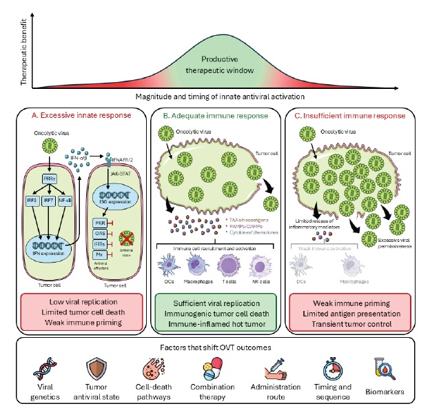
This commentary is intended as a focused conceptual perspective rather than a systematic review of innate antiviral signaling in OVT. Although the antiviral and immunostimulatory functions of IFN and ISG pathways have been extensively reviewed, less attention has been given to how the timing, magnitude, and cellular context of these responses determine whether OV infection becomes abortive, cytolytic, inflammatory, or therapeutically productive. We therefore propose the “productive immune window” as a framework for understanding how viral replication kinetics, tumor-intrinsic antiviral competence, innate inflammatory signaling, and antitumor immune priming converge to shape OVT outcomes.
Tumor-intrinsic antiviral immunity as a barrier to viral oncolysis
Tumor-cell permissiveness is a critical early determinant of OVT efficacy. Many OVs exploit cancer-associated defects in antiviral sensing, IFN signaling, protein translation control, or cell-death regulation to replicate preferentially in malignant cells. However, these defects are neither uniform across tumor types nor consistent among cells within the same tumor. Some cancer cells retain robust antiviral programs that can rapidly restrict viral gene expression, replication, and spread before sufficient oncolysis occurs. Thus, tumor-intrinsic antiviral immunity acts as an early gatekeeper that determines whether infection becomes abortive, productive, or therapeutically useful.
Type I IFNs are central to this gatekeeping function. IFNs are a group of cytokines classified into three major families: type I, type II, and type III. While type I IFNs, particularly IFN-α/β, can be produced by most nucleated cells, IFN-γ is primarily secreted by activated T cells and NK cells, and type III IFNs are especially important at epithelial barriers and in selected antigen-presenting cells [8]. In the context of OVT, type I IFN signaling is particularly relevant because many tumor cells can detect viral infection and respond by inducing an antiviral state. Viral PAMPs, including viral nucleic acids and replication intermediates, are sensed by pattern-recognition receptors (PRRs) such as RIG-I, MDA5, endosomal Toll-like receptors, and, for DNA viruses, cGAS-STING and related DNA-sensing pathways [10,13]. Activation of these pathways stimulates IRF3/IRF7- and NF-κB-dependent transcriptional programs that induce IFN-α/β and inflammatory mediators. Secreted type I IFNs then signal through their heterodimeric cell-surface receptor to activate JAK/STAT pathways and induce hundreds of IFN-stimulated genes (ISGs), establishing an antiviral state in infected and neighboring cells [8,10].
ISG products restrict viral replication at multiple stages of the viral life cycle. Protein kinase R (PKR), encoded by EIF2AK2, is activated by viral double-stranded RNA and phosphorylates eIF2α, thereby suppressing cellular and viral protein translation [14]. Interestingly, PKR has been shown to play a double role in cancer cells, where it not only limits the oncolytic potential of viruses but also increases the immune resistance of cancer cells through increased TGF-β signaling [15]. The OAS/RNase L pathway provides another important antiviral mechanism induced by IFNs. The 2′-5′-oligoadenylate synthetase (OAS) family of enzymes detect viral dsRNA and generate oligoadenylates that activate RNase L, resulting in degradation of viral and cellular RNAs and, in some settings, amplification of PRR signaling [10]. Pharmacological inhibition of this pathway has been reported to enhance the oncolytic activity of vesicular stomatitis virus in resistant tumor models, illustrating how intact ISG programs can limit OV efficacy [15,16]. Additional ISGs, including Mx proteins, IFITs, ISG15, and others, can restrict viral replication, translation, assembly, or trafficking, although their direct contributions to OVT remain to be fully addressed [17–19].
At first glance, studies showing that suppression of PKR or OAS/RNase L can enhance OV replication may appear to conflict with studies showing that innate immune activation contributes to antitumor efficacy [20]. These findings can be reconciled by distinguishing early tumor-intrinsic antiviral restriction from later immune-stimulatory inflammation. In models where intact PKR or RNase L activity prevents sufficient viral gene expression, transient inhibition of these pathways can increase viral replication and cytolysis [15]. However, when viral infection proceeds far enough to release tumor antigens, PAMPs, DAMPs, and inflammatory mediators, innate sensing becomes beneficial by promoting antigen presentation and immune-cell recruitment [21,22]. Thus, the same pathway may be detrimental or beneficial depending on the timing of activation, the OV platform, the baseline antiviral state of the tumor, and whether therapeutic outcome is measured as viral replication, direct cytotoxicity, or durable antitumor immunity [23].
Importantly, antiviral resistance can be present before infection occurs. Tumors with high basal IFN tone or constitutive ISG expression may enter treatment already poised to restrict viral replication [23]. Following infection, paracrine IFN signaling can further induce ISGs in surrounding tumor and stromal cells, creating a local antiviral “firebreak” that limits intratumoral viral spread. These mechanisms help explain why tumors with intact or elevated antiviral signaling, including some glioblastomas and other difficult-to-treat malignancies, can show resistance to OVT despite being biologically aggressive [24]. Therefore, tumor-intrinsic antiviral immunity can impose a major barrier to viral oncolysis by restricting viral entry-dependent replication, protein synthesis, spread, and productive cytotoxicity. However, when infection proceeds far enough to generate antigen release and inflammation, these same pathways can also provide the immune-activating signals that support the therapeutic benefits discussed below.
Innate immune activation as a driver of therapeutic benefit
Once sufficient infection and oncolysis have occurred, innate antiviral immunity can become essential for therapeutic efficacy. In this context, oncolytic viruses function not only as cytolytic agents, but also as inflammatory stimuli that convert tumor cell death into an immunologically productive event [1,9]. Viral infection and tumor cell lysis release TAAs, DAMPs, viral PAMPs, cytokines, and chemokines that reshape the TME, recruit innate immune cells, promote antigen uptake and presentation, and help convert immunogenic cell death into adaptive antitumor responses [25,26]. Thus, the therapeutic value of OVT may depend not simply on how efficiently a virus replicates within tumor cells, but on whether infection sufficiently inflames the tumor and engages the host immune system.
A key feature distinguishing OVT from many other forms of tumor cell death is that viral replication provides an intrinsic source of immune stimulation. Viral nucleic acids and replication intermediates are detected by PRRs, ultimately leading to the induction of IFNs and inflammatory mediators that amplify local immune activation [10,27]. While these responses may further limit viral replication, they also recruit dendritic cells (DCs), macrophages, natural killer (NK) cells, and T cells to sites of infection and tumor destruction [27]. In this sense, antiviral signaling can act as an endogenous adjuvant, helping the immune system interpret tumor cell death as a danger-associated process requiring coordinated innate and adaptive immune responses.
This inflammatory environment can determine whether tumor cell debris is cleared silently or converted into an antigenic source for adaptive immunity. Viral oncolysis releases TAAs and neoantigens in the presence of signals that promote the recruitment, activation, and maturation of antigen-presenting cells. Macrophages and DCs can take up dying tumor cells, process tumor-derived material, and present antigens to T cells in draining lymph nodes or within tertiary lymphoid-like structures in the TME [28]. Cross-presenting DCs are particularly important because they link extracellular tumor antigen uptake to CD8+ T-cell priming [9,25,29]. In parallel, virus-induced cytokines and chemokines support NK cell activation, enhance T-cell infiltration, and promote cytotoxic effector responses. Together, these processes can convert local tumor cell destruction into an immunologically productive event, linking antigen release and innate inflammation to adaptive antitumor immunity [30].
The cumulative effect of these immune events is the potential conversion of poorly inflamed “cold” tumors into more immunologically active “hot” tumors. Cold tumors are typically characterized by limited T-cell infiltration, reduced antigen presentation, impaired inflammatory signaling, an immunosuppressive TME enriched in regulatory T cells, myeloid-derived suppressor cells, M2-like macrophages, and inhibitory cytokines [31]. By contrast, productive OV infection can increase inflammation, induce chemokines that recruit effector immune cells, and promote the infiltration and activation of DCs, macrophages, NK cells, and tumor-specific T cells [1,26]. This “heating” provides a rationale for combining OVT with immune checkpoint blockade and other immunotherapies, particularly in tumors otherwise poorly responsive to T-cell-directed treatments [32].
Translational studies support this immune-priming model. In patients with high-grade glioma or brain metastases, intravenous oncolytic reovirus led to detectable tumor infection, increased cytotoxic T-cell infiltration, upregulation of IFN-regulated gene expression, and induction of the PD-1/PD-L1 axis, supporting combination with PD-1 blockade [33]. Similarly, in a neoadjuvant model of triple-negative breast cancer, OV treatment before surgical resection sensitized otherwise refractory tumors to immune checkpoint blockade and prevented relapse in most treated animals [34]. Together, these studies suggest that OVT can function as a TME-conditioning strategy that increases the likelihood that checkpoint blockade will encounter an inflamed, antigen-rich, and T-cell-infiltrated tumor. Thus, the therapeutic value of innate antiviral immunity lies not only in detecting viral infection, but in bridging local oncolysis with systemic antitumor immunity.
Viral genetics and cellular context as determinants of innate immune balance
If tumor-intrinsic immunity helps determine whether infection is restricted or immunologically productive, viral genetics help determine how strongly that response is triggered, evaded, or amplified. Current OVT strategies include both DNA and RNA viruses from diverse taxonomic families. As such, currently explored OVs differ in genome organization, replication strategy, tumor tropism, capacity for genetic engineering, sensitivity to innate immune restriction, and ability to encode immune-modulatory payloads. For example, T-VEC is an engineered herpes simplex virus expressing granulocyte-macrophage colony-stimulating factor (GM-CSF), illustrating how OVs can be designed to couple tumor-selective replication with enhanced immune-cell recruitment and antigen presentation [4,5,35]. This strategy has been extended across multiple platforms, including other herpes simplex viruses engineered to co-express GM-CSF and IL-12, and recombinant adenoviruses and reoviruses expressing GM-CSF [36–39]. Together, these examples demonstrate that viral genetics can influence OVT outcomes through intrinsic differences in replication and immune sensing, and through deliberate engineering to amplify antitumor immunity.
Importantly, these engineering strategies illustrate that increasing immune stimulation is not always equivalent to improving therapeutic efficacy. The effect of a viral transgene depends on how it alters viral replication, infected-cell death, innate immune sensing, and immune-cell recruitment within a specific tumor context. For example, GM-CSF can enhance antigen-presenting cell recruitment and activation, but differences in how GM-CSF is encoded or expressed may also influence viral fitness, cytotoxicity, immunogenic cell death, and the magnitude or quality of antitumor immunity [36]. Thus, viral design choices can shift the therapeutic “set point” of OVT toward greater replication, stronger inflammation, enhanced immune priming, or, in some settings, premature antiviral restriction. These examples reinforce the need to view OV engineering as a strategy for tuning the balance between direct oncolysis and immune activation rather than simply maximizing either one.
Viral tuning, however, is not limited to engineered transgenes or rationally designed deletions. Naturally occurring viral diversity can also alter how an OV interacts with tumor-intrinsic antiviral defenses, including differences in receptor engagement, replication kinetics, IFN induction, immune evasion, and cell-death programs. This is particularly relevant for viruses with segmented genomes, where reassortment can generate progeny with new combinations of viral genes and phenotypes. In this context, reovirus provides a useful case study for how natural genetic variation can reshape the balance between replication, innate immune activation, and tumor cell killing.
The clinically developed oncolytic reovirus pelareorep, previously known as Reolysin®, is derived from the naturally occurring serotype 3 Dearing (T3D) strain, which has been widely used because of its benign clinical profile and preferential cytotoxicity toward transformed cells [40]. However, reliance on T3D also raises an important question: whether other reovirus genotypes may be better suited for specific tumor contexts. T3D is a potent inducer of IFNs and IFN-stimulated gene (ISG) expression in several systems, in part because it lacks the M1 gene-associated repression of IFN signaling described for the reovirus type 1 Lang strain [41–43]. More recent work demonstrates that even closely related T3D laboratory strains can differ in replication kinetics, IFN induction, host gene expression, and oncolytic potency [44]. These observations support the idea that subtle viral genetic differences can substantially alter the innate immune landscape of infected tumor cells. Consistent with this model, hybrid reassortant reoviruses generated from genetically distinct parental strains have shown enhanced infectivity and cytotoxicity in triple-negative breast cancer cells [45] and, in our recent work, in human fibrosarcoma and diverse epithelial cancer cell lines [46,47]. Thus, for viruses with segmented genomes, genetic reassortment provides a natural strategy for exploring how viral strain, tumor cell type, infection kinetics, and cell-death pathways interact to determine whether infection is primarily restricted, cytolytic, inflammatory, or therapeutically productive.
At the same time, reassortment should be viewed as a discovery and optimization strategy rather than an immediately straightforward path to clinical translation. Reassortant OVs would require rigorous evaluation of genetic stability, lot-to-lot reproducibility, scalable manufacturing, viral fitness, biodistribution, shedding, and safety in immunocompetent systems [48,49]. These considerations are especially important for live, replication-competent biological products, for which product identity, purity, potency, stability, and transmission risk must be carefully controlled. Thus, while reassortment can reveal viral gene combinations that improve infection, cytotoxicity, or immune activation in preclinical models, clinical development would require demonstrating that these phenotypes are stable, manufacturable, and safe across relevant tumor and host contexts.
Importantly, strain-dependent effects are unlikely to be universal across tumor types, and our recent work further supports this hypothesis [47]. The same viral genotype may produce distinct outcomes depending on tumor-cell receptor availability, entry efficiency, viral protein synthesis, basal IFN tone, inducibility of antiviral genes, and the cell-death pathways available in a given cancer cell. In this sense, viral genetics and cellular context should be viewed as interacting determinants of OVT outcome rather than independent variables. A reovirus variant that replicates rapidly and induces robust cytotoxicity in one tumor setting may be restricted by early IFN activation in another, whereas a more inflammatory but less cytolytic infection may be valuable if it enhances antigen presentation and immune-cell recruitment. Defining this balance will require comparing viral strain performance across tumor models while measuring not only viral replication and cell viability, but also IFN induction, inflammatory gene expression, immunogenic cell death, and immune activation. Transcriptomic and immunologic comparisons of host responses induced by distinct oncolytic reoviruses across cancer cell types may help identify virus–tumor pairings optimized for direct oncolysis, immune priming, or rational combination therapy.
Toward rational tuning of innate immunity in oncolytic virotherapy
Operationally, the productive immune window could be defined by paired measurements of viral replication and immune activation over time (Table 1). On the viral side, useful parameters include the fraction of infected tumor cells, intracellular viral RNA or protein accumulation, infectious viral yield, intratumoral spread, and the rate of viral clearance [12]. On the host-response side, relevant parameters include the magnitude and timing of IFN-α/β production, ISG induction, inflammatory cytokine and chemokine expression, antigen-presentation markers, and recruitment or activation of DCs, NK cells, and tumor-specific T cells [11,50]. Importantly, these variables should be interpreted kinetically rather than as static endpoints. Early and high-magnitude IFN/ISG induction may suppress viral amplification before sufficient tumor cell killing occurs, whereas delayed or intermediate innate activation may permit productive infection while preserving the inflammatory cues needed for antigen presentation and adaptive immune priming [12,50].
With these parameters in mind, combination strategies should be viewed as tools for shaping the timing, magnitude, and consequences of innate antiviral immunity. Chemotherapy and radiation therapy can increase tumor stress, antigen release, vascular permeability, and immunogenic cell death, potentially creating a tumor environment that is both more permissive to OV activity and more inflammatory [1,51]. Similarly, immune checkpoint inhibitors, adoptive cell therapies, and cytokine-based approaches may capitalize on OV-induced antigen release, inflammatory signaling, and immune-cell recruitment. In this framework, combination therapy is not simply intended to increase tumor cell killing through additive cytotoxicity, but to coordinate viral replication, innate immune sensing, tumor-cell death, and adaptive immune priming. The therapeutic goal is therefore to identify combinations that move tumors toward a productive immune state in which viral infection is sufficient to inflame and reduce tumor burden while preserving the immune signals required for durable antitumor immunity.
The effectiveness of these combinations is likely to depend strongly on therapeutic timing and sequence. Administering chemotherapy or radiation before OVT may increase tumor-cell stress, antigen availability, vascular access, or susceptibility to viral replication [52,53], whereas delivering an OV before immune checkpoint blockade may first convert a poorly inflamed tumor into a more antigen-rich and T-cell-infiltrated lesion [9,33,34]. Conversely, suppressing antiviral signaling too early may enhance viral replication but blunt the inflammatory cues required for antigen presentation and immune priming. The same intervention may therefore produce different outcomes depending on whether it is used to prepare the tumor for infection, extend viral replication, amplify immune activation, or sustain adaptive antitumor responses after viral clearance. Rational OVT combinations should therefore be evaluated not only by which agents are paired, but by when each component is delivered relative to viral infection and the evolving innate immune response [9,54].
Rational tuning of innate immunity will also require biomarkers that define the antiviral and immune state of each tumor before treatment. Baseline IFN signaling, ISG expression, PRR expression or activity, antigen-presentation capacity, and defects in JAK/STAT signaling may influence whether a tumor is permissive to viral replication or rapidly restricts infection [8,23]. At the same time, the immune contexture of the TME, including T-cell infiltration, DC abundance, PD-L1 expression, suppressive myeloid populations, and cytokine profiles, may determine whether viral infection is likely to generate productive antitumor immunity (Table 1). For example, tumors with high IFN activity may be relatively resistant to viral spread but poised for inflammatory immune activation, whereas tumors with impaired antiviral signaling may support greater viral replication but require additional strategies to promote antigen presentation and T-cell priming. Incorporating such biomarkers into preclinical studies and clinical trial design could help match viral platforms and therapeutic combinations to tumor-specific vulnerabilities rather than treating OVT as a one-size-fits-all intervention.
|
Category |
Candidate measurable parameter |
Interpretation |
Validation status and limitations |
|
Viral delivery |
Viral genome or protein detection in tumor biopsy; infectious viral recovery; viral shedding |
Confirms whether non-response reflects delivery failure or true tumor resistance |
Clinically feasible but inconsistently incorporated |
|
Viral replication and spread |
Viral RNA/protein kinetics; infectious titer; infected-cell fraction; spatial spread |
Defines whether infection is abortive, productive, or rapidly cleared |
Strong preclinical use but limited standardized clinical use |
|
Tumor-intrinsic antiviral state |
Basal IFN and ISG expression; PRR expression; JAK/STAT activity; genetic IFN-pathway defects |
Predicts whether tumor cells restrict or permit OV replication |
Supported by preclinical and limited clinical studies |
|
Early innate activation |
Levels of IFN-α/β, IFN-γ, CXCL9/10, CCL5, IL-6, TNF, and ISG induction |
Captures magnitude and timing of inflammatory conversion |
Increasingly measurable by RNA-sequencing, NanoString, and/or cytokine profiling |
|
Antigen presentation |
Levels of HLA/MHC-I, β2M, TAP1/2, and dendritic cell activation markers |
Indicates whether tumor debris can support adaptive priming |
Biologically strong but needs OV-specific validation |
|
Immune cell context |
Immune cell composition including CD8+ T cells, NK cells, DCs, macrophage phenotype, Tregs, and MDSCs |
Predicts whether OV-induced inflammation becomes antitumor immunity or immune suppression |
Strong immunotherapy relevance, with OV-specific validation emerging |
|
Clinical response and toxicity |
Objective response rate, progression-free survival, overall survival, durable response, flu-like symptoms, inflammatory toxicity, grade ≥3 adverse events |
Links biological window to therapeutic index |
Available in trials but often not biomarker-integrated |
|
Abbreviations: RNA: Ribonucleic Acid; IFN: Interferon; ISG: Interferon-Stimulated Gene; PRR: Pattern-Recognition Receptor; JAK/STAT: Janus Kinase/Signal Transducer and Activator of Transcription; CXCL: C-X-C Motif Chemokine Ligand; CCL: C-C Motif Chemokine Ligand; IL: Interleukin; TNF: Tumor Necrosis Factor; HLA: Human Leukocyte Antigen; MHC-I: Major Histocompatibility Complex Class I; β2M: β2-Microglobulin; TAP: Transporter Associated with Antigen Processing; CD8: Cluster of Differentiation 8; NK: Natural Killer; Treg: Regulatory T cell; MDSC: Myeloid-Derived Suppressor Cell |
|||
Clinically, defining this window will require distinguishing biological resistance from delivery failure. Lack of response may reflect insufficient viral access to tumor tissue, rapid neutralization or clearance, poor intratumoral spread, intact tumor-intrinsic antiviral defenses, immunosuppressive myeloid or stromal barriers, or failure to generate adaptive immune priming [55,56]. These mechanisms have different therapeutic implications: delivery failure may require alternative routes of administration or carrier strategies, whereas antiviral restriction may require transient modulation of IFN/ISG pathways, and immune-excluded tumors may require combinations that enhance antigen presentation, T-cell recruitment, or checkpoint responsiveness [55]. Clinical experience with pelareorep illustrates this challenge, as pelareorep plus carboplatin and paclitaxel did not improve progression-free survival in metastatic pancreatic adenocarcinoma despite evidence of immunomodulatory activity and biomarker associations [57]. Clinical trial designs should therefore incorporate paired pre-treatment and on-treatment biopsies, viral detection assays, blood-based cytokine and immune profiling, toxicity monitoring, and response endpoints that capture both local oncolysis and systemic immune activation [58,59]. Evidence supporting these candidate biomarker categories and clinical endpoints is drawn from recent systematic reviews and meta-analyses of OVT efficacy, safety, and predictive biomarkers [59–61], and is summarized in Table 1.
Finally, efforts to modulate innate immunity must account for the risk of suppressing protective antitumor responses. Transient inhibition of IFN signaling or downstream antiviral pathways may enhance viral replication in tumors with strong antiviral defenses, but broad or prolonged suppression could impair antigen presentation, DC maturation, NK-cell activation, T-cell priming, and the inflammatory remodeling needed for systemic immunity [8,62]. Conversely, strategies that intensify innate immune activation may improve immune recruitment and checkpoint responsiveness but also risk premature viral clearance or inflammatory toxicity. The central challenge is therefore not to eliminate innate antiviral immunity, but to tune its timing, magnitude, and cellular targets.
Conclusions and Future Directions
Oncolytic virotherapy occupies a unique position at the intersection of virology, tumor biology, and cancer immunology. Its therapeutic activity depends not only on whether a virus can infect and kill malignant cells, but also on how the host interprets that infection. Innate antiviral immunity is central to this process. The same pathways that detect viral infection and restrict viral spread can also provide the inflammatory signals needed to recruit immune cells, promote antigen presentation, and convert local tumor destruction into systemic antitumor immunity [8]. Thus, innate immunity should not be viewed simply as a barrier to be overcome, nor as a response to be maximized without restraint.
A useful model is that successful OVT requires a productive balance between viral replication and innate immune activation (Figure 1). Excessive early antiviral restriction may prevent viral dissemination, reduce tumor cell killing, and limit antigen release. Conversely, insufficient innate immune activation may allow local viral replication but fail to generate the inflammatory context needed for durable immune priming [8]. The optimal therapeutic window likely lies between these extremes: sufficient viral replication to infect, inflame, and reduce tumor burden, coupled with sufficient innate immune sensing to recruit and educate antitumor immunity before viral clearance or immunosuppressive feedback dominates.
Future OVT strategies should therefore move beyond a one-size-fits-all approach and toward rational matching of viral platforms, tumor contexts, and combination therapies. Viral genetics, whether through natural strain variation, reassortment, or deliberate engineering, can shape replication kinetics, IFN sensitivity, cell-death pathways, and immune activation. At the same time, tumor-intrinsic variables such as receptor availability, basal IFN and ISG profiles, antigen-presentation capacity, and cell-death competence can determine whether infection becomes abortive, cytolytic, inflammatory, or immunologically productive. Integrating these variables into preclinical testing and clinical trial design may help identify virus–tumor pairings that are optimized for direct oncolysis, immune priming, or combination therapy.
Future studies should therefore pair viral replication measurements with time-resolved immune profiling rather than evaluating either endpoint in isolation. Preclinical studies should compare OV platforms and viral variants across tumor models with distinct basal IFN/ISG states, while measuring viral growth, IFN and ISG induction, antigen presentation, immunogenic cell death, and immune-cell recruitment (Table 1). Clinically, trials should incorporate biomarker-guided stratification, paired biopsies, viral detection assays, blood-based immune profiling, and rational sequencing of combination therapies. Such studies would allow the productive immune window to be tested as a measurable therapeutic state rather than treated only as a conceptual model.
Combination approaches will be especially important for placing tumors within this productive immune window, but they must be guided by timing and mechanism. Transient modulation of antiviral signaling may improve viral replication in some settings, but excessive suppression could compromise antigen presentation, DC activation, NK-cell function, and T-cell priming [62]. Conversely, strategies that intensify inflammation may improve immune recruitment and checkpoint responsiveness but risk premature viral clearance or toxicity.
Ultimately, the goal of OVT should not be maximal viral replication or maximal innate immune activation in isolation. Rather, the field should aim to define and manipulate the conditions under which viral infection, tumor cell death, innate immune sensing, and adaptive immune priming can optimally occur. Understanding this balance will be essential for improving response consistency, designing more effective combinations, and realizing the full potential of OVs as cancer immunotherapies.
Acknowledgements
This work was supported by institutional faculty support from Elon University to E.E.R.-S. A.T.R. received financial support from the Elon Undergraduate Research Program, the North Carolina Independent Colleges and Universities, and the North Carolina Academy of Science.
Declarations
The authors declare that no conflicts of interest exist.
References
2. Yu W, Fang H. Clinical trials with oncolytic adenovirus in China. Curr Cancer Drug Targets. 2007;7(2):141–8.
3. Frampton JE. Teserpaturev/G47Δ: first approval. BioDrugs. 2022;36(5):667–72.
4. Andtbacka RH, Kaufman HL, Collichio F, Amatruda T, Senzer N, Chesney J, et al. Talimogene laherparepvec improves durable response rate in patients with advanced melanoma. J Clin Oncol. 2015;33(25):2780–8.
5. Macedo N, Miller DM, Haq R, Kaufman HL. Clinical landscape of oncolytic virus research in 2020. J Immunother Cancer. 2020;8(2).
6. Alam SMS, Tasnim M, Amin T, Islam S. A new era in oncology: clinical insights into the application of oncolytic viruses. Cancer Control. 2025;32:10732748251382295.
7. Appleton E, Chiocca EA, Ungerechts G, Melcher A, Vile R. Oncolytic viruses as anticancer agents: clinical progress and remaining challenges. Lancet. 2025;406(10509):1295–312.
8. Li Q, Tan F, Wang Y, Liu X, Kong X, Meng J, et al. The gamble between oncolytic virus therapy and IFN. Front Immunol. 2022;13:971674.
9. Russell L, Peng KW, Russell SJ, Diaz RM. Oncolytic viruses: priming time for cancer immunotherapy. BioDrugs. 2019;33(5):485–501.
10. Stetson DB, Medzhitov R. Type I interferons in host defense. Immunity. 2006;25(3):373–81.
11. Guo E, Dobrovolny HM. Mathematical modeling of oncolytic virus therapy reveals role of the immune response. Viruses. 2023;15(9):1812.
12. Mahasa KJ, Eladdadi A, de Pillis L, Ouifki R. Oncolytic potency and reduced virus tumor-specificity in oncolytic virotherapy. A mathematical modelling approach. PLoS One. 2017;12(9):e0184347.
13. McFadden MJ, Gokhale NS, Horner SM. Protect this house: cytosolic sensing of viruses. Curr Opin Virol. 2017;22:36–43.
14. Gal-Ben-Ari S, Barrera I, Ehrlich M, Rosenblum K. PKR: a kinase to remember. Front Mol Neurosci. 2018;11:480.
15. Jha BK, Dong B, Nguyen CT, Polyakova I, Silverman RH. Suppression of antiviral innate immunity by sunitinib enhances oncolytic virotherapy. Mol Ther. 2013;21(9):1749–57.
16. Jha BK, Polyakova I, Kessler P, Dong B, Dickerman B, Sen GC, et al. Inhibition of RNase L and RNA-dependent protein kinase (PKR) by sunitinib impairs antiviral innate immunity. J Biol Chem. 2011;286(30):26319–26.
17. Vladimer GI, Górna MW, Superti-Furga G. IFITs: emerging roles as key anti-viral proteins. Front Immunol. 2014;5:94.
18. Betancor G. You shall not pass: mx2 proteins are versatile viral inhibitors. Vaccines (Basel). 2023;11(5):930.
19. Aref S, Castleton AZ, Bailey K, Burt R, Dey A, Leongamornlert D, et al. Type 1 interferon responses underlie tumor-selective replication of oncolytic measles virus. Mol Ther. 2020;28(4):1043–55.
20. Gujar S, Pol JG, Kim Y, Lee PW, Kroemer G. Antitumor benefits of antiviral immunity: an underappreciated aspect of oncolytic virotherapies. Trends Immunol. 2018;39(3):209–21.
21. Lichty BD, Breitbach CJ, Stojdl DF, Bell JC. Going viral with cancer immunotherapy. Nat Rev Cancer. 2014;14(8):559–67.
22. Bartlett DL, Liu Z, Sathaiah M, Ravindranathan R, Guo Z, He Y, et al. Oncolytic viruses as therapeutic cancer vaccines. Mol Cancer. 2013;12(1):103.
23. Kurokawa C, Iankov ID, Anderson SK, Aderca I, Leontovich AA, Maurer MJ, et al. Constitutive interferon pathway activation in tumors as an efficacy determinant following oncolytic virotherapy. J Natl Cancer Inst. 2018;110(10):1123–32.
24. Coates K, Nibbs RJ, Fraser AR. Going viral: targeting glioblastoma using oncolytic viruses. Immunother Adv. 2025;5(1):ltaf024.
25. Svensson-Arvelund J, Cuadrado-Castano S, Pantsulaia G, Kim K, Aleynick M, Hammerich L, et al. Expanding cross-presenting dendritic cells enhances oncolytic virotherapy and is critical for long-term anti-tumor immunity. Nat Commun. 2022;13(1):7149.
26. Gujar S, Pol JG, Kroemer G. Heating it up: Oncolytic viruses make tumors 'hot' and suitable for checkpoint blockade immunotherapies. Oncoimmunology. 2018;7(8):e1442169.
27. McNab F, Mayer-Barber K, Sher A, Wack A, O'Garra A. Type I interferons in infectious disease. Nat Rev Immunol. 2015;15(2):87–103.
28. Gujar SA, Lee PW. Oncolytic virus-mediated reversal of impaired tumor antigen presentation. Front Oncol. 2014;4:77.
29. Fu C, Jiang A. Dendritic cells and CD8 T cell immunity in tumor microenvironment. Front Immunol. 2018;9:3059.
30. Russell SJ, Barber GN. Oncolytic viruses as antigen-agnostic cancer vaccines. Cancer Cell. 2018;33(4):599–605.
31. Wu B, Zhang B, Li B, Wu H, Jiang M. Cold and hot tumors: from molecular mechanisms to targeted therapy. Signal Transduction and Targeted Therapy. 2024;9(1):274.
32. Liu YT, Sun ZJ. Turning cold tumors into hot tumors by improving T-cell infiltration. Theranostics. 2021;11(11):5365–86.
33. Samson A, Scott KJ, Taggart D, West EJ, Wilson E, Nuovo GJ, et al. Intravenous delivery of oncolytic reovirus to brain tumor patients immunologically primes for subsequent checkpoint blockade. Sci Transl Med. 2018;10(422):eaam7577.
34. Bourgeois-Daigneault MC, Roy DG, Aitken AS, El Sayes N, Martin NT, Varette O, et al. Neoadjuvant oncolytic virotherapy before surgery sensitizes triple-negative breast cancer to immune checkpoint therapy. Sci Transl Med. 2018;10(422):eaao1641.
35. Kaufman HL, Shalhout SZ, Iodice G. Talimogene laherparepvec: moving from first-in-class to best-in-class. Front Mol Biosci. 2022;9:834841.
36. Cao H, Ye J, Li X, Si Y, Jin X, Xiong S, et al. A novel GM-CSF-encoding oncolytic adenovirus induces profound autophagy and promotes viral replication to enhance anti-tumor efficacy. Cancer Gene Therapy. 2025;32(11):1276–91.
37. Xiao H, Yin Q, Liu J, Hu H, Li J, Wang M, et al. Oncolytic HSV-1 expressing GM-CSF and IL-12 enhances anti-tumor efficacy in immunocompetent murine melanoma model. Virol Sin. 2025;40(6):977–88.
38. Malhotra S, Kim T, Zager J, Bennett J, Ebright M, D'Angelica M, et al. Use of an oncolytic virus secreting GM-CSF as combined oncolytic and immunotherapy for treatment of colorectal and hepatic adenocarcinomas. Surgery. 2007;141(4):520–9.
39. Kemp V, van den Wollenberg DJM, Camps MGM, van Hall T, Kinderman P, Pronk-van Montfoort N, et al. Arming oncolytic reovirus with GM-CSF gene to enhance immunity. Cancer Gene Ther. 2019;26(9-10):268–81.
40. Müller L, Berkeley R, Barr T, Ilett E, Errington-Mais F. Past, present and future of oncolytic reovirus. Cancers (Basel). 2020;12(11):3219.
41. Irvin SC, Zurney J, Ooms LS, Chappell JD, Dermody TS, Sherry B. A single-amino-acid polymorphism in reovirus protein μ2 determines repression of interferon signaling and modulates myocarditis. J Virol. 2012;86(4):2302–11.
42. Zurney J, Kobayashi T, Holm GH, Dermody TS, Sherry B. Reovirus mu2 protein inhibits interferon signaling through a novel mechanism involving nuclear accumulation of interferon regulatory factor 9. J Virol. 2009;83(5):2178–87.
43. Rivera-Serrano EE, Fritch EJ, Scholl EH, Sherry B. A cytoplasmic rna virus alters the function of the cell splicing protein SRSF2. J Virol. 2017;91(7):e02488–16.
44. Mohamed A, Smiley JR, Shmulevitz M. Polymorphisms in the most oncolytic reovirus strain confer enhanced cell attachment, transcription, and single-step replication kinetics. J Virol. 2020;94(4):e01937–19.
45. Rodríguez Stewart RM, Berry JTL, Berger AK, Yoon SB, Hirsch AL, Guberman JA, et al. Enhanced killing of triple-negative breast cancer cells by reassortant reovirus and topoisomerase inhibitors. J Virol. 2019;93(23):e01411–19.
46. Dagli CV, Hutchinson JR, Efremov AK, Rivera-Serrano EE. Identification of reovirus reassortants with enhanced oncolytic activity in human fibrosarcoma cells. Virus Genes. 2026 May 9.
47. Hutchinson JR, Carter OM, Dagli CV, Rivera-Serrano EE. Reovirus reassortants reveal context-dependent oncolytic phenotypes across epithelial cancer cell lines. Viral Immunology. 2026:08828245261464827.
48. Ungerechts G, Bossow S, Leuchs B, Holm PS, Rommelaere J, Coffey M, et al. Moving oncolytic viruses into the clinic: clinical-grade production, purification, and characterization of diverse oncolytic viruses. Mol Ther Methods Clin Dev. 2016;3:16018.
49. Li Y, Qin X, Liang C, Wang L. Progress in the research and development of oncolytic virus therapies. Front Pharmacol. 2026;17:1751206.
50. Ambegoda P, Wei HC, Jang SR. Oncolytic viral therapy under type I interferon regulation: mathematical modeling and analysis. Math Biosci. 2026;396:109677.
51. Du W, Na J, Zhong L, Zhang P. Advances in preclinical and clinical studies of oncolytic virus combination therapy. Front Oncol. 2025;15:1545542.
52. Nguyen A, Ho L, Wan Y. Chemotherapy and oncolytic virotherapy: advanced tactics in the war against cancer. Front Oncol. 2014;4:145.
53. Jhawar SR, Wang SJ, Thandoni A, Bommareddy PK, Newman JH, Marzo AL, et al. Combination oncolytic virus, radiation therapy, and immune checkpoint inhibitor treatment in anti-PD-1-refractory cancer. J Immunother Cancer. 2023;11(7):e006780.
54. Thomas RJ, Bartee E. The use of oncolytic virotherapy in the neoadjuvant setting. J Immunother Cancer. 2022;10(4):e004462.
55. Guo Y, Huang C, Xu L, Xuan Y, Wang P. Advances in oncolytic virus delivery strategies and challenges in clinical translation. Mater Today Bio. 2026;39:103316.
56. Alwithenani A, Hengswat P, Chiocca EA. Oncolytic viruses as cancer therapeutics: from mechanistic insights to clinical translation. Mol Ther. 2025;33(5):2217–28.
57. Noonan AM, Farren MR, Geyer SM, Huang Y, Tahiri S, Ahn D, et al. Randomized phase 2 trial of the oncolytic virus pelareorep (reolysin) in upfront treatment of metastatic pancreatic adenocarcinoma. Mol Ther. 2016;24(6):1150–8.
58. Lauer UM, Beil J. Oncolytic viruses: challenges and considerations in an evolving clinical landscape. Future Oncol. 2022.
59. Lau P, Liang L, Chen X, Zhang J, Liu H. Comparative safety and efficacy of oncolytic virotherapy for the treatment of individuals with malignancies: a systematic review, meta-analysis, and Bayesian network meta-analysis. EClinicalMedicine. 2025;86:103362.
60. El-Tanani M, Rabbani SA, Patni MA, Babiker R, Satyam SM, Rangraze IR, et al. Efficacy, Safety and Predictive Biomarkers of Oncolytic Virus Therapy in Solid Tumors: A Systematic Review and Meta-Analysis. Vaccines (Basel). 2025;13(10):1070.
61. Xu JZ, Sun JX, Liu CQ, Pan RY, Zeng N, Zhang SH, et al. Efficacy of oncolytic virus in the treatment of intermediate-to-advanced solid tumors: a systematic review and meta-analysis. J Virol. 2025;99(7):e0064025.
62. El-Sayes N, Walsh S, Vito A, Reihani A, Ask K, Wan Y, et al. IFNAR blockade synergizes with oncolytic VSV to prevent virus-mediated PD-L1 expression and promote antitumor T cell activity. Mol Ther Oncolytics. 2022;25:16–30.