

Abstract
Pre-eclampsia (PE) and eclampsia remain important causes of maternal and perinatal morbidity in West Africa, but the regional evidence on their hematological features is fragmented. This structured narrative review searched PubMed/MEDLINE, Google Scholar, African Journals Online, medical theses, and the digital repositories of Université Cheikh Anta Diop and Université Assane Seck de Ziguinchor through 20 July 2026. West African original studies were prioritized; international guidelines and high-quality reviews were used for definitions and contextual comparison. Available regional studies are mostly small, single-center, cross-sectional or retrospective. They suggest that thrombocytopenia, anemia, and coagulation abnormalities are more frequent in severe or complicated disease, but reported associations with platelet indices, inflammatory ratios, antiphospholipid antibodies, and inherited thrombophilia are inconsistent. A full blood count with platelet count is central to assessment. Tests for hemolysis and coagulation should be guided by severity, suspected hemolysis, bleeding, placental abruption, or disseminated intravascular coagulation. Current evidence does not support routine D-dimer, platelet-index, inflammatory-ratio, antiphospholipid-antibody or inherited-thrombophilia panels for every patient with PE. Multicenter prospective studies using standard definitions and clinically relevant outcomes are required to identify markers that improve care in West African settings.
Keywords
Pre-eclampsia, Eclampsia, Hematological aspects, Thrombocytopenia, Hemostasis, Antiphospholipid antibodies, Thrombophilia, HELLP syndrome, Maternal–fetal complications, West Africa
Introduction
Pre-eclampsia (PE) is a multisystem pregnancy disorder, and eclampsia is one of its most severe neurological complications. Hypertensive disorders of pregnancy continue to contribute substantially to maternal and perinatal morbidity, particularly where antenatal detection, referral, blood products and critical care are constrained [1–7]. West African reports describe a persistent burden of severe PE, eclampsia, placental abruption and hemolysis, elevated liver enzymes, and low platelet count (HELLP) syndrome, but estimates vary by setting and study design.
Hematological abnormalities are clinically relevant because endothelial injury can activate platelets and coagulation, while severe disease may cause thrombocytopenia, microangiopathic hemolysis and consumption of coagulation factors [8–20]. Nevertheless, a plausible mechanism does not by itself establish the diagnostic or prognostic value of a laboratory marker. Regional studies are heterogeneous, often observational, and sometimes examine related obstetric conditions rather than PE itself.
This review therefore aims to synthesize the hematological evidence on PE and its complications in West Africa, distinguish regional findings from international evidence, appraise the main methodological limitations, and propose a pragmatic laboratory approach for settings with variable resources. The review also identifies priorities for research on antiphospholipid antibodies and inherited thrombophilia.
Pre-eclampsia (PE) is a multisystem pregnancy disorder, and eclampsia is one of its most severe neurological complications. Hypertensive disorders of pregnancy continue to contribute substantially to maternal and perinatal morbidity, particularly where antenatal detection, referral, blood products and critical care are constrained [1–7]. West African reports describe a persistent burden of severe PE, eclampsia, placental abruption and hemolysis, elevated liver enzymes, and low platelet count (HELLP) syndrome, but estimates vary by setting and study design.
Hematological abnormalities are clinically relevant because endothelial injury can activate platelets and coagulation, while severe disease may cause thrombocytopenia, microangiopathic hemolysis and consumption of coagulation factors [8–20]. Nevertheless, a plausible mechanism does not by itself establish the diagnostic or prognostic value of a laboratory marker. Regional studies are heterogeneous, often observational, and sometimes examine related obstetric conditions rather than PE itself.
This review therefore aims to synthesize the hematological evidence on PE and its complications in West Africa, distinguish regional findings from international evidence, appraise the main methodological limitations, and propose a pragmatic laboratory approach for settings with variable resources. The review also identifies priorities for research on antiphospholipid antibodies and inherited thrombophilia.
Methods
This article is a structured narrative review rather than a systematic review. PubMed/MEDLINE, Google Scholar, African Journals Online, medical-thesis sources, and the digital repositories of Université Cheikh Anta Diop and Université Assane Seck de Ziguinchor were searched from database inception to 20 July 2026. The focused hematology synthesis prioritized publications from 2008 onwards, while earlier landmark West African reports and international mechanistic or diagnostic sources were retained when needed.
Search terms combined “pre-eclampsia” or “eclampsia” with “West Africa” or individual country names and the terms “hematology”, “platelet”, “thrombocytopenia”, “coagulation”, “hemolysis”, “HELLP”, “antiphospholipid”, “factor V Leiden”, “prothrombin G20210A”, “protein C”, “protein S”, “sickle cell disease” and “pregnancy loss”. English- and French-language full texts were considered.
Original studies were included when they reported West African data on PE, eclampsia, relevant obstetric complications, or hematological markers. Regional systematic reviews, guidelines and international high-quality reviews were used for context when direct West African evidence was absent. Duplicate reports, studies without extractable outcomes and publications unrelated to pregnancy hypertension or their hematological differential diagnoses were excluded. Country, year, design, sample size, principal findings, and major limitations were extracted. Because populations, definitions, assays and outcomes were too heterogeneous for valid pooling, findings were synthesized narratively and interpreted according to design, sample size, presence of a comparator, consistency, and directness to West African PE.
Generalities on Pre-eclampsia and Its Complications
Definitions
Pre-eclampsia
Current International Society for the Study of Hypertension in Pregnancy (ISSHP) recommendations define PE as new-onset hypertension (systolic blood pressure ≥140 mmHg and/or diastolic blood pressure ≥90 mmHg) after 20 weeks of gestation together with proteinuria, maternal organ dysfunction or uteroplacental dysfunction. Proteinuria is therefore not mandatory when other qualifying features are present, and the amount of proteinuria alone does not define severity [18,21,22]. Severe hypertension (≥160/110 mmHg) requires urgent confirmation and management.
Eclampsia
Eclampsia is the occurrence of generalized seizures in a woman with PE when no alternative neurological cause explains them. It may occur antepartum, intrapartum or postpartum [21,23–27].
Complications of pre-eclampsia
Complications reflect progressive maternal organ or uteroplacental dysfunction. The most clinically relevant events are summarized in Table 1 [28–41].
|
Maternal |
Fetal and neonatal |
|
Eclampsia; HELLP syndrome; placental abruption; disseminated intravascular coagulation; stroke; pulmonary oedema; acute kidney injury; hepatic hematoma or rupture; maternal death. |
Fetal growth restriction; prematurity; low birth weight; fetal compromise; stillbirth; neonatal respiratory and hematological morbidity; perinatal death. |
|
Abbreviations: HELLP: Hemolysis, Elevated Liver Enzymes and Low Platelet Count |
|
Etiology
PE is multifactorial. Established associations include nulliparity, previous or family history of PE, multifetal pregnancy, chronic hypertension, chronic kidney disease, diabetes, obesity, autoimmune disease including antiphospholipid syndrome, and conception using assisted reproductive technology [1,21,22,42–44]. Sickle cell disease is associated with adverse pregnancy outcomes and may increase PE risk, but effect estimates are not uniform across genotypes and settings [45–49]. Biological susceptibility is shared across populations; differences in observed consequences also reflect case ascertainment, access to care and referral delay.
Pathophysiology
The prevailing model links abnormal placentation to placental hypoperfusion, oxidative and inflammatory stress, anti-angiogenic signaling and systemic endothelial dysfunction. This framework explains hypertension, organ dysfunction and activation of platelets and coagulation, but individual pathways vary between early- and late-onset disease [8–16].
Defective trophoblastic invasion
Incomplete remodeling of spiral arteries preserves a high-resistance uteroplacental circulation. Resulting placental stress promotes the release of mediators that affect the maternal vasculature [8,9,13–15].
|
Aspect |
Higher-resource settings |
Sub-Saharan African settings |
|
Underlying risk factors |
Maternal, placental, metabolic, vascular and genetic risk factors. |
The same biological risk factors apply; the distribution of chronic hypertension, metabolic disease, haemoglobinopathies and access to prevention varies by setting. |
|
Case detection |
Earlier antenatal detection and broader access to laboratory and imaging assessment. |
Late booking, incomplete surveillance and referral barriers can increase the proportion of severe presentations. |
|
Consequences |
Fetal growth restriction, indicated preterm birth, HELLP syndrome, abruption and later maternal cardiovascular risk. |
The same complication spectrum, often with higher case fatality where emergency care, blood products or neonatal support are limited. |
|
Evidence base |
Larger cohorts, registries and validated prediction studies are more available. |
Predominantly hospital-based observational studies with heterogeneous definitions and limited multicenter validation. |
|
This comparison describes evidence and health-system context; it should not be interpreted as a biological dichotomy between populations. Abbreviation: HELLP, hemolysis, elevated liver enzymes and low platelet count. |
||
Tissue hypoxia and oxidative stress
Intermittent placental perfusion can generate oxidative stress and amplify maternal inflammation. These mechanisms have been associated with endothelial injury and hemostatic activation, although circulating markers are not yet validated for routine diagnosis in West Africa [10–13,50,51].
Endothelial dysfunction
Systemic endothelial dysfunction alters vascular tone and exposes procoagulant surfaces. Platelet activation and thrombin generation may be accompanied by enhanced consumption, fibrinolytic imbalance and, in severe disease, microangiopathic hemolysis or disseminated intravascular coagulation [10,13,14,19]. The sequence is summarized in Figure 1.
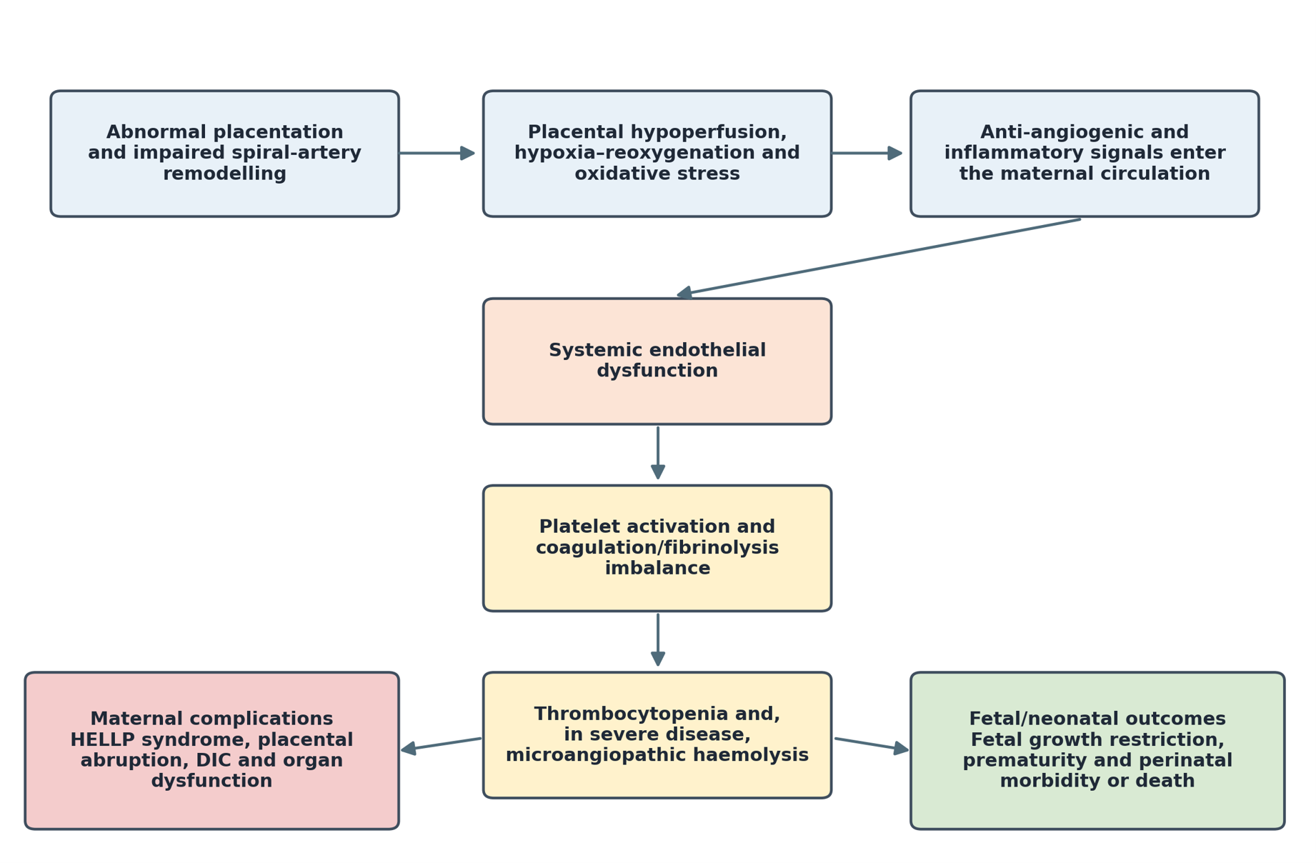
Figure 1. Proposed link between placental dysfunction, endothelial injury, hemostatic abnormalities and maternal–fetal complications. The arrows represent a conceptual pathway and not a validated diagnostic algorithm. Abbreviations: DIC: Disseminated Intravascular Coagulation; HELLP: Hemolysis, Elevated Liver Enzymes and Low Platelet Count.
Clinical Signs
PE may be asymptomatic and detected during blood-pressure or laboratory surveillance. Warning features include severe hypertension, persistent severe headache, visual symptoms, epigastric or right-upper-quadrant pain, dyspnea, reduced urine output and signs of fetal compromise. Seizures suggest eclampsia after alternative causes have been considered [17,18,21,22,52]. Clinical severity should be assessed together with maternal laboratory and fetal findings.
Biological Signs
Laboratory assessment evaluates proteinuria and maternal organ involvement. A full blood count (FBC) with platelet count, serum creatinine and liver enzymes are central. Where hemolysis is suspected, lactate dehydrogenase (LDH), bilirubin, haptoglobin where available and a peripheral blood film are useful. Prothrombin time (PT), international normalized ratio, activated partial thromboplastin time (aPTT) and fibrinogen are particularly relevant when bleeding, placental abruption, severe hepatic dysfunction or disseminated intravascular coagulation is suspected [19–22]. Neither proteinuria ≥5 g/24 h nor an isolated D-dimer value should be used as a stand-alone criterion of severe PE.
Consequences
Consequences for the mother
Maternal complications arise from severe hypertension, endothelial injury, microangiopathy, ischemia and hemostatic consumption. Their frequency in West African series is influenced by referral pathways and access to timely obstetric and critical care [28–37].
HELLP syndrome
HELLP syndrome combines hemolysis, elevated liver enzymes and a low platelet count. Diagnostic thresholds vary, but a platelet count below 100 × 109/L is commonly used for complete HELLP syndrome. Trends in platelets, LDH, bilirubin, aminotransferases, and the blood film are more informative than a single isolated value [33,53,54].
Subcapsular liver hematoma
This rare complication should be considered when severe right-upper-quadrant or epigastric pain accompanies PE or HELLP syndrome. Urgent imaging and multidisciplinary management are required [33,34].
Subcapsular liver hematoma
Capsular rupture is an obstetric emergency associated with intra-abdominal bleeding and hemodynamic instability. It may require massive-transfusion support and surgical or interventional-radiology expertise [33,34].
Hepatic infarction
Hepatic infarction is uncommon and is usually reported with severe PE or HELLP syndrome. Diagnosis relies on compatible symptoms, liver-test abnormalities and imaging [33,34].
Acute kidney injury
Acute kidney injury may accompany severe PE, placental abruption, HELLP syndrome, hemorrhage or pre-existing renal disease. Serial creatinine and urine-output assessment are more useful than proteinuria quantity alone [20,21,35].
Pulmonary edema
Pulmonary edema reflects endothelial permeability, altered oncotic pressure, cardiac dysfunction and sometimes excessive fluid administration. It is a severe feature requiring urgent management [21,33,36].
Abruptio placentae (retroplacental hematoma)
Placental abruption can cause substantial thrombin generation, falling fibrinogen, thrombocytopenia and disseminated intravascular coagulation. Senegalese reports document its prognostic importance, but detailed serial coagulation data remain limited [37,38,55–57].
Consequences for the fetus
Acute fetal distress
Uteroplacental insufficiency may produce acute or chronic fetal compromise. The consequences include fetal growth restriction, indicated preterm birth and fetal death [16,39–41].
Prematurity
Prematurity is birth before 37 completed weeks. In PE, it may follow spontaneous disease or medically indicated delivery for maternal or fetal safety [39–41,55,58–60].
Intrauterine growth restriction (IUGR)
The contemporary term fetal growth restriction (FGR) describes failure to reach growth potential. In PE it usually reflects placental insufficiency and is associated with prematurity, low birth weight and perinatal morbidity [40,41].
Intrauterine fetal death (IUFD)
Severe placental insufficiency, abruption or acute maternal deterioration may lead to fetal death. Definitions vary by gestational-age and birth-weight threshold, which complicates comparison between studies [40,41,58,61].
Consequences for the newborn
Most neonatal consequences are mediated by placental insufficiency, growth restriction and prematurity rather than by PE-specific congenital anomalies [39–41,58].
Long-term consequences
A history of PE is associated with later chronic hypertension, cardiovascular disease, stroke, kidney disease and metabolic risk. Postpartum counselling and long-term primary-care follow-up are therefore important [21,22,61,62]. Children born after pregnancies complicated by PE may also have consequences related to prematurity or growth restriction, but regional longitudinal data are sparse.
|
Domain |
Commonly reported consequences |
|
Growth, respiratory and metabolic |
Fetal growth restriction, low birth weight, prematurity, respiratory morbidity, hypoglycemia and temperature instability. |
|
Hematological, neurological and survival |
Thrombocytopenia, neutropenia or anemia may occur; severe prematurity or fetal compromise can require neonatal care and is associated with perinatal mortality. |
State of Knowledge in West Africa on Pre-eclampsia
Socio-demographic profiles
West African studies frequently report primigravidity, limited antenatal booking and extremes of maternal age among women presenting with PE or eclampsia [30,43,55,56,63–65]. Age findings are not uniform: some hospital-based reports emphasize younger women, whereas the 15-year Maiduguri series reported that 55.2% of eclampsia cases were older than 35 years [65]. A sub-Saharan African meta-analysis found associations with primigravidity and several clinical risk factors, but pooled estimates were heterogeneous [42]. These patterns should not be interpreted causally because referral, parity distribution and case definitions differ across studies.
Main etiologies
Regional studies identify the same broad risk domains reported internationally: chronic hypertension, kidney disease, diabetes, obesity, sickle cell disease, multifetal pregnancy, and previous or family history of PE [42–44,65–73]. However, most data are observational and support association rather than individual causal attribution.
Sickle cell disease and pre-eclampsia
Sickle cell disease is prevalent in parts of West Africa and pregnancy is high risk. International and African syntheses have associated sickle cell disease—particularly severe genotypes—with PE, anemia, thrombosis, preterm birth, and fetal growth restriction [45–49]. The multinational meta-analysis included 21 studies and a very large comparator population, but most women were enrolled in high-income settings; its effect estimates therefore require local validation. A Ghanaian prospective study of 150 women with PE linked hemoglobin variants and oxidative-stress measures with outcomes but lacked a normotensive comparator [51].
Metabolic disorders and pre-eclampsia
Obesity and pre-existing or gestational diabetes have been associated with PE through vascular, inflammatory and metabolic pathways [59,67–69]. African meta-analyses show substantial variation in gestational-diabetes prevalence and diagnostic criteria. Consequently, regional associations should be interpreted with attention to screening practices and confounding rather than presented as proof of a single mechanism.
Hypertension and pre-eclampsia
Chronic hypertension is an established risk factor for superimposed PE and adverse pregnancy outcome [43,44,70,71]. It must be distinguished from the new-onset hypertension that forms part of the PE definition. Studies that describe “hypertension” without separating pre-existing disease from the diagnostic blood-pressure rise of PE risk circular interpretation.
Idiopathic pre-eclampsia
Because PE is a syndrome with multiple pathways, the absence of a recognized risk factor does not establish a distinct idiopathic subtype. Regional studies rarely apply biomarker-defined phenotypes, and unmeasured chronic hypertension, kidney disease or metabolic risk may remain. Future work should use standard early- versus late-onset and maternal–placental phenotypes [21,44,71–73].
Multiple pregnancies and the incidence of pre-eclampsia
Multifetal pregnancy is associated with a higher PE risk, probably in part because of greater placental mass. The association appears in regional and international studies, although West African estimates are imprecise and may be influenced by referral patterns [21,42,63].
Family history of pre-eclampsia
A family or personal history of PE has been associated with recurrence or future disease [42,44,65,72]. This supports a familial component but does not yet identify a clinically useful genetic screening panel for West African populations.
Spontaneous and/ or recurrent miscarriages and pre-eclampsia
Recurrent pregnancy loss and severe placenta-mediated complications may raise clinical suspicion of antiphospholipid syndrome (APS). In Northern Nigeria, Abdullahi et al. found antiphospholipid antibodies (aPL) in 14.1% of 85 women with recurrent fetal loss and 4.7% of 85 controls [74]. This was not a PE cohort, and the finding should not be extrapolated to universal aPL screening in PE. International evidence supports targeted evaluation based on clinical history [75,76].
Hematological Aspects in West Africa (2008–2025)
The most reproducible hematological abnormality in severe PE is a falling platelet count. Anemia may coexist because of nutritional deficiency, hemoglobinopathy, hemorrhage or microangiopathic hemolysis; its mechanism must therefore be established rather than attributed automatically to PE. Nigerian studies have reported differences in platelet count or indices, but results are not consistent across cohorts [50,77,78]. A 2023 international meta-analysis confirmed lower platelet counts in PE on average, while also showing heterogeneity that limits the use of a single threshold as a diagnostic test [79].
HELLP syndrome
Direct West African data describing complete serial hematological profiles in HELLP syndrome are scarce. The international syndrome definition and management literature supports assessment of platelets, blood-film evidence of microangiopathy, LDH, bilirubin, and aminotransferases [33,53,54,80]. A low platelet count alone is not specific, and alternative diagnoses such as gestational thrombocytopenia, immune thrombocytopenia, thrombotic microangiopathy, infection and acute fatty liver of pregnancy may require consideration.
Abruptio placentae (retroplacental hematoma)
Senegalese studies document the clinical burden of placental abruption but provide limited serial information on fibrinogen and D-dimer [37,55,56]. In suspected abruption, platelet count, PT, aPTT, and fibrinogen should be interpreted together with bleeding, hemodynamic status, and trends. Normally early result does not exclude evolving consumption.
Coagulation profiles
Physiological pregnancy is hypercoagulable, while uncomplicated PE may have normal routine coagulation times. Nigerian data from Ekun et al. showed longer PT and aPTT with lower platelet counts in 49 women with PE than in 50 controls [50], but the small cross-sectional design and possible differences in disease severity limit generalization. Routine coagulation screening in every uncomplicated case has uncertain yield; testing is most valuable when disease is severe, bleeding is present, abruption or liver dysfunction is suspected, or an invasive procedure is planned [13,19–22].
The place of antiphospholipid antibodies (aPL) in the occurrence of pre-eclampsia and its complications in West Africa
The laboratory aPL panel comprises lupus anticoagulant, anticardiolipin antibodies, and anti-β2-glycoprotein I antibodies. APS requires compatible clinical features and persistent laboratory positivity; the 2023 American College of Rheumatology/European Alliance of Associations for Rheumatology (ACR/EULAR) criteria are classification criteria and should not replace clinical judgment [76]. Direct West African PE evidence is very limited. The Nigerian recurrent fetal-loss study [74] and international meta-analysis of placenta-mediated complications [75] support further study but not testing every woman with PE. Testing is more defensible when the history suggests APS, such as thrombosis, recurrent pregnancy morbidity or selected severe early placenta-mediated complications, with specialist interpretation and repeat confirmation when required.
Thrombotic disease and pre-eclampsia in West Africa
Pregnancy, PE and immobility can increase venous thromboembolism risk, but this does not establish inherited thrombophilia as a cause of PE [12,14,81–84]. In Nigeria, Okoye et al. found no association between protein C or protein S deficiency and PE in a cross-sectional comparison of 90 cases and 90 controls [85]. In Ghana, Ababio et al. reported an association between factor V Leiden and PE in 81 women [86], but the small exploratory sample and absence of independent regional replication preclude routine population screening. Factor V Leiden, prothrombin G20210A, protein C, protein S and antithrombin testing should be reserved for clinically selected patients after consideration of timing, anticoagulation, and pregnancy-related changes.
Critical synthesis of regional evidence
The studies summarized in Table 4 do not support a single regional hematological signature. Adeyemo et al. and Ekun et al. reported platelet or coagulation differences [50,77], whereas Nwagha et al. found no significant difference in platelet count or indices and inadequate specificity for inflammatory ratios [78]. Okoye et al. did not confirm an association with protein C or protein S deficiency [85], and the factor V Leiden signal reported by Ababio et al. remains unreplicated [86]. The aPL study by Abdullahi et al. addressed recurrent fetal loss rather than PE [74]. These inconsistencies likely reflect small samples, different phenotypes and gestational ages, single time-point measurements, assay variation and incomplete control of confounding.
|
Location/year |
First author [ref.] |
Design and sample |
Main findings |
Key limitations/interpretation |
|
Senegal, 2021 |
Coly MN [56] |
Mixed retrospective/prospective hospital study; n=74 |
Exploratory associations between ABO group and eclampsia or placental abruption; anemia and thrombocytopenia were not associated with ABO group. |
Small single-center sample; mixed design; multiple comparisons; limited adjustment. |
|
Nigeria, 2022 |
Adeyemo MA [77] |
Case–control; n=80 (40 PE, 40 controls) |
Platelet count/indices and erythrocyte sedimentation rate differed between groups; authors proposed usefulness in assessment. |
Small cohort; single center; cross-sectional sampling; diagnostic performance not validated. |
|
Nigeria, 2016 |
Abdullahi ZG [74] |
Analytical cross-sectional; n=170 (85 recurrent fetal loss, 85 controls) |
aPL prevalence was 14.1% in recurrent fetal loss and 4.7% in controls. |
Not a PE cohort; older testing framework; persistence and PE-specific value not established. |
|
Nigeria, 2023 |
Geidam AD [65] |
15-year retrospective review; n=761 eclampsia cases |
Primigravidity and lack of antenatal booking were common; substantial maternal and perinatal morbidity. |
Retrospective records; referral bias; limited detailed hematological-marker data. |
|
Nigeria, 2017 |
Okoye HC [85] |
Cross-sectional comparison; n=180 (90 PE, 90 controls) |
Protein C measures did not differ materially; free protein S was higher in PE; deficiencies were not associated with PE. |
Single time point; physiological pregnancy changes affect protein S; no causal inference. |
|
Nigeria, 2018 |
Ekun OA [50] |
Comparative cross-sectional; n=99 (49 PE, 50 controls) |
Lower platelets and antioxidant markers and longer PT/aPTT were reported in PE. |
Small sample; severity mix and confounding; assays not linked to prospective outcomes. |
|
Ghana, 2019 |
Ababio GK [86] |
Prospective follow-up; n=81 |
Factor V Leiden polymorphism was reported in association with PE and selected outcomes. |
Exploratory small sample; high reported allele frequency requires independent genotyping replication. |
|
Ghana, 2023 |
Abdul G [51] |
Multicenter prospective study; n=150 women with PE |
Hemoglobin variants and oxidative-stress biomarkers were associated with selected adverse outcomes. |
No normotensive comparator; purposive enrolment; associations need external validation. |
|
Nigeria, 2025 |
Nwagha TU [78] |
Comparative cross-sectional; n=90 (45 PE, 45 controls) |
RDW and NLR were higher and PLR lower in PE; platelet count/indices did not differ. NLR AUC was 0.82 but specificity was 22.2%. |
Small sample; cross-sectional; low specificity; authors did not support stand-alone diagnostic use. |
|
Sub-Saharan Africa, 2020 (context) |
Meazaw MW [42] |
Systematic review/meta-analysis; 51 studies, 25,789 women |
Synthesized demographic, obstetric and medical factors associated with PE/eclampsia. |
Mostly observational; high heterogeneity; English-only; only 16 studies entered meta-analysis. |
|
Multinational, 2016 (context) |
Boafor T [49] |
Systematic review/meta-analysis; 21 studies, 26,349 women with sickle cell disease |
Sickle cell disease was associated with increased PE and other adverse pregnancy outcomes. |
Most participants came from high-income settings; genotype and study-quality heterogeneity limit directness. |
|
Abbreviations: ABO: ABO Blood-group System; aPL: Antiphospholipid Antibodies; aPTT: Activated Partial Thromboplastin Time; AUC: Area Under the Receiver-operating Characteristic Curve; NLR: Neutrophil-to-Lymphocyte Ratio; PE: Pre-Eclampsia; PLR: Platelet-to-Lymphocyte Ratio; PT: Prothrombin Time; RDW: Red-Cell Distribution Width |
||||
Accordingly, platelet count and tests directed at hemolysis or consumptive coagulopathy have clear clinical roles, while platelet indices, inflammatory ratios, D-dimer, aPL and inherited-thrombophilia panels remain context-specific or investigational for PE risk stratification. No included West African study demonstrated that routine use of an expanded biomarker panel improves maternal or neonatal outcomes.
Practical laboratory guidance in West African settings
Laboratory requests should answer a clinical question, use serial trends when disease is evolving, and be adapted to local turnaround time. Table 5 separates core assessment from targeted or research tests [21,22,76,78,79].
|
Clinical situation |
Suggested tests |
Established or plausible value |
Important cautions |
|
All women with suspected or diagnosed PE |
FBC with platelet count; creatinine; AST/ALT; proteinuria assessment. |
Detects thrombocytopenia and maternal organ dysfunction; establish a baseline for trends. |
Normal single results do not exclude deterioration. Interpret anemia by cause. |
|
Suspected HELLP syndrome or hemolysis |
Repeat FBC/platelets; blood film; LDH; bilirubin; haptoglobin where available; AST/ALT. |
Assesses platelet consumption and microangiopathic hemolysis. |
No single test is diagnostic. Consider thrombotic microangiopathy and other differentials. |
|
Bleeding, abruption, severe liver disease, shock or planned urgent procedure |
PT/international normalized ratio; aPTT; fibrinogen; FBC; blood group and compatibility testing. |
Assesses evolving consumption and supports transfusion planning. |
Use serial results and clinical bleeding. Early coagulation tests may still be normal. |
|
Possible APS based on thrombosis or qualifying pregnancy morbidity |
Lupus anticoagulant; anticardiolipin IgG/IgM; anti-β2-glycoprotein I IgG/IgM. |
Supports APS evaluation when the clinical history is compatible. |
Not a universal PE screen. Persistent positivity generally requires confirmation ≥12 weeks apart. |
|
Personal venous thromboembolism or strong family history |
Specialist-directed factor V Leiden, prothrombin G20210A, antithrombin, protein C and protein S testing. |
May inform selected thrombosis evaluation. |
Not routine for PE alone; pregnancy, acute thrombosis and anticoagulation affect results. |
|
Exploratory risk stratification |
D-dimer, mean platelet volume and other platelet indices, RDW, NLR and PLR. |
Research signals exist in small studies. |
Insufficient regional validation, specificity or outcome benefit for routine PE diagnosis. |
|
Abbreviations: aPTT: Activated Partial Thromboplastin Time; APS: Antiphospholipid Syndrome; AST: Aspartate Aminotransferase; ALT: Alanine Aminotransferase; FBC: Full Blood Count; HELLP: Hemolysis, Elevated Liver Enzymes and Low Platelet Count; Ig: Immunoglobulin; LDH: Lactate Dehydrogenase; NLR: Neutrophil-to-Lymphocyte Ratio; PE: Pre-Eclampsia; PLR: Platelet-to-Lymphocyte Ratio; PT: Prothrombin Time; RDW: Red-cell Distribution Width |
|||
Conclusion
PE and its complications remain major maternal and perinatal problems in West Africa. Available evidence suggests that platelet count, evidence of hemolysis and coagulation abnormalities are most clinically relevant when disease is severe or complicated. However, the regional literature is dominated by small observational studies and does not establish routine diagnostic or prognostic value for platelet indices, inflammatory ratios, D-dimer, aPL or inherited-thrombophilia panels in all women with PE. A pragmatic approach should prioritize an FBC with platelet count and organ-function tests, then add hemolysis and coagulation studies according to the clinical presentation. Expanded testing should be targeted and interpreted with obstetric and hematology expertise.
Study limitations
This is a narrative review and does not claim the exhaustiveness of a PRISMA-conformant systematic review. Scopus and Web of Science were not searched, and grey-literature retrieval may be incomplete. No formal risk-of-bias instrument or quantitative meta-analysis was applied. Regional studies used heterogeneous PE definitions, laboratory methods, sampling times and outcomes; many were small, single-center and cross-sectional or retrospective. Some contextual evidence came from sub-Saharan African or international studies and may not be directly transferable to West Africa. These limitations reduce certainty and require cautious interpretation.
Perspectives and next steps
Future research should use multicenter prospective designs, standard ISSHP phenotypes, appropriate normotensive and disease-control groups, serial sampling and pre-specified maternal and neonatal outcomes. Diagnostic-accuracy, incremental-value, and cost-effectiveness analyses are needed before new markers enter routine care. A planned doctoral program could evaluate aPL—lupus anticoagulant, anticardiolipin, and anti-β2-glycoprotein I antibodies—together with factor V Leiden and prothrombin G20210A in carefully selected women with severe PE, eclampsia or recurrent pregnancy morbidity. Repeating aPL testing, quality-controlled genotyping, and transparent handling of ancestry, anticoagulation, and gestational age would be essential. The aim should be a validated, affordable clinical strategy rather than a broad unselected panel.
Declarations
Declaration of competing interest
The authors declare that they have no known competing financial interests or personal relationships that could have appeared to influence the work reported in this article.
Funding
This research did not receive any specific grant from funding agencies in the public, commercial, or not-for-profit sectors.
Author contributions
Mame Ngoné Coly: Conceptualization, literature review, Writing – original draft, Writing – review and editing. Macoura Gadji: Supervision, Writing – review and editing. Demba Macalou, Mamadou Wagué Guèye and Sérigne Modou Kane Guèye: Validation, Writing – review and editing. All authors read and approved the final version.
Data availability
No new datasets were generated or analyzed during the current review.
References
2. Cissé CT, Faye Dieme ME, Ngabo D, Mbaye M, Diagne PM, Moreau JC. Indications thérapeutiques et pronostic de l'éclampsie au CHU de Dakar [Therapeutics indications and prognosis of eclampsia at Dakar University Teaching Hospital]. J Gynecol Obstet Biol Reprod (Paris). 2003;32(3 Pt 1):239–45.
3. Diouf AA, Diallo M, Mbaye M, Sarr SD, Faye-Diémé ME, Moreau JC, et al. Profil épidémiologique et prise en charge de l'éclampsie au Sénégal: à propos de 62 cas [Epidemiological profile and management of eclampsia in Senegal: about 62 cases]. Pan Afr Med J. 2013 Nov 6; 16:83. French.
4. Cissé CT, Ewagnignon E, Hojeige A, Diadhiou F. Eclampsie en milieu africain: épidémiologie et pronostic au CHU de Dakar. La Semaine des hôpitaux de Paris. 1997;73(33-34):1062–7.
5. Boulanger H, Flamant M. Avancées récentes dans la compréhension de la physiopathologie de la prééclampsie et conséquences thérapeutiques potentielles [New insights in the pathophysiology of preeclampsia and potential therapeutic implications]. Nephrol Ther. 2007 Dec;3(7):437–48.
6. United States Agency for International Development. Prévention, détection et prise en charge de la pré-éclampsie et de l’éclampsie: guide de prise en charge. Washington, DC: USAID; 2011.
7. Djigandé N. Pré éclampsie et éclampsie dans le centre de sante de référence de Koutiala: Aspects epidemio cliniques thérapeutiques et pronostic (Doctoral dissertation). Bamako: Université des Sciences, des Techniques et des Technologies de Bamako; 2020.
8. Tsatsaris V, Fournier T, Winer N. Physiopathologie de la prééclampsie [Pathophysiology of preeclampsia]. J Gynecol Obstet Biol Reprod (Paris). 2008 Feb;37(1):16–23.
9. Lecarpentier E, Fournier T, Guibourdenche J, Tsatsaris V. Physiopathologie de la pré-éclampsie [Pathophysiology of preeclampsia]. Presse Med. 2016 Jul-Aug;45(7-8 Pt 1):631–7.
10. Perry KG Jr, Martin JN Jr. Abnormal hemostasis and coagulopathy in preeclampsia and eclampsia. Clin Obstet Gynecol. 1992 Jun;35(2):338–50.
11. Roberts JM, Hubel CA. Oxidative stress in preeclampsia. Am J Obstet Gynecol. 2004 May;190(5):1177–8.
12. Verspyck E, Marpeau L. Thrombophilies et pathologies vasculaires placentaires. Revue de la littérature [Thrombophilias and vascular placental pathology. A survey of the literature]. Rev Med Interne. 2005 Feb;26(2):103–8.
13. Ducloy-Bouthors AS. Hémostase et prééclampsie [Clotting disorders and preeclampsia]. Ann Fr Anesth Reanim. 2010 May;29(5):e121–34.
14. Christelle T, Masson L, Vermersch C, Houfflin-Debarge V, Bauters A, Ducloy-Bouthors AS. Hypercoagulabilité et prééclampsie: profil thromboélastométrique. Anesth Réanim. 2015;1:A76.
15. Tesse A, Meziani F, David E, Carusio N, Kremer H, Schneider F, et al. Microparticles from preeclamptic women induce vascular hyporeactivity in vessels from pregnant mice through an overproduction of NO. Am J Physiol Heart Circ Physiol. 2007 Jul;293(1):H520–5.
16. Uszyński M, Uszyński W. Coagulation and fibrinolysis in amniotic fluid: physiology and observations on amniotic fluid embolism, preterm fetal membrane rupture, and pre-eclampsia. Semin Thromb Hemost. 2011 Mar;37(2):165–74.
17. Abass AE, Abdalla R, Omer I, Ahmed S, Khalid A, Elzein H. Evaluation of platelets count and indices in pre-eclampsia compared to normal pregnancies. IOSR J Dent Med Sci. 2016 Jul;1(15):5–8.
18. Lazo-Vega L, Toledo-Jaldin L, Badner A, Barriga-Vera JL, Castro-Monrroy M, Euser AG, et al. ACOG and local diagnostic criteria for hypertensive disorders of pregnancy (HDP) in La Paz-El Alto, Bolivia: A retrospective case-control study. Lancet Reg Health Am. 2022 May; 9:100194.
19. Trably C, Rudigoz RC, Dubernard G, Huissoud C. Les troubles biologiques au cours des états pré-éclamptiques: aspects physiopathologiques et cliniques. Revue francophone des laboratoires. 2010 Apr 1;2010(421):43–50.
20. Winer N, Caroit Y, Esbelin J, Philippe HJ. Prééclampsie: Evaluation du pronostic maternel et fœtal, biologique, clinique, signes de gravité, indication des transferts. J Gynecol Obstet Biol Reprod. 2010 Dec;39:S1–342.
21. Magee LA, Brown MA, Hall DR, Gupte S, Hennessy A, Karumanchi SA, et al. The 2021 International Society for the Study of Hypertension in Pregnancy classification, diagnosis & management recommendations for international practice. Pregnancy Hypertens. 2022 Mar; 27:148–69.
22. National Institute for Health and Care Excellence. Hypertension in pregnancy: diagnosis and management (NG133). London: NICE; 2019. Updated 17 April 2023. Available from: https://www.nice.org.uk/guidance/ng133
23. Lipstein H, Lee CC, Crupi RS. A current concept of eclampsia. Am J Emerg Med. 2003 May;21(3):223–6.
24. Laghzaoui M. Epidémiologie, morbidité, mortalité de la pré éclampsie et de léclampsie. J Gynecol Obstet Biol Reprod. 2010;39(Suppl 2):S1–342.
25. Bedel B, Cartron G, Vayssière C, Parant O. A propos d'un cas d'éclampsie tardive du post-partum [Late post-partum eclampsia: a case report]. Gynecol Obstet Fertil. 2010 Jan;38(1):45–7.
26. Valentin M, Ducarme G, Wernet A, Mantz J, Luton D. Eclampsie retardée: leçons à tirer d'un cas survenu à j11 post-partum [Delayed presentation of eclampsia: lessons from a case diagnosed at Day 11]. Gynecol Obstet Fertil. 2008 Jun;36(6):641–3.
27. Sabiri B, Moussalit A, Salmi S, El Youssoufi S, Miguil M. L'éclampsie du post-partum: épidémiologie et pronostic [Post-partum eclampsia: epidemiology and prognosis]. J Gynecol Obstet Biol Reprod (Paris). 2007 May;36(3):276–80.
28. Nguefack CT, Priso EB, Ekane GH, Tsabze LF, Njamen TN, Kamgaing J, et al. Complications et prise en charge de la prééclampsie sévère et de l’éclampsie à l’hôpital général de Douala. Rev Med Pharm. 2015;5(1):483-90.
29. Sibai BM. Diagnosis and management of gestational hypertension and preeclampsia. Obstet Gynecol. 2003 Jul;102(1):181–92.
30. Diouf AA. Accident vasculaire cérébral hémorragique compliquant une crise d’éclampsie au centre hospitalier national de Pikine (CHNP): à propos d’un cas. JOURNAL DE LA SAGO (Gynécologie–Obstétrique et Santé de la Reproduction). 2020;21(2).
31. Diarra N. Aspects épidémio-cliniques et pronostic materno-foetal de l’éclampsie dans le service de gynécologie-obstétricale du CHU Kati Secteur d’intérêt: gynécologie-obstétricale (Doctoral dissertation). Bamako: Université des Sciences, des Techniques et des Technologies de Bamako; 2020.
32. Otiobanda GF. Pré éclampsie et ses complications: quelle prise en charge en Afrique. Cames. 2013;1:46–50.
33. Gedik E, Yücel N, Sahin T, Koca E, Colak YZ, Togal T. Hemolysis, elevated liver enzymes, and low platelet syndrome: Outcomes for patients admitted to intensive care at a tertiary referral hospital. Hypertens Pregnancy. 2017 Feb;36(1):21–9.
34. Société Française d’Anesthésie et de Réanimation. Réanimation des formes graves de pré-éclampsie: conférence d’experts. Paris: Elsevier; 2000.
35. Diallo T, Amiel I, Lira E, Borie C, Skhiri A, Hilly J, et al. Hématome sous-capsulaire du rein compliquant une prééclampsie sévère: cas clinique et revue de la littérature [Sub-capsular renal hematoma during severe preeclampsia: clinical case and review of the literature]. Ann Fr Anesth Reanim. 2014 Sep-Oct;33(9-10):536–9.
36. Nwobodo EI, Ahmed Y. Maternal mortality associated with eclampsia in Sokoto, Nigeria. Orient Journal of Medicine. 2011;23(1-4):49–53.
37. Biaye B, Gassama O, Gueye MD, Diallo M, Niass A, Cisse M, et al. Epidemiology and Prognosis of Retro placental Hematoma in a Maternity Ward at a Regional Hospital Center in Southern Senegal. Open J Obstet Gynecol. 2019 Feb 1;9(02):149–57.
38. Qin ZJ, Xu Y, Du Y, Chen YL, Sun L, Zheng A. Intrauterine Hematoma in the First Trimester and Pregnancy Complications: A Systematic Review and Meta-Analysis. Front Med (Lausanne). 2022 Jun 17; 9:892146.
39. Niokhor DF, Bintou DF, Lamine T, Yaya K, Demba BI, Abouba B, et al. Evaluation De La Prematurite Superieure Ou Egale A 32 Semaine D’amenorhée A L’hopital Regional De Ziguinchor Au Sud Du Senegal (Afrique De L’ouest). European Scientific Journal, ESJ. 2017;13(36):325.
40. Smiti Y, El Haddad H, El Bouti A, Hniad A, Slaoui A, Kharbach A, et al. Complications materno-fœtale de la pré-éclampsie: étude rétrospective (à propos de 136 cas). PAMJ Clinical Medicine. 2021 Dec 10;7(25).
41. Boiro D, Faye PM, Gueye M, Sow A, Dieng A, Ndongo AA, et al. La pré-éclampsie: quelles complications chez le nouveau-né?. Journal de Pédiatrie et de Puériculture. 2018 Dec 1;31(6):282–6.
42. Meazaw MW, Chojenta C, Muluneh MD, Loxton D. Systematic and meta-analysis of factors associated with preeclampsia and eclampsia in sub-Saharan Africa. PLoS One. 2020 Aug 19;15(8):e0237600.
43. Musa J, Mohammed C, Ocheke A, Kahansim M, Pam V, Daru P. Incidence and risk factors for pre-eclampsia in Jos Nigeria. Afr Health Sci. 2018 Sep;18(3):584–95.
44. Jikamo B, Adefris M, Azale T, Alemu K. Incidence, trends and risk factors of preeclampsia in sub-Saharan Africa: a systematic review and meta-analysis. PAMJ-One Health. 2023 May 2;11(1).
45. Noubiap JJ, Temgoua MN, Tankeu R, Tochie JN, Wonkam A, Bigna JJ. Sickle cell disease, sickle trait and the risk for venous thromboembolism: a systematic review and meta-analysis. Thromb J. 2018 Oct 4;16:27.
46. Mbodji A. Identification des facteurs de risque de complications maternelles et fœtales chez des patientes atteintes de syndrome drépanocytaire majeur: à propos de 198 cas. Mémoire pour l’obtention du diplôme de sage-femme. Université Paris Descartes. 2012 Apr 13.
47. Galiba Atipo Tsiba FO, Itoua C, Ehourossika C, Ngakegni NY, Buambo G, Potokoue Mpia NSB, et al. Pregnancy Outcomes among Patients with Sickle Cell Disease in Brazzaville. Anemia. 2020 Sep 15; 2020:1989134.
48. Togo AY. Le trait drépanocytaire augmente le risque de la pré-éclampsie et aggrave son pronostic: Propos de l’analyse d’une base de données obstétricales du CHU Gabriel Touré (Doctoral dissertation). Bamako: Université des Sciences, des Techniques et des Technologies de Bamako; 2019.
49. Boafor TK, Olayemi E, Galadanci N, Hayfron-Benjamin C, Dei-Adomakoh Y, Segbefia C, et al. Pregnancy outcomes in women with sickle-cell disease in low and high income countries: a systematic review and meta-analysis. BJOG. 2016 Apr;123(5):691–8.
50. Ekun OA, Ogidi NO, Lawal RA, Ogunmuyiwa OA, Umewune MC, Adefolaju FO, et al. Interrelationship Between Markers of Oxidative Stress, Inflammation and Hematological Parameters Among Preeclamptic Nigerian Women. Med Sci Monit Basic Res. 2018 Dec 17; 24:225–31.
51. Abdul G, Osei-Wusu W, Asare GA, Daud S, Opoku S, Tamakloe VCKT, et al. Biomarkers of oxidative stress and its nexus with haemoglobin variants and adverse foeto-maternal outcome among women with preeclampsia in a Ghanaian population: a multi-centre prospective study. PLoS One. 2023;18(3):e0283638.
52. Winer N, Tsasaris V. État des connaissances: prise en charge thérapeutique de la prééclampsie. La revue Sage-femme. 2008 Mar 1;7(1):27–37.
53. Sibai BM. Diagnosis, controversies, and management of the syndrome of hemolysis, elevated liver enzymes, and low platelet count. Obstet Gynecol. 2004 May;103(5 Pt 1):981–91.
54. Haram K, Svendsen E, Abildgaard U. The HELLP syndrome: clinical issues and management. A Review. BMC Pregnancy Childbirth. 2009 Feb 26;9:8.
55. Thiam O, Mbaye M, Diouf AA, Touré FB, Gueye M, Niang M, et al. Aspects épidémiologiques, pronostiques et thérapeutiques de l'hématome retro placentaire (HRP) dans une maternité de référence en zone rurale [Epidemiological, prognostic and therapeutic aspects of retro placental hematoma (HRP) in a referral maternity in rural area]. Pan Afr Med J. 2014 Jan 14; 17:11.
56. Coly MN, Makalou D, Faye CI. Association entre le Groupe Sanguin, le Profil Hématologique et la Survenue de l’Éclampsie et de l’Hématome Rétroplacentaire dans un Groupe de Femmes Sénégalaises. Health Sci Dis. 2021;22(2):80–4.
57. Saquib S, Hamza LK, AlSayed A, Saeed F, Abbas M. Prevalence and its feto-maternal outcome in placental abruption: A retrospective study for 5 years from Dubai Hospital. Dubai Medical Journal. 2020 Apr 16;3(1):26–31.
58. Zounfa SN. La prééclampsie: profil épidémio-clinique et pronostique dans les centres hospitaliers de Ziguinchor. Université Assane Seck de Ziguinchor; 2020.
59. Azeez TA, Abo-Briggs T, Adeyanju AS. A systematic review and meta-analysis of the prevalence and determinants of gestational diabetes mellitus in Nigeria. Indian J Endocrinol Metab. 2021 May-Jun;25(3):182–90.
60. Tyas BD, Lestari P, Aldika Akbar MI. Maternal Perinatal Outcomes Related to Advanced Maternal Age in Preeclampsia Pregnant Women. J Family Reprod Health. 2019 Dec;13(4):191–200.
61. Berkane N. Définitions et conséquences des hypertensions artérielles de la grossesse [Gestational hypertensions: definitions and consequences in outcome of pregnancy]. Ann Fr Anesth Reanim. 2010 Mar;29(3):e1–6.
62. Osoti AO, Page ST, Richardson BA, Guthrie BL, Kinuthia J, Polyak SJ, et al. Postpartum metabolic syndrome and high-sensitivity C-reactive protein after gestational hypertension and pre-eclampsia. Int J Gynaecol Obstet. 2020 Dec;151(3):443–49.
63. Goita S. Aspect Epidemio-Clinique de la pré-éclampsie au centre de santé de Référence de Nioro du Sahel (Doctoral dissertation). Bamako: Université des Sciences, des Techniques et des Technologies de Bamako; 2021.
64. Ahenkorah B, Sakyi S, Helegbe G, Owiredu E, Ofosu W, Der E, et al. Prevalence and risk factors associated with preeclampsia, low birth weight and postpartum hemorrhage in Northern Ghana. Research Square. 2020. doi:10.21203/rs.3.rs-109200/v1.
65. Geidam AD, Atterwahmie A, Usman A, Idrisa A. Prevalence, Risk Factors, Maternal and Perinatal Outcome of Patients with Eclampsia in University of Maiduguri Teaching Hospital, Maiduguri, Nigeria: A 15-Year Retrospective Review. West Afr J Med. 2023 Jan 30;40(1):97–103.
66. Nakimuli A, Chazara O, Byamugisha J, Elliott AM, Kaleebu P, Mirembe F, et al. Pregnancy, parturition and preeclampsia in women of African ancestry. Am J Obstet Gynecol. 2014 Jun;210(6):510–20.e1.
67. Muche AA, Olayemi OO, Gete YK. Prevalence and determinants of gestational diabetes mellitus in Africa based on the updated international diagnostic criteria: a systematic review and meta-analysis. Arch Public Health. 2019 Aug 6; 77:36.
68. Mwanri AW, Kinabo J, Ramaiya K, Feskens EJ. Gestational diabetes mellitus in sub-Saharan Africa: systematic review and metaregression on prevalence and risk factors. Trop Med Int Health. 2015 Aug;20(8):983–1002.
69. Yang Y, Wu N. Gestational Diabetes Mellitus and Preeclampsia: Correlation and Influencing Factors. Front Cardiovasc Med. 2022 Feb 16;9:831297.
70. Thiam M, Goumbala M, Gning SB, Fall PD, Cellier C, Perret JL. Pronostic maternel et foetal de l'association hypertension et grossesse en Afrique sub-saharienne (Sénégal) [Maternal and fetal prognosis of hypertension and pregnancy in Africa (Senegal)]. J Gynecol Obstet Biol Reprod (Paris). 2003 Feb;32(1):35–8.
71. Fikadu K, G/Meskel F, Getahun F, Chufamo N, Misiker D. Determinants of pre-eclampsia among pregnant women attending perinatal care in hospitals of the Omo district, Southern Ethiopia. J Clin Hypertens (Greenwich). 2021 Jan;23(1):153–62.
72. Roberts JM, Cooper DW. Pathogenesis and genetics of pre-eclampsia. Lancet. 2001 Jan 6;357(9249):53–6.
73. Salifou K, Obossou AAA, Vodouhè M, Agbanda LM, Sidi R, Hounkponou F, et al. Factors associated with the occurrence of retroplacental hematoma in the departmental maternity hospital of Borgou. J Preg Neonatal Med. 2019;3(1):8–12.
74. Abdullahi ZG, Abdul MA, Aminu SM, Musa BO, Amadu L, Jibril el-BM. Antiphospholipid antibodies among pregnant women with recurrent fetal wastage in a tertiary hospital in Northern Nigeria. Ann Afr Med. 2016 Jul-Sep;15(3):133–7.
75. Abou-Nassar K, Carrier M, Ramsay T, Rodger MA. The association between antiphospholipid antibodies and placenta mediated complications: a systematic review and meta-analysis. Thromb Res. 2011 Jul;128(1):77–85.
76. Barbhaiya M, Zuily S, Naden R, Hendry A, Manneville F, Amigo MC, et al. The 2023 ACR/EULAR Antiphospholipid Syndrome Classification Criteria. Arthritis Rheumatol. 2023 Oct;75(10):1687–702.
77. Adeyemo MA, Salawu L, Makinde ON, Mabayoje VO. Platelet Indices and Erythrocyte Sedimentation Rate are useful Parameters in the Assessment of a Cohort of Nigerian Women with Preeclampsia. West Afr J Med. 2022 Dec 29;39(12):1273–9.
78. Nwagha TU, Okoye HC, Enebe JT, Izuka EO, Ekwuazi KE, Onyebuchukwu CQ, et al. Assessment of some hematologic markers of Systemic Inflammatory Response (SIR) as eliable biomarkers in detecting preeclampsia; a comparative cross-sectional study in Enugu, Nigeria. Afr Health Sci. 2025 Sep;25(3):51–61.
79. Woldeamanuel GG, Tlaye KG, Wu L, Poon LC, Wang CC. Platelet count in preeclampsia: a systematic review and meta-analysis. Am J Obstet Gynecol MFM. 2023 Jul;5(7):100979.
80. Adeyeye OV, Ebubechukwu NV, Olanrewaju OF, Eniayewun AG, Nwuta C, Effiong FB, et al. Management and Prevention of Pre-Eclampsia in Nigeria. Healthcare (Basel). 2023 Jun 23;11(13):1832.
81. Gilabert J, Fernandez JA, España F, Aznar J, Estelles A. Physiological coagulation inhibitors (protein S, protein C and antithrombin III) in severe preeclamptic states and in users of oral contraceptives. Thromb Res. 1988 Feb 1;49(3):319–29.
82. Pourrat O, Deconinck M, Duchemin J, Brossard A, Desseauve D, Pierre F. Évaluation du bilan thrombophilique héréditaire pratiqué en consultation de médecine interne de la grossesse: étude rétrospective de 336 dossiers. La Revue de Médecine Interne. 2014 Dec 1;35: A61–2.
83. Ezigbo ED, Nwagha TU, Nwuzor VC. Prevalence of venous thromboembolism risk factors in hospitalized patients at the University of Nigeria Teaching Hospital, Enugu Nigeria. Afr Health Sci. 2022 Jun;22(2):736–44.
84. Fall M. Thrombose veineuse cérébrale en tout début de grossesse avec déficit en protéine S: une association assez rarement diagnostiquée en zone tropicale: à propos d’un cas. PAMJ Clinical Medicine. 2016 Apr 27;23(243).
85. Okoye HC, Eweputanna LI, Okpani AO, Ejele OA. Associations between pre-eclampsia and protein C and protein S levels among pregnant Nigerian women. Int J Gynaecol Obstet. 2017 Apr;137(1):26–30.
86. Ababio GK, Adu-Bonsaffoh K, Abindau E, Narh G, Tetteh D, Botchway F, et al. Effects of factor v Leiden polymorphism on the pathogenesis and outcomes of preeclampsia. BMC Med Genet. 2019 Nov 27;20(1):189.