

Abstract
Background: Heart Failure with preserved ejection fraction (HFpEF) accounts for up to half of all heart failure and is increasing in prevalence. Unlike heart failure with reduced ejection fraction (HFrEF), HFpEF lacks a defining systolic abnormality, making diagnosis challenging—particularly when comorbidities mimic or confound the presentation.
Objective: To provide general internists with a practical framework for integrating clinical judgement, laboratory data, and echocardiographic tools to diagnose and manage HFpEF.
Methods: Current guidelines and key literature on HFpEF pathophysiology, diagnostic algorithms, and echocardiographic parameters were synthesized. A hypothetical case illustrates diagnostic and management decision points; all clinical data are fictional.
Conclusion: Earlier recognition of HFpEF—using validated scoring systems, natriuretic peptides, targeted echocardiographic tools, and multidisciplinary collaboration—combined with guideline directed therapy can improve outcomes.
Keywords
HFpEF, Diastolic dysfunction, Global longitudinal strain, Left atrial reservoir strain, Right ventricular systolic pressure, HFA-PEFF, HF2PEF, Echocardiography
|
HYPOTHETICAL CASE - PRESENTATION |
|
Patient Profile A 62-year-old African American male with a history of carpal tunnel syndrome, alcohol use disorder, obesity (BMI 36), hypertension (HTN), type 2 diabetes mellitus (DM), atrial fibrillation (AF), and chronic kidney disease (CKD) stage 3b presents with progressive exertional dyspnea over four months. He reports orthopnea, lower extremity swelling, and a 15-pound weight gain, along with numbness in his feet. |
|
Vital Signs
|
|
Key Exam Findings
|
|
Key Laboratory & Imaging
|
|
Transthoracic Echocardiogram (TTE)
|
|
Current Medications
This case illustrates the diagnostic complexity of HFpEF. The patient has multiple HFpEF risk factors and classical heart failure signs, but additional workup is warranted before attributing everything to HFpEF — discussed in detail in sections below. |
Introduction
HFpEF accounts for approximately half of all heart failure cases, and its prevalence is rising with the aging population [1]. Unlike HFrEF, which is defined by reduced systolic function, HFpEF is diagnosed clinically: patients must have heart failure symptoms (exertional dyspnea, edema, orthopnea), preserved ejection fraction (EF) ≥ 50% and evidence of elevated LV filling pressures via echocardiography, natriuretic peptides, or invasive hemodynamics. The diagnosis is challenging because multiple cardiac and non-cardiac conditions mimic HFpEF, and specific subtypes (e.g., infiltrative disease, cardiotoxicity) require targeted workup where early diagnosis can alter the disease course [1,4]. This review summarizes current diagnostic algorithms and echocardiographic tools to help general internists recognize HFpEF, distinguish it from mimickers, and initiate appropriate management.
Methods
This narrative review synthesizes current guidelines, landmark trials, and key literature on HFpEF pathophysiology, diagnostic algorithms, echocardiographic parameters, and guideline-directed medical therapy. Priority was given to clinical practice guidelines from the AHA/ACC/HFSA, ACC Expert Consensus Decision Pathways, and the American Society of Echocardiography. A hypothetical case is used for educational purposes in a case-based review format. All patient data are fictional. The case is introduced at the beginning of the review and revisited at the diagnosis and management sections.
Pathophysiology
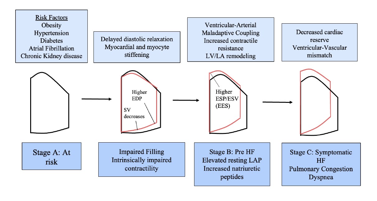
The 2022 AHA/ACC guidelines define four stages of heart failure:
- Stage A (At risk): Risk factors present (obesity, hypertension, diabetes, chronic kidney disease, cardiotoxin exposure) but no symptoms or structural disease.
- Stage B (Pre-Heart Failure): Patients are asymptomatic, but they demonstrate structural abnormalities (e.g., chamber enlargement or hypertrophy, ventricular or atrial strain, wall motion abnormalities), increased filling pressures at rest or with stress, and elevated cardiac biomarkers.
- Stage C (Symptomatic heart failure): Structural disease with current or prior symptoms.
- Stage D (Advanced heart failure): Refractory symptoms despite optimized therapy
The core pathophysiology involves a stiff, poorly relaxing left ventricle that cannot fill adequately at normal pressures [7]. Four interrelated mechanisms drive this process [8,11]:
- Impaired relaxation: Delayed LV relaxation reduces early diastolic filling. End-diastolic pressures increase while left atrial and pulmonary capillary wedge pressures may remain normal. The left atrium compensates with stronger late-diastolic contraction, but this mechanism fails during stress or exercise, leading to underfilling and eventually elevated left atrial pressures with LA remodeling [25]. Mild slowing of LV relaxation occurs with normal aging and should not be equated with HFpEF in asymptomatic patients [8,13,25].
- Myocardial stiffening: Chronic inflammation and fibrosis—driven by diabetes, hypertension, and neurohumoral activation—increase chamber stiffness, raising filling pressures. Patients with smaller left ventricles and low afterload may have a “normal” EF despite intrinsically impaired contractility—a key reason EF alone is insufficient to assess cardiac function.
- Maladaptive ventricular-arterial coupling: Aging and hypertension stiffen the arterial system, increasing ventricular workload. Maladaptive coupling leads to increased contractile resistance, causing the heart in turn to work harder to maintain an adequate cardiac output (Figure 1). Small changes in volume status can produce large blood pressure swings, particularly during exercise.
- Limited cardiac reserve: HFpEF is fundamentally a disorder of exercise intolerance. The heart cannot augment output appropriately during exertion due to chronotropic incompetence and inadequate contractile reserve, accentuated in patients with obesity, who have higher circulating blood volume and greater myocardial demand [24].
Clinical implication for the internist
These mechanisms explain why HFpEF patients can be asymptomatic at rest but develop dyspnea with exertion—and why resting echocardiographic parameters may be normal in early disease.
Diagnostic Approach to HFpEF
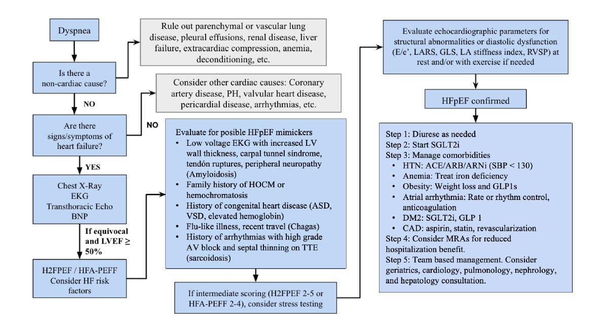
When patients present with heart failure signs and symptoms, providers should exclude cardiac and non-cardiac mimickers of heart failure, employ diagnostic scoring systems, and use echocardiographic imaging to assess structural abnormalities and diastolic dysfunction. Mimickers include parenchymal or vascular lung disease, anemia, neuromuscular disease, renal insufficiency, coronary ischemia, pericardial disease, valvular damage, arrhythmias, infiltrative disorders (e.g. amyloidosis, sarcoidosis), genetic storage disorders (e.g. Fabry disease), and high output heart states.
HFpEF is a clinical diagnosis requiring heart failure symptoms, LVEF ≥ 50%, and at least one of the following [5,8,13]:
- Abnormal diastolic parameters (e.g., E/E’ ≥ 15, which is an echocardiogram estimation of left ventricular filling pressures)
- Structural heart disease which includes LA or LV dilation, LV hypertrophy, and/or moderate to severe valvular disease.
These findings are corroborated by elevated natriuretic peptides (Ambulatory: BNP ≥ 35 or NT-proBNP ≥ 125; hospitalized: BNP ≥ 100 or NT-proBNP ≥ 300) or objective evidence of congestion either at rest or with exercise. Approximately 35% of patients with HFpEF have elevated filling pressures only during exercise, making resting studies insufficient to rule out the diagnosis [19]. A normal resting echocardiogram does not rule out HFpEF; additional stress testing should be considered. Importantly, diastolic dysfunction alone does not equate HFpEF—asymptomatic patients with diastolic dysfunction should be categorized as at risk or pre-heart failure.
BNP and NT-proBNP support the diagnosis and severity of heart failure, but have important limitations in HFpEF [1,29]. Approximately 29% of patients with catheterization-confirmed HFpEF have BNP < 100 pg/mL, and levels are disproportionately lower in patients with obesity [1,5,26]. Conversely, BNP can be elevated in non-heart failure conditions (atrial fibrillation, renal failure, pulmonary disease, etc) [1,19]. A normal BNP does not rule out HFpEF. Prognostically, natriuretic peptides predict hospitalizations and mortality, even in patients with GFR < 60 ml/min/1.73 m [26–28,30].
Diagnostic scoring systems for HFpEF include:
- HF2PEF score: weighted score ranging from 0 to 9 based on clinical variables, such as hypertension, body mass index, atrial fibrillation, pulmonary hypertension, elderly age, and filling pressures. Scores 0–2 indicate low probability, 2–5 intermediate probability requiring exercise testing, and >5 showing high probability for HFpEF [8].
- HFA-PEFF score: Stepwise algorithm in the ambulatory setting, requiring echocardiography and natriuretic peptide scores, functional testing including diastolic stress tests or right heart catheterization, and special imaging, biopsy, or genetic testing. Scores 5 or greater are diagnostic for HFpEF [20].
- Exercise Stress Echocardiography (ESE score) [17]: Incorporation of resting Left Atrial Reservoir Strain (LARS) <20%, exercise septal E/e’ ratio >13, and increase in B lines with exercise as independent predictors of HFpEF. Scoring ranges from 0 to 5, with 1–2 points having 60–80% probability of HFpEF, and ≥3 having near 100% probability of HFpEF.
When scores indicate high probability of HFpEF (>90%), formal filling pressure confirmation is practically not required—all three scoring systems already incorporate filling pressure proxies [20].
Diagnostic echocardiogram parameters for HFpEF include:
1. Left ventricular diastolic dysfunction [8,13]: By ASE criteria, diastolic dysfunction is present when more than half of the following are abnormal:
- Average E/e’ >14
- Septal e’ <7 cm/s or lateral e’ <10 cm/s
- TR velocity >2.8 m/s
- LA volume index >34 ml/m2
Diastolic dysfunction in the absence of symptoms does not equate to HFpEF. Asymptomatic individuals should be categorized into at-risk or pre-heart failure depending on structural or biomarker abnormalities.
2. LV global longitudinal strain [3,8]: GLS measures myocardial fiber shortening during systole, detecting intrinsic systolic impairment despite a normal EF. This is particularly useful in patients with pulmonary congestion where a primary myocardial cause is uncertain. As diastolic dysfunction often progresses into systolic dysfunction, an abnormal GLS (less negative than -16%) in dyspneic patients suggests underlying LV filling abnormalities.
3.Left atrial reservoir strain (LARS) [12]: LARS measures total LA deformation at end-systole via speckle tracking echocardiography and outperforms E/e’, GLS, and LA volume in differentiating HFpEF from noncardiac dyspnea. The 2025 ASE guidelines establish resting LARS <18% as the optimal cutoff for elevated LV filling pressures [8]. Exercise testing can unmask abnormal LA strain in patients with normal resting values [14]. The LA stiffness index (E/e' ÷ LARS) has higher diagnostic utility for HFpEF than either parameter alone [8].
4.Right ventricular systolic pressure (RVSP) [8–10]: RVSP noninvasively estimates pulmonary artery systolic pressures (PASP) via TR jet velocity with a modified Bernoulli equation. In the absence of primary pulmonary or vascular disease, elevated RVSP reflects chronic left-sided filling pressure elevation. RVSP >35 mmHg was present in 83% of HFpEF patients in a community-based study and was higher than preclinical hypertensive heart disease, supporting its prognostic value [10]. RVSP can identify higher risk patients who may benefit from more aggressive therapy [9] and can guide volume management in low-resource settings when right heart catheterization is unavailable.
Practical Guidance for Ordering Echocardiography
- When ordering a TTE for suspected HFpEF, specifically request diastolic function assessment, including E/e’ ratio, LA volume index, TR velocity, and (if available), GLS and LA reservoir strain. These measurements can be technically difficult to obtain, however, and some sonographers may not be skilled enough to capture this data.
- If resting parameters are normal but clinical suspicion remains (H2FPEF score 2–5), consider stress echocardiography to unmask exercise-induced elevation of filling pressures (exercise E/e’ >14–15) (Table 1).
- If diagnosis remains uncertain after noninvasive testing, refer to cardiology for invasive hemodynamic assessment.
|
Parameter |
Description |
Abnormal Cutoff |
Interpretation |
Limitations |
|
E/e′ Ratio |
Ratio of early mitral inflow velocity (E) to tissue Doppler annular velocity (e′). |
Average >14; Septal ≥15; Lateral ≥13 |
Higher values suggest elevated LA filling pressures. |
Inaccurate with mitral annular calcification, prosthetic valves, pericardial disease, or regional wall motion abnormalities. Sensitivity only 34–60% at rest. |
|
e′ Velocity |
Tissue Doppler measure of myocardial relaxation and recoil. |
Septal <7 cm/s; Lateral <10 cm/s |
Reduced e′ indicates impaired LV relaxation — the earliest sign of diastolic dysfunction. |
Age-dependent (decreases with age); affected by regional wall motion abnormalities and annular calcification. |
|
LA Reservoir Strain (LARS) |
Total LA deformation during ventricular systole measured via speckle-tracking echocardiography. |
<18% suggests elevated filling pressures; <23% is abnormal |
Outperforms E/e′ in discriminating HFpEF from noncardiac dyspnea; detects early LA dysfunction. |
Requires speckle-tracking software; sensitivity ~56% (specificity 94%). Normal values can occur in HFpEF. |
|
LV Global Longitudinal Strain (GLS) |
LV myocardial fiber shortening during systole; detects intrinsic systolic impairment despite normal EF. |
Less negative than −16%; borderline −16% to −18% |
Detects subtle systolic impairment despite preserved EF; prognostic value. |
Technically difficult; vendor variability exists. |
|
LA Stiffness Index |
LA compliance ratio: E/e′ ÷ LARS. |
Higher values = stiffer LA |
Better diagnostic performance than LARS alone; correlates with exercise intolerance. |
Combines limitations of both E/e′ and LARS. |
|
Exercise E/e′ |
Filling pressure response to exertion during exercise stress echocardiography (ESE). |
>14–15 during exercise |
Unmasks HFpEF when resting parameters are normal; improves sensitivity to ~90%. |
Technically difficult; requires experienced operator and dedicated protocol. |
|
TR Velocity / RVSP |
Estimates pulmonary artery systolic pressure (PASP) via TR jet and modified Bernoulli equation. |
TR velocity >2.8 m/s; RVSP >35 mmHg |
Elevated values suggest pulmonary hypertension from elevated left-sided pressures. |
Requires adequate TR jet signal; can be elevated from primary pulmonary disease; underestimates in severe TR. |
|
E/e′ and e′ evaluate elevated filling pressures and impaired LV relaxation. Decreased strain pattern suggests high filling pressures and impaired intrinsic contractility. LA stiffness index combines E/e′ and LARS and correlates with exercise intolerance. RVSP suggests chronic pulmonary hypertension from longstanding passive congestion. TR = tricuspid regurgitation; LARS = left atrial reservoir strain; GLS = global longitudinal strain. |
||||
|
CASE APPLICATION — Diagnosis |
|
The patient's H2FPEF score is 94.1%, with symptoms, elevated BNP, structural heart disease (concentric LV hypertrophy, LA dilation), and elevated filling pressures (E/e′ 18, TR jet velocity 3.4 m/s, GLS -12%) confirming HFpEF. However, several findings demand additional workup before attributing the full picture to HFpEF alone. |
|
1. Cardiac Amyloidosis (High Suspicion) Cardiac amyloidosis should be strongly considered given peripheral neuropathy, carpal tunnel syndrome, low-voltage ECG with pseudo-infarct pattern (Q waves in V1-V3), concentric LV hypertrophy, thickened interatrial septum, and GLS with apical sparing. Workup includes Tc-99m pyrophosphate scintigraphy, serum/urine free light chains and immunofixation, and cardiology referral for advanced imaging (cardiac MRI or PET/CT) or biopsy if needed. Genetic testing for hereditary transthyretin amyloidosis should be considered given his clinical profile and African American ancestry [18]. |
|
2. Pulmonary Hypertension Pulmonary Hypertension (RVSP 56 mmHg, TAPSE 14 mm) may reflect Group 2 disease from chronic left-sided congestion, Group 3 disease from likely obstructive sleep apnea, or both. A normal pulmonic valve gradient makes pulmonic stenosis unlikely, localizing the elevated RVSP to left-sided or pulmonary vascular disease. A right heart catheterization is indicated to distinguish pre- vs. post-capillary pulmonary hypertension, which is critical before considering advanced therapies. |
|
3. Liver Disease Elevated transaminases, low albumin, coagulopathy, and thrombocytopenia suggest hepatic dysfunction, from passive congestion, alcohol use, metabolic associated steatohepatitis, or a combination. The AST/ALT ratio >1 raises concern for alcohol-related liver disease or advanced fibrosis/cirrhosis. A serum-ascites albumin gradient (SAAG), ascitic fluid protein, and noninvasive fibrosis assessment should be obtained. Hepatology consultation is warranted. If decompensated cirrhosis and portopulmonary hypertension are confirmed, pulmonary arterial hypertension (PAH) targeted therapies should be initiated with goal of achieving transplant-eligible hemodynamics. Screening for esophageal varices, hepatocellular carcinoma, hepatopulmonary syndrome, and cirrhotic cardiomyopathy are also warranted. Obtain GGT; if normal, carbohydrate-deficient transferrin can screen for chronic alcohol use. |
|
4. Cardiorenal Syndrome Evaluate urine sodium (UNa) and fractional excretion of sodium (FENa) to differentiate prerenal from intrinsic renal or hepatorenal physiology |
|
5. Other Contributors to Dyspnea Bilateral pleural effusions (right > left) may represent hepatic hydrothorax secondary to ascites rather than cardiac congestion alone. AF with rapid ventricular response (RVR), suspected obesity hypoventilation syndrome (OHS), and restrictive lung disease are also possible contributors that should be evaluated in detail. |
Management of HFpEF
Management of HFpEF encompasses diagnostic monitoring, imaging, hemodynamic assessment, and pharmacologic therapy. Current practice guideline recommendations include [1]:
Natriuretic peptides
- Screening BNP for evaluation of dyspnea (Class I), serial BNP for chronic HFpEF risk stratification (Class I), and admission BNP for prognosis in acute decompensation (Class I).
- STOP HF trial showed reduction of LV dysfunction and new onset heart failure with routine BNP screening.
- Pre-discharge BNP for trajectory assessment (Class II, LOE A)
Echocardiography and imaging
- Initial TTE for suspected cardiopulmonary dyspnea (Class I). Repeat echocardiography for clinical change or after initiation of guideline-directed medical therapy (GDMT) (Class I).
- EF reassessment timing: 40 days post-MI, 90 days post-revascularization, 90 days post-GDMT initiation
- Cardiac MRI or CT when echocardiography is inadequate for EF assessment (Class I).
Hemodynamic monitoring
- Right heart catheterization for persistent or worsening symptoms despite medical therapy (Class IIb). Routine pulmonary artery (PA) catheter monitoring has not shown mortality benefit (ESCAPE, GUIDE-HF), though the CHAMPION trial demonstrated a 28% reduction in HF hospitalizations with implantable PA monitoring, although this was a nonblinded study.
- CardioMEMS (Class IIb) may benefit patients with recurrent admissions but requires dedicated infrastructure.
Exercise and functional capacity testing
- Cardiopulmonary Exercise testing and 6-minute walking tests (Class I) are useful to evaluate for candidacy for advanced HF therapies (LVAD, transplantation).
HF with improved ejection fraction
- Continue GDMT indefinitely, even if asymptomatic (Class I). Withdrawal of GDMT in patients whose LVEF had improved to ≥ 50% resulted in relapse of cardiomyopathy in 40% of patients [21].
Additional screening
- Patients at risk for sleep apnea should undergo formal sleep assessment (Class II, LOE A)
- First degree relatives of patients with genetic or inherited cardiomyopathies should undergo genetic screening and counseling
Clinical judgment remains paramount, particularly in resource-limited settings where PA monitoring is not routinely available.
Guideline-Directed Medical Therapy for Established HFpEF
Patients with established HFpEF should undergo workup for underlying etiology (ischemic, infiltrative, toxic, infectious, etc.) while initiating GDMT. The 2023 ACC Expert Consensus Decision Pathway recommends SGLT2 inhibitors for all HFpEF patients without contraindications, with additional consideration of MRAs and ARNi particularly in patients with LVEF at the lower end of the preserved range. The 2025 AHA/ACC hypertension guidelines recommend RAAS inhibitors to target SBP <130 mmHg in HFpEF and advise against beta-blockers for blood pressure management in HFpEF given negative chronotropic effects.
Key Pharmacologic Considerations:
- Diuretics (Class I): OPTIMIZE-HF showed reduced 30-day mortality and HF hospitalization with discharge diuretics. Loop diuretics are preferred, and thiazides can be added for refractory edema.
- SGLT2 inhibitors (Class II, LOE A): A meta-analysis of EMPEROR-Preserved and DELIVER showed a consistent 20% reduction in composite of HF hospitalization and CV death (HR 0.80; 95% CI 0.73-0.87), though the benefit was driven primarily by reduced hospitalizations rather than CV mortality. SGLT2 inhibitors are especially beneficial for diabetic management and CKD [2,22].
- MRAs (Class II, LOE B): The TOPCAT trial showed a modest reduction in HF hospitalizations with spironolactone, though the primary composite endpoint was not met and geographic cohort heterogeneity raised questions about adherence and HFpEF diagnosis [19,37]. FINEARTS-HF showed finerenone reduced composite HF events and CV death, although also driven by hospitalization reduction, with higher hyperkalemia rates versus placebo [35]. Monitor potassium closely, especially with concurrent RAAS inhibitors and/or reduced eGFR [31].
- ARNi (Class II, LOE B): PARAGON-HF did not meet primary endpoint overall, but subgroup analysis showed benefit with LVEF 40-55%. ARBS have not shown benefit with cardiovascular mortality, all-cause mortality, or HF hospitalizations [23].
- Beta Blockers: No HFpEF specific benefit. Consider in patients with history of myocardial infarction and/or atrial fibrillation for rate control [1,2].
- GLP1 receptor agonists: STEP-HFpEF showed meaningful improvements in symptoms, functional capacity, and weight loss in patients with obesity and HFpEF [35,36]. Consider in patients with obesity, diabetes, and CKD.
- Comorbidity Management: HFpEF is a systemic disease with multiple phenotypes (obesity related, ischemic, vascular, fibrotic). Management requires treating underlying contributors, including AF, obesity, hypertension, diabetes, anemia, and sleep apnea, as emphasized in the 2023 ACC Expert Consensus Decision Pathway [2].
- When to refer to cardiology: Equivocal, diagnostic uncertainty, need for stress echocardiography or invasive hemodynamics, evaluation for specific etiologies (amyloidosis, infiltrative disease), or advanced HF therapies. Additionally, because coronary disease is common in HFpEF patients, revascularization can be considered to improve cardiac function and survival [24].
|
CASE APPLICATION – Management |
|
This patient's dyspnea is multifactorial, requiring simultaneous management of HFpEF, liver disease, CKD, and AF — while actively pursuing workup for cardiac amyloidosis. Cases with cardiac amyloidosis should be managed by expert cardiologists at specialized centers. The following medication changes are recommended:
|
Discussion and Conclusion
This case-based review illustrates that HFpEF diagnosis requires integration of clinical context, natriuretic peptides, validated scoring systems, and targeted echocardiographic parameters including GLS, LA reservoir strain, and RVSP. HFpEF frequently coexists with conditions that mimic or compound the presentation—including liver disease, pulmonary hypertension, and infiltrative cardiomyopathy—making systematic evaluation of mimickers essential before attributing the clinical picture to HFpEF alone. For the general internist, the most impactful practice changes are:
- Maintain clinical suspicion for HFpEF even when BNP and resting echo are unremarkable; request diastolic function testing and strain imaging and consider stress echocardiography when resting studies are equivocal.
- Initiate SGLT2 inhibitors early in confirmed HFpEF and address modifiable comorbidities.
- Recognize red flags for specific etiologies—particularly cardiac amyloidosis—that require targeted workup and referral. Early and accurate phenotyping and target therapies can meaningfully change outcomes.
HFpEF is a systemic disease that benefits from multidisciplinary collaboration among internists, cardiologists, hepatologists, nephrologists, and pulmonologists. Early recognition, accurate phenotyping, and timely initiation of guideline-directed therapy can meaningfully improve outcomes.
Conflict of Interest/Disclosures
None.
References
2. Kittleson MM, Panjrath GS, Amancherla K, Davis LL, Deswal A, Dixon DL, et al. 2023 ACC Expert Consensus Decision Pathway on Management of Heart Failure With Preserved Ejection Fraction: A Report of the American College of Cardiology Solution Set Oversight Committee. J Am Coll Cardiol. 2023 May 9;81(18):1835–78.
3. Janwanishstaporn S, Cho JY, Feng S, Brann A, Seo JS, Narezkina A, et al. Prognostic Value of Global Longitudinal Strain in Patients With Heart Failure With Improved Ejection Fraction. JACC Heart Fail. 2022 Jan;10(1):27–37.
4. Shah SJ, Borlaug BA, Kitzman DW, McCulloch AD, Blaxall BC, Agarwal R, et al. Research Priorities for Heart Failure With Preserved Ejection Fraction: National Heart, Lung, and Blood Institute Working Group Summary. Circulation. 2020 Mar 24;141(12):1001–26.
5. Bozkurt B, Coats AJS, Tsutsui H, Abdelhamid CM, Adamopoulos S, Albert N, et al. Universal definition and classification of heart failure: a report of the Heart Failure Society of America, Heart Failure Association of the European Society of Cardiology, Japanese Heart Failure Society and Writing Committee of the Universal Definition of Heart Failure: Endorsed by the Canadian Heart Failure Society, Heart Failure Association of India, Cardiac Society of Australia and New Zealand, and Chinese Heart Failure Association. Eur J Heart Fail. 2021 Mar;23(3):352–80.
6. Bozkurt B. Pre-Heart Failure: An Important Opportunity to Prevent a Deadly Disease. JACC Heart Fail. 2023 Aug;11(8 Pt 1):1027–31.
7. Zile MR, Baicu CF, Gaasch WH. Diastolic heart failure--abnormalities in active relaxation and passive stiffness of the left ventricle. N Engl J Med. 2004 May 6;350(19):1953–9.
8. Nagueh SF, Sanborn DY, Oh JK, Anderson B, Billick K, Derumeaux G, et al. Recommendations for the Evaluation of Left Ventricular Diastolic Function by Echocardiography and for Heart Failure With Preserved Ejection Fraction Diagnosis: An Update From the American Society of Echocardiography. J Am Soc Echocardiogr. 2025 Jul;38(7):537–69.
9. Kotrri G, Youngson E, Fine NM, Howlett JG, Lyons K, Paterson DI, et al. Right Ventricular Systolic Pressure Trajectory as a Predictor of Hospitalization and Mortality in Patients With Chronic Heart Failure. CJC Open. 2023 Jun 3;5(9):671–9.
10. Lam CS, Roger VL, Rodeheffer RJ, Borlaug BA, Enders FT, Redfield MM. Pulmonary hypertension in heart failure with preserved ejection fraction: a community-based study. J Am Coll Cardiol. 2009 Mar 31;53(13):1119–26.
11. Sharma K, Kass DA. Heart failure with preserved ejection fraction: mechanisms, clinical features, and therapies. Circ Res. 2014 Jun 20;115(1):79–96.
12. Reddy YNV, Obokata M, Egbe A, Yang JH, Pislaru S, Lin G, et al. Left atrial strain and compliance in the diagnostic evaluation of heart failure with preserved ejection fraction. Eur J Heart Fail. 2019 Jul;21(7):891–900.
13. Mitter SS, Shah SJ, Thomas JD. A Test in Context: E/A and E/e' to Assess Diastolic Dysfunction and LV Filling Pressure. J Am Coll Cardiol. 2017 Mar 21;69(11):1451–64.
14. Thomas L, Marwick TH, Popescu BA, Donal E, Badano LP. Left Atrial Structure and Function, and Left Ventricular Diastolic Dysfunction: JACC State-of-the-Art Review. J Am Coll Cardiol. 2019 Apr 23;73(15):1961–77.
15. Cannata A, McDonagh TA. Heart Failure with Preserved Ejection Fraction. N Engl J Med. 2025 Jan 9;392(2):173–84.
16. Campbell P, Rutten FH, Lee MM, Hawkins NM, Petrie MC. Heart failure with preserved ejection fraction: everything the clinician needs to know. Lancet. 2024 Mar 16;403(10431):1083–92.
17. Kagami K, Harada T, Yuasa N, Tani Y, Murakami F, Saito Y, et al. A scoring system for diagnosing heart failure with preserved ejection fraction based on exercise echocardiography. Eur Heart J Cardiovasc Imaging. 2025 Apr 30;26(5):866–75.
18. Kittleson MM, Maurer MS, Ambardekar AV, Bullock-Palmer RP, Chang PP, Eisen HJ, et al. Cardiac Amyloidosis: Evolving Diagnosis and Management: A Scientific Statement From the American Heart Association. Circulation. 2020 Jul 7;142(1):e7–22.
19. Redfield MM, Borlaug BA. Heart Failure With Preserved Ejection Fraction: A Review. JAMA. 2023 Mar 14;329(10):827–38.
20. Egashira K, Sueta D, Komorita T, Yamamoto E, Usuku H, Tokitsu T, et al. HFA-PEFF scores: prognostic value in heart failure with preserved left ventricular ejection fraction. Korean J Intern Med. 2022 Jan;37(1):96–108.
21. Halliday BP, Wassall R, Lota AS, Khalique Z, Gregson J, Newsome S, et al. Withdrawal of pharmacological treatment for heart failure in patients with recovered dilated cardiomyopathy (TRED-HF): an open-label, pilot, randomised trial. Lancet. 2019 Jan 5;393(10166):61–73.
22. Vaduganathan M, Docherty KF, Claggett BL, Jhund PS, de Boer RA, Hernandez AF, et al. SGLT-2 inhibitors in patients with heart failure: a comprehensive meta-analysis of five randomised controlled trials. Lancet. 2022 Sep 3;400(10354):757–67.
23. Martin N, Manoharan K, Thomas J, Davies C, Lumbers RT. Beta-blockers and inhibitors of the renin-angiotensin aldosterone system for chronic heart failure with preserved ejection fraction. Cochrane Database Syst Rev. 2018 Jun 28;6(6):CD012721.
24. Borlaug BA, Sharma K, Shah SJ, Ho JE. Heart Failure With Preserved Ejection Fraction: JACC Scientific Statement. J Am Coll Cardiol. 2023 May 9;81(18):1810–34.
25. Sagmeister P, Rosch S, Fengler K, Kresoja KP, Gori T, Thiele H, et al. Running on empty: Factors underpinning impaired cardiac output reserve in heart failure with preserved ejection fraction. Exp Physiol. 2025 May;110(5):694–707.
26. Anjan VY, Loftus TM, Burke MA, Akhter N, Fonarow GC, Gheorghiade M, et al. Prevalence, clinical phenotype, and outcomes associated with normal B-type natriuretic peptide levels in heart failure with preserved ejection fraction. Am J Cardiol. 2012 Sep 15;110(6):870–6.
27. Santaguida PL, Don-Wauchope AC, Ali U, Oremus M, Brown JA, Bustamam A, et al. Incremental value of natriuretic peptide measurement in acute decompensated heart failure (ADHF): a systematic review. Heart Fail Rev. 2014 Aug;19(4):507–19.
28. Chen LJ, Hung CL, Yeh HI, Jeng MJ, Su CH, Wu TY, et al. The utilization and prognostic impact of B-type Natriuretic Peptide in hospitalized acute decompensated heart failure in an Asian population. BMC Cardiovasc Disord. 2016 Sep 9;16(1):178.
29. Linssen GCM, Jaarsma T, Hillege HL, Voors AA, van Veldhuisen DJ. A comparison of the prognostic value of BNP versus NT-proBNP after hospitalisation for heart failure. Neth Heart J. 2018 Oct;26(10):486–92.
30. Anwaruddin S, Lloyd-Jones DM, Baggish A, Chen A, Krauser D, Tung R, et al. Renal function, congestive heart failure, and amino-terminal pro-brain natriuretic peptide measurement: results from the ProBNP Investigation of Dyspnea in the Emergency Department (PRIDE) Study. J Am Coll Cardiol. 2006 Jan 3;47(1):91–7.
31. Zannad F, McMurray JJ, Krum H, van Veldhuisen DJ, Swedberg K, Shi H, et al. Eplerenone in patients with systolic heart failure and mild symptoms. N Engl J Med. 2011 Jan 6;364(1):11–21.
32. Pitt B, Zannad F, Remme WJ, Cody R, Castaigne A, Perez A, et al. The effect of spironolactone on morbidity and mortality in patients with severe heart failure. Randomized Aldactone Evaluation Study Investigators. N Engl J Med. 1999 Sep 2;341(10):709–17.
33. Pitt B, Pfeffer MA, Assmann SF, Boineau R, Anand IS, Claggett B, et al. Spironolactone for heart failure with preserved ejection fraction. N Engl J Med. 2014 Apr 10;370(15):1383–92.
34. Solomon SD, McMurray JJV, Vaduganathan M, Claggett B, Jhund PS, Desai AS, et al. Finerenone in Heart Failure with Mildly Reduced or Preserved Ejection Fraction. N Engl J Med. 2024 Oct 24;391(16):1475–85.
35. Kosiborod MN, Abildstrøm SZ, Borlaug BA, Butler J, Rasmussen S, Davies M, et al. Semaglutide in Patients with Heart Failure with Preserved Ejection Fraction and Obesity. N Engl J Med. 2023 Sep 21;389(12):1069–84.
36. Thomas J, Dagan M, Wang B, Gutman S, Kaye DM. Mechanisms of GLP-1 Receptor Agonists in HFpEF: Exploring Weight-Dependent and Independent Drivers of Therapeutic Benefit. Circ Heart Fail. 2026 May;19(5):e013279.
37. Anand IS, Claggett B, Liu J, Shah AM, Rector TS, Shah SJ, et al. Interaction Between Spironolactone and Natriuretic Peptides in Patients With Heart Failure and Preserved Ejection Fraction: From the TOPCAT Trial. JACC Heart Fail. 2017 Apr;5(4):241–52.