

Abstract
Background: Increased CD8+ T cells in the tumor has shown improved response to therapy and prognosis in breast cancer. Cytotoxic chemotherapies increase immune recognition of the tumor but the effect of chemotherapy on the tumor immune environment is a balance between immune activation and systemic immune suppression. The rexinoic acid receptor agonist bexarotene previously was shown to increase intratumoral CD8+ T cells in TgMMTV-neu transgenic mouse tumors. We therefore determined whether bexarotene could enhance the anti-tumor efficacy of common breast cancer chemotherapies and whether the immune role of bexarotene was contributing to the anti-tumor benefit.
Methods: In two immune competent mouse mammary tumor models, TgMMTV-neu and C3(1)Tag mice, spontaneous tumors were treated with bexarotene and chemotherapy (doxorubicin and paclitaxel in both models, cyclophosphamide in TgMMTV-neu mice, and cisplatin in C3(1)Tag mice) and the tumors were evaluated for CD8 and CD4 T cells. Immune cell populations that express the RXRα receptor were evaluated in human peripheral blood mononuclear cells (PBMC) and the direct role of bexarotene on human dendritic cell populations was evaluated by flow cytometry.
Results: Bexarotene enhanced tumor growth inhibition with all chemotherapies in TgMMTV-neu mice, decreasing growth by 98.1% with doxorubicin, 88% with cyclophosphamide, and 89% with paclitaxel. In C3(1)Tag mice bexarotene and cisplatin decreased tumor growth by 99%. Bexarotene doubled the incidence of CD8+ T cells in the tumors of TgMMTV-neu mice and 5.7 fold increase in C3(1)Tag mice. RXRα receptors were expressed highest in human monocytic (mDC, 33.1±5.7%) and plasmocytic dendritic cells (pDCs 38.6±4.8%), and in 25±4.4% macrophages. Bexarotene activated type I mDCs and pDCs that released IL-1β and TNFα type I cytokines.
Conclusions: Bexarotene enhances the anti-tumor efficacy of cytotoxic chemotherapy in transgenic mouse mammary tumor models and increases intratumoral CD8+ T cells. Bexarotene directly activates type I mDCs and pDCs.
Keywords
Bexarotene, Dendritic cells, Breast cancer, Macrophages
Background
As a whole, breast tumors have fewer immune cells infiltrating the tumor than more immune active cancers including melanoma and lung cancer [1]. In breast cancer tumor infiltrating lymphocytes (TILs) analysis, the immune infiltrate is found mostly in the stroma surrounding the tumor rather than infiltrating the tumor. In the aggressive triple negative breast cancer subtype, each decile increase in percent stromal TILs prior to therapy is associated with improved overall and disease free survival [2]. However, even in the triple negative subtype only 20% of tumors are immune high (>50% TILs) while the majority have low TIL infiltrate [3]. Therapies are needed to increase immune infiltrate in most breast cancers to get similar prognostic benefit. Chemotherapy, which is cytotoxic to CD8+ T cells, can have multiple immunostimulatory roles that impact the tumor immune environment. These include decreasing regulatory T cells and regulatory cytokines, increasing immunogenic cell death, increasing immune recognition of the tumor, and activating anti-tumor innate immune stress responses [4,5]. Many chemotherapies have been shown to enhance the anti-tumor immune response and impact the tumor immune environment. Three of the chemotherapies used predominantly in breast cancer therapy have demonstrated improved immune function. Doxorubicin has been shown to deplete myeloid derived suppressor cells (MDSC) and enhance CD8 T cell infiltration in mice and increases T cell cytotoxicity and inflammatory genes in triple negative breast cancer patients [6–8]. Paclitaxel has been shown to shift macrophage profiles to M1 macrophages through TLR4 signaling in a triple negative mouse model [9]. Cyclophosphamide has been shown to increase regulatory T cell apoptosis in mouse models of breast cancer, but the immune function differs between low and high doses of cyclophosphamide [10–12]. These immune functions all have to be considered with the balance of systemic immune suppression from the cytotoxic effect of chemotherapy therefore finding additional therapies that can enhance anti-tumor immunity may improve the immune stimulatory properties and eventual clinical efficacy of chemotherapy.
The rexinoid family are vitamin A derivatives and a nuclear receptor family that homo- or heterodimerize on retinoic acid responsive elements as ligand-dependent transcription factors to regulate multiple cancer signaling pathways [13,14]. Rexinoids have been associated with multiple cell functions including proliferation, differentiation, modifying the immune system, and apoptosis [15,16]. Bexarotene is a synthetic rexinoic acid receptor agonist that has an FDA approved indication in cutaneous T cell lymphoma therapy [17]. Furthermore, it has efficacy in breast cancer, in 100 metastatic hormone receptor positive patients that were resistant to hormone therapy there was a clinical response in 20% of patients receiving bexarotene as monotherapy but the majority of responses were stable disease [18]. In mouse models, bexarotene was shown to have a role in decreasing proliferation by decreasing cyclin D1 activity through inducing the transcriptional repressor DEC2 [19]. Bexarotene was also shown to have an immune role, enhancing the efficacy of a HER2- IGFBP2-IGF1R peptide vaccine by preventing invasive cancer in 87% of mice as compared to 67% of mice with vaccine alone (HR 18.1 95% CI 6.4 to 63.2, p<0.0001). This was associated with an increased intratumoral CD8+ T cell infiltrate in the tumors that developed in mice treated with bexarotene alone as compared to adjuvant controls (P=0.015) and in mice treated with bexarotene and the HER2-IGFBP2-IGF1R vaccine as compared the vaccine alone (P=0.0014) [21]. Similarly, the rexinoid receptor agonist MSU-42001 has been shown to modulate CD8+ T cell expression and reduce tumor burden in a neurofibromatosis mouse cancer model with or without a MEK inhibitor [20]. We therefore wanted to determine if the addition of bexarotene increased intratumoral CD8+ T cells as compared to chemo alone.
Rexinoids have been previously shown to modulate the tumor immune environment, particularly impacting the innate immune response, in mouse mammary tumor models. The rexinoic acid receptor LG100268 showed to decrease MDSC and M2 (CD206 positive) macrophages in the MMTV-PyMT triple negative transgenic mouse mammary tumor model thus increasing the CD8/CD4 ratio in the tumor [22]. Activating antigen presenting cells (APCs) and the innate immune environment can induce a more anti-tumor immune environment allowing for more CD8+ T cell infiltration and enhancing the function of chemotherapy. This study was to evaluate whether bexarotene enhances the efficacy of chemotherapy and determine how bexarotene directly impacts the immune response in human immune cells.
Methods
Maintenance of the TgMMTV-neu mice
TgMMTV-neu mice were obtained from Jackson Laboratories and maintained under strict inbreeding conditions and the colony was re-tested for expression of the neu transgene annually. All mice were bred and maintained under specific pathogen free conditions based on the University of Washington institutional animal care and use committee (IACUC) protocol.
Tumor evaluation in the transgenic mice
Mice were enrolled into the study upon development of measurable, spontaneous, subcutaneous breast tumors. Mice were randomly allocated to treatment groups. Therapy started when the tumor reached ~100 mm3 and was continued for four weeks unless the tumors were ulcerated or tumor volume exceeded 10% of overall mouse body weight. Sample size was calculated as 5 per group based on the power calculation of mean difference of 2 with 2 sided α=0.05 with power of 80%. Mice were examined for tumor development, and tumor growth was measured three times per week from six weeks of age until sacrifice. Tumor volumes were calculated from raw measurements by [(length) x (width) x (depth) x (π/6)] and are presented as an average of the three weekly measurements. Multiple tumors were tracked independently. Tumor growth rates were calculated by determining the change in sum of tumor volumes between subsequent measurements.
Dosing and delivery of bexarotene and chemotherapy
The chemotherapies were given intravenously through the tail vein weekly. Doxorubicin was dosed at 5 mg/kg, cyclophosphamide was dosed at 100 mg/kg, and Paclitaxel was dosed at 10 mg/kg infused weekly. Control mice had 50 μL PBS infused weekly. Bexarotene was dissolved in sesame oil and given 5 days a week by oral gavage at 50 mg/kg and control mice were given 100 μL of sesame oil 5 days per week by oral gavage.
Evaluation of the tumor immune infiltrates
At sacrifice, half of the largest measurable tumor was processed for flow cytometry. For flow cytometry evaluation, immune cells were isolated from the spleen through a 70 μm cell strainer and in the tumor immune cells were enriched through FICOL gradient. Fresh cells were stained with a live/dead Pacific Blue fixable viability dye and PE CD3, PE Cy-5 CD4, and PE Cy-7 CD8. All antibodies were from ebioscience. These cells were analyzed by flow cytometry on a FACSCanto flow cytometer. Flow analysis was blinded to treatment arm. Flow cytometry gating strategy was shown in Supplemental Figure 5.
RXR expression in human PBMC
Human peripheral blood mononuclear cells (PBMC) from women without breast cancer (n=15) were used from the University of Washington Cancer Vaccine Institute repository (University of Washington Human Subject Division STUDY0001570). 2×10⁶ cells were stained with the surface monoclonal antibodies and prepared for the intracellular RXR stains using the FOXP3 intracellular stain kit (eBioscience), fixed with 1.6% paraformaldehyde, measured on a BD FacsCANTO II flow cytometer, and evaluated on FlowJo software. RXRα expression was measured using a FITC RXRα monoclonal antibody (Millipore) in immune cell populations PE Cy7 CD3+ PECy5 CD8+ T-cells, PECy7 CD3+ PE CD4+ T-cells, PE CD56+ NK T-cells, PECy7 CD20+ B-cells, PECy5 CD11c+ APC HLADR+ PECy7 LIN- monocytic dendritic cells, PE CD123+ APC HLADR+ PECy7 LIN- plasmocytic dendritic cells, PE CD14+ APC HLADR+ macrophages, PE CD11b+ PECy7 LIN- APC HLADR- monocytic MDSC, and APC HLADR- PE CD14+ PECy5 CD33+ PE CD11b+ plasmocytic myeloid derived dendritic cells. All flow cytometry antibodies were from ebioscience (ThermoFisher Scientific). Live cell populations were selected using a Pacific Blue fixable viability dye (ebioscience). Fluorescence minus one controls were used in panel development. Flow cytometry gating strategy was shown in Supplemental Figure 6.
Activation of antigen presenting cells by bexarotene
PBMC from women without breast cancer (n=8) were used from the University of Washington Cancer Vaccine Institute repository. 1.5´106 human PBMC per sample were plated in 48 well tissue culture plates with 0, 5, 10, and 20 µM of bexarotene diluted in RPMI T-cell media. As bexarotene needed 10% DMSO to go into solution, 0 µM is RPMI with 10% DMSO. The bexarotene stock was 10 mM diluted into DMSO. The cells were incubated at 37°C for 48 hours and then collected, washed, and stained for FITC CD40 and PE CD11c+ APC HLADR+ PECy7 LIN- monocytic dendritic cells, PE CD123+ APC HLADR+ PECy7 LIN- plasmocytic dendritic cells, and PE CD14+ APC HLADR+ macrophages. All antibodies were from ebioscience (ThermoFisher Scientific). Fluorescence minus one controls were used in panel development. The cells were stained with antibodies, fixed with 1.6% paraformaldehyde, measured on a BD Facs CANTO II flow cytometer, and evaluated on FlowJo software. Flow cytometry gating strategy was shown in Supplemental Figure 7.
Evaluation of cytokine release by bexarotene
Supernatant from the above 48 hour experiments were collected and immediately frozen at -80°C. Ten cytokines were evaluated by a multiplex magnetic fluorescent bead assay (Invitrogen, Carlsbad CA). The assay was read on a LiquidChip instrument (Qiagen, Valencia CA).
Evaluation of bexarotene in human dendritic cell maturation
Three independent human PBMC donors were evaluated. 100´106 cells were thawed in Iscove’s modified Dulbecco’s media (IMDM) supplemented with 10% FBS and selected for CD14+ monocytes using autoMACS (Miltenyi Biosciences, CA). The cells were then suspended in media that contained IL4 (Peprotech, 20 ng/mL) and GM-CSF (Peprotech, 50 ng/mL) and stored at 37°C at 5% CO2 with media changed every 2 days. On day 6 immature dendritic cells were harvested and counted, and half the cells were maintained as immature dendritic cells with continued IL-4 and GM-CSF supplemented media until day 8. The other half of the cells were matured using IL-1β (Peprotech, 25 ng/mL), TNF-α (Peprotech, 50 ng/mL), IFN-α (Peprotech, 3X10³ IU/mL), IFN-γ (Peprotech, 100 ng/mL), Poly I:C (Peprotech, 20 µg/mL), IL-2 (Peprotech, 5 ng/mL), and IL-7 (Peprotech, 10 ng/mL) [23,24]. All cells were harvested and evaluated at day 8 using the Cytoflex flow cytometer (Beckman Coulter). In flow cytometry, all cells were stained for far blue live/dead, CD56 BV510, HLA-DR BV605, CD123 BV650, CD16 BV785, CD33 PE-CF594, CD15 PerCP-Cy5.5, CD11c PE-Cy7, CD14 Alexa700, CD3 APC-H7, CD19 APC-H7, CD20 APC-H7, CD11b BV421, and CD40 PE (all antibodies BD Biosciences). Fluorescence minus one controls were used in panel development. Flow cytometry was run on the BD Fortessa and data was analyzed in FlowJo 10. Flow cytometry gating strategy was shown in Supplemental Figure 7.
Statistical analysis
Graphs, t-tests, and ANOVA comparisons were completed using GraphPad Prism v5.03 software. A One-Way repeated measures ANOVA with Tukey’s post-test was used for comparisons of tumor growth differences in mice and a two-Way ANOVA with Bonferroni’s post-test was used for grouped comparisons. Significance was considered at p<0.05 for all statistical tests.
Results
Bexarotene enhances the anti-tumor effect with chemotherapy in two mouse mammary tumor models
In TgMMTV-neu mice with spontaneous established tumors ~100 mm3 (n=5), the addition of bexarotene to doxorubicin, cyclophosphamide, or paclitaxel reduced tumors significantly more than the chemotherapies alone. Treating the mice with bexarotene for four weeks reduced tumor size by 65.9% compared to PBS treated mice (p=0.003, mean of bexarotene treated tumors were 149.6±41.8 mm3 as compared to 438.3±86.2 mm3 in PBS treated tumors). Doxorubicin reduced tumor size by 81.7% compared to PBS treated mice (p=0.0005, mean of doxorubicin treated tumors were 80.11±24.0 mm3 compared to PBS) while mice treated with bexarotene and doxorubicin reduced tumor size by 98.1% (p<0.0001 compared to PBS and p=0.02 compared to doxorubicin therapy alone, tumors were 8.2±2.4 mm3, Figure 1A). While cyclophosphamide did not decrease tumor size as compared to PBS controls (p=0.9), cyclophosphamide and bexarotene decreased tumor growth by 87.7% (p=0.009 compared to PBS and p=0.02 compared to cyclophosphamide, mean tumor size was 59.3±17.8 mm3, Figure 1B). Paclitaxel decreased TgMMTV-neu tumor growth by 57% as compared to PBS controls (p=0.02, mean size of tumor 176.1±28.2 mm3) and bexarotene and paclitaxel further decreased tumor growth by 88.5% (p=0.02, mean size of tumor 50.4±5.6 mm3, Figure 1C). Individual mouse growth curves for all treatment groups were shown in Supplemental Figure 3.
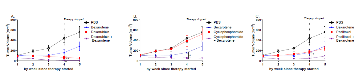
Figure 1. Decreased tumor growth in mice that received chemo and concurrent bexarotene in TgMMTV-neu mice. TgMMTV-neu female mice (n=5 mice/group A-C) were treated with weekly chemotherapy (red), daily bexarotene (blue), chemotherapy and bexarotene (purple), or control (PBS, black) once the mice had spontaneously developed 100 mm3 palpable breast tumors for 4 weeks. A shows doxorubicin chemotherapy, B shows cyclophosphamide chemotherapy, and C shows paclitaxel chemotherapy in TgMMTV-neu mice. * p< 0.05 ** p<0.01.
In C3(1) Tag mice with spontaneous tumors ~50 mm3, treatment with four weeks of bexarotene could inhibit tumor growth by 73.4% (p=0.02, mean size of PBS treated tumors is 912.4±200.1 mm3 and mean size of bexarotene treated tumors is 243.0±32.7 mm3, Supplemental Figure1). However, Bexarotene only enhanced tumor inhibition with cisplatin, cisplatin decreased tumor growth by 86.2% (mean tumor with cisplatin was 125.7±84.20 mm3) while cisplatin and bexarotene decreased tumor growth by 98.6% (p=0.02, mean tumor with bexarotene and cisplatin was 12.20±6.0 mm3) (Supplemental Figure 1B). Bexarotene, doxorubicin, and bexarotene with doxorubicin had similar tumor inhibition but bexarotene did not enhance tumor inhibition of doxorubicin (Supplemental Figure 1A). Paclitaxel and bexarotene did not further inhibit tumor growth as compared to bexarotene alone (Supplemental Figure 1C). Individual mouse growth curves for all treatment groups were shown in Supplemental Figure 4. As bexarotene function differed between chemotherapies and mouse models, we suspected that the role of bexarotene was beyond its impact on cell proliferation [19] and therefore wanted to evaluate whether the immune function of bexarotene may be contributing to the tumor inhibition.
Bexarotene increases CD8 T cells and decreases CD4 T cells in the tumors
To evaluate the impact of bexarotene on the tumor immune environment, we evaluated T cells in the tumor at 4 weeks of therapy by flow cytometry. In TgMMTV-neu mice, there was a significant increase in CD3+ CD8+ and a decrease in CD3+CD4+ populations in the tumor of mice treated with 4 weeks of bexarotene or any of the chemo as compared to PBS or oil controls. However, bexarotene with the chemotherapies did not either increase CD8+ T cells or decrease CD4+ T cells as compared to chemotherapy or bexarotene alone (Figures 2A–2F). Treatment with bexarotene doubled CD3+CD8+ T cells as compared to PBS (p=0.02 mean CD3+CD8+ T cells with PBS 21.0±6.4% and mean CD3+CD8+ T cells with bexarotene 52.2±2.4%). However, while treatment with doxorubicin increased CD3+CD8+ T cells as compared to PBS (mean doxorubicin treated CD3+CD8+ T cells 69.7%±0.1 (p=0.01), treatment with bexarotene and doxorubicin had significantly less CD8+ T cells than doxorubicin alone (28.8±4.3% CD3+CD8+ T cells, p=0.03, Figure 2A) despite improved tumor inhibition. Treatment with cyclophosphamide (CD3+CD8+ T cells 47.3±4.6%, p=0.03) and cyclophosphamide and bexarotene (CD3+CD8+ T cells 35.7±9.4%, p=0.04) both increased CD8+ T cells in the tumor but addition of bexarotene did not have more CD8+ T cells compared to cyclophosphamide alone (Figure 2B). Treating TgMMTV-neu mice with paclitaxel or paclitaxel and bexarotene increased CD3+CD8+ T cells but there was no increase with paclitaxel plus bexarotene as compared to paclitaxel alone (52.4±4.3% CD3+CD8+ T cells with paclitaxel, p=0.005 and 49.7±4.3% CD3+CD8+ T cells with paclitaxel and bexarotene, p=0.01, Figure 2C). Similarly, both bexarotene and chemotherapy increased CD3+CD8+ T cells in the C3(1)Tag mouse tumors both there was only increase in CD8+ T cells with doxorubicin and bexarotene (p=0.004) but none of the other chemotherapies (Supplemental Figure 2).
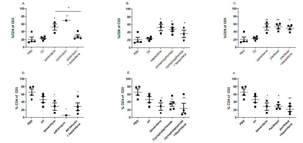
Figure 2. Increased CD8+ immune infiltrate and decreased CD4+ immune infiltrate with chemo and concurrent bexarotene in TgMMTV-neu. Evaluating the tumors at sacrifice by flow cytometry showed that there were increased CD3+CD8+ T cells (A, B, and C) and decreased CD3+CD4+ T cells (D, E, and F) by flow cytometry in chemotherapy, bexarotene, or chemotherapy and bexarotene in TgMMTV-neu mice (n=5/treatment group). A and D are with doxorubicin chemotherapy, B and F are with cyclophosphamide chemotherapy, and C and E are with paclitaxel chemotherapy. **** p<0.0001, *** p<0.001, ** p<0.01, and * p<0.05.
While increasing CD8+ T cells, bexarotene, chemotherapy, and bexarotene with chemotherapy also decreased CD4+ T cells. In TgMMTV-neu tumors, there was a 3.7 fold decrease in CD3+CD4+ T cells when treated with bexarotene as compared to PBS controls (20.5±8.7% in tumors treated with bexarotene as compared to 65.7±8.2% in PBS controls, p=0.05). Doxorubicin decreased CD3+CD4+ T cells in the tumor to 5.8±0.01% (p=0.05) but the combination of doxorubicin and bexarotene did not decrease CD3+CD4+ T cells more than doxorubicin alone to 27.9±9.7% (p=0.05) (Figure 2D). Cyclophosphamide treated tumors had decreased CD3+CD4+ T cells in the tumor to 29.2±4.3% (p=0.01) but the combination of cyclophosphamide and bexarotene decreased CD3+CD4+ T cells in the tumor to 12.3±3.1 (p=0.001, Figure 2E). Treatment of tumors with paclitaxel decreased CD3+CD4+ T cells to 29.0±4.0% (p=0.02) but the paclitaxel and bexarotene decreased CD3+CD4+ T cells in the tumor to 21.9±6.3% (p=0.004, Figure 2F). Bexarotene enhances anti-tumor impact with chemotherapy, yet while it increases CD8+ T cells and decreases CD4+ T cell the impact is increased over chemotherapy alone therefore we wanted to better understand possible roles of bexarotene on the human immune system.
In human PBMC RXR receptors are highly expressed in dendritic cells and macrophages
As bexarotene could increase CD8+ infiltrate in tumors and had anti-tumor function with chemotherapy in the mouse, we wanted to determine the immune cell populations that had highest RXRα expression in human PBMC. RXRα had the highest expression in ~30% of human APCs, 24.9±4.4% of macrophages (CD14+HLADR+), 38.6±4.8% of plasmacytoid dendritic cells (pDC), and 33.1±5.7% of monocytoid dendritic cells (mDC) (Figure 3A). The CD4+CD3+ T cells had 4.5±1.1% of the cells expressing RXR and CD8+CD3+ T cells had 3.1±0.6% of the cells expressing RXR. NK cells (CD56+CD16+ cells) had 0.18±0.03% cells expressing RXR, B cells (CD20+ cells) had 0.6±0.2% cells expressing RXR, and granulocytic MDSC cells (LIN-CD14+CD33+HLADR-) had 0.05±0.02% cells expressing RXR. Monocytic MDSC (LIN-CD11b+HLADR-CD33+ cells) had 5.9±2.2% cells expressing RXR.
Increasing doses of bexarotene increase type I dendritic cells in human PMBC
As bexarotene was expressed in APCs, we wanted to determine how increasing doses of bexarotene could impact the APCs. Treating human PBMC with 0 μM, 5 μM, 10 μM, and 20 μM doses of bexarotene increased CD40+ activation of pDC and mDC but not macrophages (Figures 3B–3D). 20 μM of bexarotene increased activated mDC (4.9±0.8% LIN-CD11c+HLADR+CD40+ cells/LIN-CD11c+ cells with 20 μM bexarotene and 0.8±0.2% with 0 μM bexarotene p=0.01) (Figure 3B). Both 10 μM and 20 μM of bexarotene increased CD40+ pDC activation as compared to 0 μM of bexarotene (9.4±2.2% LIN-CD11c+HLADR+CD40+ cells/LIN-CD11c+ HLADR+ cells with 10 μM bexarotene p=0.03, 8.7±1.9% LIN-CD11c+HLADR+CD40+ cells/LIN-CD11c+HLADR+ cells with 20 μM bexarotene p=0.05, and 3.1±1.2% with 0 µM bexarotene) (Figure 3C). There was no significant increase in activated macrophages with 20 µM of bexarotene (p=1.0) (Figure 3D). We further evaluated the cytokines released with increasing doses of bexarotene. Evaluating 10 different cytokines, we found that there were increased type I cytokines IL-1β (p=0.04 between 0 and 20 µM bexarotene Figure 4A) and TNFα (p=0.03 between 0 and 20 µM bexarotene Figure 4B) but not type II cytokines including IL-10 (p=0.99) and IL-4 (p=0.97) (Figures 4C and 4D).
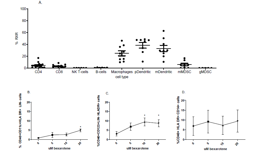
Figure 3. RXR is expressed primarily in antigen presenting cells in human PBMC and bexarotene increases CD40 expression in both monocytic and plasmocytic dendritic cells. 2×10⁶ peripheral blood mononuclear cells from women without breast cancer (n=15) were evaluated for expression of the retinoid X receptor (RXR) by flow cytometry. (A) CD4+ T cells: RXR+ CD3+CD4+, CD8+T cells: RXR+ CD3+ CD8+, NK cells: RXR+ CD56+, B cells: RXR+ CD20+, Macrophages: RXR+ HLA DR+ CD14+, Plasmacytoid dendritic cells: RXR+ LIN- HLA DR+ CD123+ (pDC), Monocytic dendritic cells: RXR+ LIN- HLA DR+ CD11c+ (mDCs), and myeloid derived suppressor cells (MDSC) Monocytic MDSC (mMDSC) RXR+HLA DR- CD14+CD11b+ and plasmocytic MDSC (pMDSC) RXR+ HLA DR- LIN- CD33+CD11b+. B, C, and D 1.5×10⁶ peripheral blood mononuclear cells from women without breast cancer (n=8) were stimulated by increasing concentrations (0 µM, 5 µM, 10 µM, and 20 µM) of bexarotene for 48 hours. Monocytoid dendritic cells (CD11c+HLA DR+ LIN-) (B) and plasmacytoid dendritic cells (CD123+ HLA DR+ LIN-) (C) have increased CD40+ expression suggesting increased activation while there was no increase of CD40+ in macrophages (CD14+ HLADR+) (D). *p<0.05.
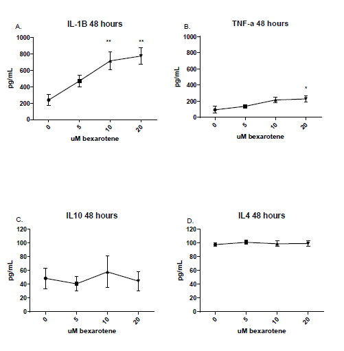
Figure 4. Bexarotene induces predominantly type I, not type II, dendritic cells. 1.5×10⁶ peripheral blood mononuclear cells from women without breast cancer (n=8) were stimulated by increasing concentrations of bexarotene for 48 hours. PBMC released increased type I IL-1β (A) and TNF-α (B) but no increase in IL-4 (C) or IL-10 (D) by multiplex cytokine assay. * p<0.05 ** p<0.01.
Increasing dosing of bexarotene induces direct activation of human mDCs
As we demonstrated that bexarotene activated type I mDCs when treating human PBMC, we wanted to show that this activation was direct. CD14+ monocytes were matured into both immature (maintained on human GM-CSF and IL4) or mature (further matured with 48 hours of with IL-1β, TNF-α, IFN-α, IFN-γ, Poly I:C, IL-2, and IL-7) mDC. The percentage of mDC expressing CD80 and CD86 did not change with doses of bexarotene (data not shown) but bexarotene did increase CD40+ expression on immature and mature mDC. For immature mDC, 10 µM bexarotene had 3.2±0.05% (p=0.04) and 20 µM bexarotene had 3.3±0.2% (p=0.05) as compared to 0 µM bexarotene which had 1.8±0.3% CD40+ HLA DR+ CD11b+ cells/CD40+ HLA DR+ cells (Figure 5A). Mature mDC had a larger proportion of mDC expressing CD40+ but this also is increased with bexarotene. 20 µM bexarotene had 35.7±0.2% (p=0.05) as compared to 0 µM bexarotene which had 14.3±2.8% CD40+ HLA DR+ CD11b+ cells/CD40+ HLA DR+ cells (Figure 5B).
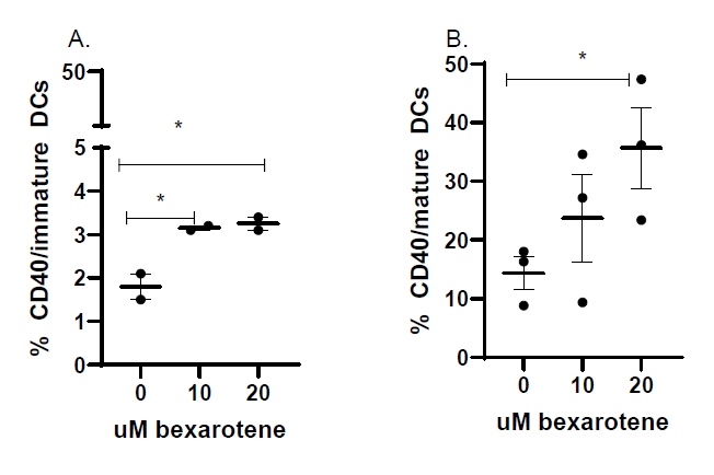
Figure 5. Bexarotene directly activates type I dendritic cells in human PBMC. CD14+ monocytes were isolated from human cryopreserved PBMC (n=3) from the Earle A. Chiles immune monitoring laboratory repository and matured with cytokines in vitro. Immature dendritic cells were maintained for 48 hours with human GM-CSF and IL4. Mature dendritic cells were matured for 48 hours with IL-1β, TNF-α, IFN-α, IFN-γ, Poly I:C, IL-2, and IL-7. Immature mDCs (A) and mature mDCs (B) were evaluated for CD40. mDCs are the LIN- CD14+ HLADR+ CD11c+ population. * p<0.05.
Discussion
Bexarotene has previously been shown to prevent tumor development and increase CD8+ T cells in TgMMTV-neu mice [19,21]. We further have demonstrated that bexarotene enhanced inhibition of tumor growth along with chemotherapy in both TgMMTV-neu and C3(1)Tag mice with established tumors (Figure 1 and Supplemental Figure 1). While bexarotene did at least double CD8+ T cells and decrease CD4+ T cells by approximately 3 fold in the tumor in both mouse models it was not higher when combined with chemotherapy as compared to bexarotene or chemotherapy alone except with doxorucibin (Figure 2 and Supplemental Figure 2). Based on our previous data on the immune environment of TgMMTV-neu and C3(1)Tag mice, we have shown that the spontaneous tumors have predominantly Th2 CD4+ T cell infiltrate therefore reduction of CD4+ T cells suggests a more antitumor proinflammatory immune response [25]. We therefore wanted to determine what direct role bexarotene was playing the immune system that could contribute to its anti-tumor immune role. In human PBMC, the RXRα receptor had the highest expression in APCs (pDCs, mDCs, and macrophages, Figure 3A). Increasing doses of bexarotene induced activated type I mDCs and pDCs but not macrophages that released increased IL-1B and TNFα (Figures 3B–3D and Figure 4). We demonstrated that bexarotene is directly activating human mDCs (Figure 5). Chemotherapies have been shown to impact the tumor immune environment but their cytotoxicity still induces systemic immune suppression. Our data suggests that bexarotene can enhance chemotherapy efficacy and increase CD8+ T cells in the tumor and based on the human data, may be through increasing type I dendritic cell activation.
Chemotherapy induces apoptosis and decreases cell proliferation of tumor cells. However chemotherapy has also been shown to have dynamic roles in immune responses in the tumor immune environment. Cyclophosphamide has been shown to reduce regulatory T cells and multiple inflammatory mediators including GM-CSF, IL-1β, IFN-γ, and TNFα [26–28]. However, cyclophosphamide also recruits MDSC to the tumor immune environment inducing immune suppression [29]. Similarly, paclitaxel has been shown to induce TNFα and activate TLR-4 in the tumor immune environment which are both associated with immune activation [29]. Paclitaxel also induces chronic inflammation driving angiogenesis and increasing immunosuppressive cytokines IL6 and IL8 [30]. Doxorubicin induces TLR4 activation and the production of inflammatory cytokines and reactive oxygenation species [31–33]. Cisplatin can induce reactive oxygenation species and induce inflammation [34]. For all of these chemotherapies, the balance of the pro and anti- inflammatory effects impacts chemotherapy efficacy and chemoresistance [35]. A therapy that can enhance the anti-tumor efficacy of chemotherapy could improve the efficacy of chemotherapy. The impact of CD8+ T cell infiltrate on anti-tumor immune function appears to be chemotherapy and mouse model specific. For example, in TgMMTV-neu mice, combining bexarotene with doxorubicin had decreased CD8+ T cell infiltrate but also improved tumor inhibition (Figure 1A and 2A). In C3(1)Tag mice, bexarotene and doxorubicin had increased CD8+ T cell infiltrate compared to doxorubicin (p=0.004) (Supplemental Figure 2A) but did not significantly improve tumor inhibition because was so effective with doxorubicin alone. (Supplemental Figure 1A). We demonstrate that bexarotene enhances anti-tumor efficacy when combined with chemotherapy (Figure 1 and Supplementary Figure 1) in two independent mouse mammary tumor models and both chemotherapy and bexarotene increase CD8+ T cells while decreasing CD4+ T cells in the tumor but the direct impact of bexarotene on the increased CD8+ T cells remains to be shown.
Bexarotene has previously been associated with an anti-tumor immune response. Bexarotene decreases malignant regulatory CD4+ T cells in cutaneous T cell lymphoma and increases CD8+ T cells in TgMMTV-neu tumors that develop during treatment with bexarotene [21,36]. The presence of T cells, particularly cytotoxic T cells, have been associated with improved prognosis across cancer types, including breast cancer [3,37]. However, the presence of cytotoxic T cells in the stroma or infiltrating the tumor are lower in untreated breast cancer than other more immune active cancer and this is believed to be because of immune suppressive populations in the tumor immune environment particularly the innate immune cells [1,38]. In human PBMC, only ~5% of peripheral CD4+ T cells and ~3% of peripheral CD3+ T cells expressed RXRα and there was no expression in NK cells or B cells. The highest expression of RXRα was in APCs (macrophages, pDC, and mDC) which were ~30% of the cells expressing RXRα (Figure 3A). Professional APCs are important to present tumor antigens to T cells in a cytokine milieu that determines whether those anti-tumor T cells to be active [39]. Increased type 1 conventional dendritic cells (mDCs) have been associated with improved prognosis across cancer types [40,41]. In breast cancer, the presence of mDC has been associated with improved survival [40]. Increasing doses of bexarotene increased activation of type I mDCs and pDCs but not macrophages (Figures 3B–3D and 4). Bexarotene was able to directly induce type I mDC (Figure 5). Bexarotene may be inhibiting tumor growth and increasing type I CD8 T cells by activating type I dendritic cells which can increase tumor-antigen specific CD8+ T cells that can infiltrate the tumor and decrease CD4+ T cells allowing those CD8+ T cells to be more active. This study did not directly show the impact of dendritic cell activation in tumor inhibition in the mouse models.
This data has demonstrated that bexarotene can directly activate human type I dendritic cells and can increase CD8+ T cells in two transgenic mouse models of breast cancer enhancing tumor inhibition with chemotherapy. One limitation of the study is that dendritic cell activation was not shown to be necessary for the anti-tumor effect in mice or humans therefore further studies are needed. There is a current trial NCT02876640 that treated patients with early breast cancer with 2 weeks of the rexinoid 9cUAB30 (a rexinoid that specifically targets endothelial cells) that will be evaluating the impact of the rexinoid on the activation of systemic dendritic cells but again does not directly evaluate the anti-tumor impact of the dendritic cells [42]. Our data support using bexarotene in combination with chemotherapy to enhance the function of the chemotherapy in breast cancer.
Conclusions
Bexarotene enhances the function of chemotherapy in breast cancer, increasing CD8+T cells and decreasing CD4+ T cells in TgMMTV-neu and C3(1)Tag transgenic mouse models. In human PBMC, RXRα is expressed highest in APCs, and can activate type I mDC and pDC. This suggests that bexarotene’s improved anti-tumor benefit with chemotherapy has an immune component that increases CD8+ T cells in the tumor. Further work is needed but this may be through activation of type I dendritic cells.
List of Abbreviations
pDCs: Plasmacytic Dendritic Cells; mDCs: Monocytic Dendritic Cells; MDSC: Myeloid Derived Suppressor Cells; FFPE: Formalin Fixed Paraffin Embedded; TILs: Tumor Infiltrating Lymphocytes
Declarations
Ethics approval
All mouse work was approved by the University of Washington Mary L. Disis IACUC and all repository clinical samples were collected from the University of Washington Cancer Vaccine Institute Repository approved by the Fred Hutchinson IRB.
Consent for publication
No personal identifying information is included in this manuscript.
Availability of data and materials
All data are available by reasonable request from Sasha Stanton MD PhD.
Competing interests
MLD has grant funding from Precigen, Veanna, Bavarian Nordic, Aston Sci. Founder of Epithany. Inventor on patents held by University of Washington. Royalties received from University of Washington. SES has research funding from Veanna and Ataraxis and drug support for a clinical trial from Canwell, Inc. LC, EG, JS, and MY have no conflicts to declare.
Funding
Research was funded by the Dolson Family Foundation Funding, Web Family Foundation, the Breast Cancer Research Fund, and the Providence Cancer Foundation. SES was supported by the NIH KL2TR000421, the Dolson Family Foundation, and the Providence Cancer Institute Foundation. MLD is supported by Helen B. Slonaker Endowed Professor for Cancer Research.
Author’s contributions
Conceptualization: SES and MLD; Data Curation: SES; Formal Analysis: LC and MY; Supervision: SES and MLD; Investigation: SES, JS, and MY; Methodology: SES; Writing original draft: SES; Reviewing and Editing: SES, MLD, and LC.
Acknowledgements
We would like to acknowledge the patients that donated their blood for analysis and Jennifer Childs and the clinical research team and immune monitoring laboratory at the Cancer Vaccine Institute at the University of Washington.
References
2. Adams S, Gray RJ, Demaria S, Goldstein L, Perez EA, Shulman LN, et al. Prognostic value of tumor-infiltrating lymphocytes in triple-negative breast cancers from two phase III randomized adjuvant breast cancer trials: ECOG 2197 and ECOG 1199. J Clin Oncol. 2014 Sep 20;32(27):2959–66.
3. Stanton SE, Adams S, Disis ML. Variation in the Incidence and Magnitude of Tumor-Infiltrating Lymphocytes in Breast Cancer Subtypes: A Systematic Review. JAMA Oncol. 2016 Oct 1;2(10):1354–60.
4. Zheng Y, Dou Y, Duan L, Cong C, Gao A, Lai Q, et al. Using chemo-drugs or irradiation to break immune tolerance and facilitate immunotherapy in solid cancer. Cell Immunol. 2015 Mar;294(1):54–9.
5. Cook AM, Lesterhuis WJ, Nowak AK, Lake RA. Chemotherapy and immunotherapy: mapping the road ahead. Curr Opin Immunol. 2016 Apr;39:23–9.
6. Alizadeh D, Trad M, Hanke NT, Larmonier CB, Janikashvili N, Bonnotte B, et al. Doxorubicin eliminates myeloid-derived suppressor cells and enhances the efficacy of adoptive T-cell transfer in breast cancer. Cancer Res. 2014 Jan 1;74(1):104–18.
7. Mattarollo SR, Loi S, Duret H, Ma Y, Zitvogel L, Smyth MJ. Pivotal role of innate and adaptive immunity in anthracycline chemotherapy of established tumors. Cancer Res. 2011 Jul 15;71(14):4809–20.
8. Voorwerk L, Slagter M, Horlings HM, Sikorska K, van de Vijver KK, de Maaker M, et al. Immune induction strategies in metastatic triple-negative breast cancer to enhance the sensitivity to PD-1 blockade: the TONIC trial. Nat Med. 2019 Jun;25(6):920–28.
9. Wanderley CW, Colón DF, Luiz JPM, Oliveira FF, Viacava PR, Leite CA, et al. Paclitaxel Reduces Tumor Growth by Reprogramming Tumor-Associated Macrophages to an M1 Profile in a TLR4-Dependent Manner. Cancer Res. 2018 Oct 15;78(20):5891–900.
10. Heylmann D, Bauer M, Becker H, van Gool S, Bacher N, Steinbrink K, et al. Human CD4+CD25+ regulatory T cells are sensitive to low dose cyclophosphamide: implications for the immune response. PLoS One. 2013 Dec 23;8(12):e83384.
11. Zhao J, Cao Y, Lei Z, Yang Z, Zhang B, Huang B. Selective depletion of CD4+CD25+Foxp3+ regulatory T cells by low-dose cyclophosphamide is explained by reduced intracellular ATP levels. Cancer Res. 2010 Jun 15;70(12):4850–8.
12. Dimeloe S, Frick C, Fischer M, Gubser PM, Razik L, Bantug GR, et al. Human regulatory T cells lack the cyclophosphamide-extruding transporter ABCB1 and are more susceptible to cyclophosphamide-induced apoptosis. Eur J Immunol. 2014 Dec;44(12):3614–20.
13. Sharma S, Shen T, Chitranshi N, Gupta V, Basavarajappa D, Sarkar S, et al. Retinoid X Receptor: Cellular and Biochemical Roles of Nuclear Receptor with a Focus on Neuropathological Involvement. Mol Neurobiol. 2022 Apr;59(4):2027–50.
14. Leal AS, Hung PY, Chowdhury AS, Liby KT. Retinoid X Receptor agonists as selective modulators of the immune system for the treatment of cancer. Pharmacol Ther. 2023 Dec;252:108561.
15. de Lera AR, Bourguet W, Altucci L, Gronemeyer H. Design of selective nuclear receptor modulators: RAR and RXR as a case study. Nat Rev Drug Discov. 2007 Oct;6(10):811–20.
16. Mangelsdorf DJ, Evans RM. The RXR heterodimers and orphan receptors. Cell. 1995 Dec 15;83(6):841–50.
17. Duvic M, Martin AG, Kim Y, Olsen E, Wood GS, Crowley CA, et al. Phase 2 and 3 clinical trial of oral bexarotene (Targretin capsules) for the treatment of refractory or persistent early-stage cutaneous T-cell lymphoma. Arch Dermatol. 2001 May;137(5):581–93.
18. Esteva FJ, Glaspy J, Baidas S, Laufman L, Hutchins L, Dickler M, et al. Multicenter phase II study of oral bexarotene for patients with metastatic breast cancer. J Clin Oncol. 2003 Mar 15;21(6):999–1006.
19. Li Y, Shen Q, Kim HT, Bissonnette RP, Lamph WW, Yan B, et al. The rexinoid bexarotene represses cyclin D1 transcription by inducing the DEC2 transcriptional repressor. Breast Cancer Res Treat. 2011 Aug;128(3):667–77.
20. Hung PY, Moerland JA, Leal AS, Aleiwi B, Ellsworth E, Clapp DW, et al. The RXR Agonist MSU-42011 Reduces Tumor Burden in a Murine Preclinical NF1-Deficient Model. Cancers (Basel). 2025 Jun 9;17(12):1920.
21. Disis ML, Gad E, Herendeen DR, Lai VP, Park KH, Cecil DL, et al. A multiantigen vaccine targeting neu, IGFBP-2, and IGF-IR prevents tumor progression in mice with preinvasive breast disease. Cancer Prev Res (Phila). 2013 Dec;6(12):1273–82.
22. Leal AS, Zydeck K, Carapellucci S, Reich LA, Zhang D, Moerland JA, et al. Retinoid X receptor agonist LG100268 modulates the immune microenvironment in preclinical breast cancer models. NPJ Breast Cancer. 2019 Nov 1;5:39.
23. Tran TA, Kim YH, Jung S, Kim IY, Moon KS, Jang WY, et al. Branched Multipeptide-combined Adjuvants Potentially Improve the Antitumor Effects on Glioblastoma. J Immunother. 2021 May 1;44(4):151–61.
24. Okada H, Kalinski P, Ueda R, Hoji A, Kohanbash G, Donegan TE, et al. Induction of CD8+ T-cell responses against novel glioma-associated antigen peptides and clinical activity by vaccinations with {alpha}-type 1 polarized dendritic cells and polyinosinic-polycytidylic acid stabilized by lysine and carboxymethylcellulose in patients with recurrent malignant glioma. J Clin Oncol. 2011 Jan 20;29(3):330–6.
25. Gad E, Rastetter L, Slota M, Koehnlein M, Treuting PM, Dang Y, et al. Natural history of tumor growth and immune modulation in common spontaneous murine mammary tumor models. Breast Cancer Res Treat. 2014 Dec;148(3):501–10.
26. Lutsiak ME, Semnani RT, De Pascalis R, Kashmiri SV, Schlom J, Sabzevari H. Inhibition of CD4(+)25+ T regulatory cell function implicated in enhanced immune response by low-dose cyclophosphamide. Blood. 2005 Apr 1;105(7):2862–8.
27. Emens LA, Asquith JM, Leatherman JM, Kobrin BJ, Petrik S, Laiko M, et al. Timed sequential treatment with cyclophosphamide, doxorubicin, and an allogeneic granulocyte-macrophage colony-stimulating factor-secreting breast tumor vaccine: a chemotherapy dose-ranging factorial study of safety and immune activation. J Clin Oncol. 2009 Dec 10;27(35):5911–8.
28. Ghiringhelli F, Menard C, Puig PE, Ladoire S, Roux S, Martin F, et al. Metronomic cyclophosphamide regimen selectively depletes CD4+CD25+ regulatory T cells and restores T and NK effector functions in end stage cancer patients. Cancer Immunol Immunother. 2007 May;56(5):641–8.
29. Edwardson DW, Boudreau J, Mapletoft J, Lanner C, Kovala AT, Parissenti AM. Inflammatory cytokine production in tumor cells upon chemotherapy drug exposure or upon selection for drug resistance. PLoS One. 2017 Sep 15;12(9):e0183662.
30. Volk-Draper L, Hall K, Griggs C, Rajput S, Kohio P, DeNardo D, et al. Paclitaxel therapy promotes breast cancer metastasis in a TLR4-dependent manner. Cancer Res. 2014 Oct 1;74(19):5421–34.
31. Apetoh L, Ghiringhelli F, Tesniere A, Obeid M, Ortiz C, Criollo A, et al. Toll-like receptor 4-dependent contribution of the immune system to anticancer chemotherapy and radiotherapy. Nat Med. 2007 Sep;13(9):1050–9.
32. Sauter KA, Wood LJ, Wong J, Iordanov M, Magun BE. Doxorubicin and daunorubicin induce processing and release of interleukin-1β through activation of the NLRP3 inflammasome. Cancer Biol Ther. 2011 Jun 15;11(12):1008–16.
33. Wang L, Chen Q, Qi H, Wang C, Wang C, Zhang J, et al. Doxorubicin-Induced Systemic Inflammation Is Driven by Upregulation of Toll-Like Receptor TLR4 and Endotoxin Leakage. Cancer Res. 2016 Nov 15;76(22):6631–42.
34. Arjumand W, Seth A, Sultana S. Rutin attenuates cisplatin induced renal inflammation and apoptosis by reducing NFκB, TNF-α and caspase-3 expression in wistar rats. Food Chem Toxicol. 2011 Sep;49(9):2013–21.
35. Behranvand N, Nasri F, Zolfaghari Emameh R, Khani P, Hosseini A, Garssen J, et al. Chemotherapy: a double-edged sword in cancer treatment. Cancer Immunol Immunother. 2022 Mar;71(3):507–26.
36. Budgin JB, Richardson SK, Newton SB, Wysocka M, Zaki MH, Benoit B, et al. Biological effects of bexarotene in cutaneous T-cell lymphoma. Arch Dermatol. 2005 Mar;141(3):315–21.
37. Sun YP, Ke YL, Li X. Prognostic value of CD8+ tumor-infiltrating T cells in patients with breast cancer: A systematic review and meta-analysis. Oncol Lett. 2022 Dec 8;25(1):39.
38. Del Prete A, Salvi V, Soriani A, Laffranchi M, Sozio F, Bosisio D, Sozzani S. Dendritic cell subsets in cancer immunity and tumor antigen sensing. Cell Mol Immunol. 2023 May;20(5):432–47.
39. Darragh LB, Karam SD. Amateur antigen-presenting cells in the tumor microenvironment. Mol Carcinog. 2022 Feb;61(2):153–64.
40. Böttcher JP, Reis e Sousa C. The Role of Type 1 Conventional Dendritic Cells in Cancer Immunity. Trends Cancer. 2018 Nov;4(11):784–92.
41. Binnewies M, Mujal AM, Pollack JL, Combes AJ, Hardison EA, Barry KC, et al. Unleashing Type-2 Dendritic Cells to Drive Protective Antitumor CD4+ T Cell Immunity. Cell. 2019 Apr 18;177(3):556–571.e16.
42. Kolesar JM, Andrews S, Green H, Havighurst TC, Wollmer BW, DeShong K, et al. A Randomized, Placebo-Controlled, Double-Blind, Dose Escalation, Single Dose, and Steady-State Pharmacokinetic Study of 9cUAB30 in Healthy Volunteers. Cancer Prev Res (Phila). 2019 Dec;12(12):903–12.