

Editorial
Nanotechnology has been promised to dentistry for nearly two decades. The premise is appealing: reducing a filler, a coating, or a remineralizing agent to the nanometer scale should improve how it performs against the biology it is meant to act on. The laboratory data are encouraging. Quaternary ammonium monomers in adhesive resin reduce Streptococcus mutans biofilm by orders of magnitude [1], nanoparticulate amorphous calcium phosphate releases calcium and phosphate to remineralize enamel in vitro [2], and silver nanoparticle coatings render titanium surfaces antibacterial in culture [3,4]. The clinically relevant question is narrower than laboratory performance: whether this evidence justifies routine use in patients. Unfortunately, there is no one-size-fits-all answer, because the three applications commonly grouped together as “dental nanotechnology” are at different stages of validation. Nanoparticle-enhanced composites, antimicrobial implant coatings, and nano-remineralization agents are considered in turn.
Nanofilled composites are already established in clinical practice, and for sound reasons. Reducing filler particle size improves polishability, wear resistance, and esthetic blend, the properties that made nanohybrid composites a restorative standard [5,6]. The unresolved questions include the newer bioactive formulations, which are engineered to release antibacterial agents or mineral ions. Here the laboratory evidence outpaces the clinical evidence. Dimethylaminododecyl methacrylate and related quaternary ammonium monomers markedly reduce cariogenic biofilm in vitro [1], and calcium phosphate nanocomposites remineralize enamel under controlled conditions [2]. Whether these materials reduce secondary caries or prolong restoration survival in patients remains untested. A 2025 systematic review found that adding antibacterial agents to bonding systems often compromises mechanical and adhesive performance, and that follow-up is too short to weigh the trade-off [7]. The randomized data that exist address narrower endpoints, such as reduced post-restorative sensitivity with a titania nanoparticle bonding agent [8]. For composites, the mechanical gains are proven, while the bioactive claims are not.
In contrast to composites, antimicrobial implant coatings remain further from clinical readiness, for a pharmacological reason. The prevalence of peri-implantitis, a common postoperative complication, is itself a strong reason to pursue a preventive coating. It affects roughly 22% of patients in one systematic review, with individual estimates from 1% to 47% depending on case definition [9]. Silver nanoparticle coatings are reliably bactericidal in vitro, but their efficacy and their cytotoxicity are controlled by the same factor: the rate of silver ion release. Rapid release clears more biofilm but also kills more host fibroblasts, while slow release preserves the cells but may not control infection [3]. The therapeutic window is therefore narrow and specific to each coating. Clinical data remain preliminary. A nanosilver-coated healing abutment suppressed plaque without harming gingival fibroblasts in one combined laboratory and clinical study [10], but no randomized trial has shown reduced peri-implantitis with a nanocoated implant, and the long-term fate of released silver in peri-implant tissue is unknown. For now, these coatings remain investigational.
Nano-remineralization agents demonstrate the strongest evidence of the three, which makes their assessment the most nuanced. Nano-hydroxyapatite acts as a calcium phosphate reservoir that remineralizes early enamel lesions. Its advantage over conventional hydroxyapatite is that nanoscale particles approximate the size of enamel apatite crystals and penetrate lesion porosities more readily, although a clinical benefit over micrometric hydroxyapatite has not been demonstrated. Unlike the other two applications, it has been tested in randomized trials and pooled in a meta-analysis. That literature is cautious. A 2022 systematic review and meta-analysis judged the trials too few, too short, and too risky to confirm that nano-hydroxyapatite prevents caries [11]. Individual trials are more favorable: a one-year randomized trial found a microcrystalline hydroxyapatite dentifrice non-inferior to fluoride against early childhood caries [12], and a nano-hydroxyapatite gel remineralized approximal lesions, though less effectively than the same gel combined with ozone [13]. The limitation is consistent across these studies. In this context, fluoride serves as an appropriate comparator because it is inexpensive and widely established as standard preventive care. Most trials also report a surrogate endpoint, surface remineralization over months, rather than cavitated caries over years. For early and white-spot lesions, nano-hydroxyapatite is the application closest to routine use. The open question is whether it offers any advantage over fluoride on definitive endpoints, not whether it works.
Across all three applications, the supporting evidence is mostly preclinical. The clinical studies are relatively small and short, and their endpoints are surrogates rather than the outcomes that matter to patients. A safety question also remains unresolved. Nanoparticle toxicity depends on particle size and shape as much as on chemical composition. The same dimensions that confer biological activity also allow these particles to cross cell membranes and potentially enter the circulation. A critical review of nanoparticles in dental materials documented cytotoxic, genotoxic, and tissue-level effects that remain poorly characterized [14]. In clinical practice, such exposure comes from processes like finishing a nanocomposite or seating a coated implant, which releases particles small enough to be inhaled or ingested [15]. Heterogeneous materials and protocols further limit comparison; the trials are difficult to pool and the products difficult to evaluate against one another.
Is nanotechnology ready for routine dental use? For one application the answer is yes; for the other two it is not yet. Nano-hydroxyapatite can be used now for early lesions, with the understanding that it complements rather than replaces fluoride. Nanofilled composites are established for their mechanical and esthetic performance, while their antibacterial and remineralizing functions remain experimental. On the other hand, antimicrobial implant coatings are not yet suitable for routine use. Comparisons across multiple studies illustrate the need for antibacterial nanocomposite materials that can prevent secondary caries and silver-nanocoated implants that can reduce peri-implantitis over extended time periods with minimal systemic toxicity. For nano-hydroxyapatite materials, further benchmarking against fluoride for the treatment of cavitated caries is needed. Across the restoration, the implant, and the enamel surface, the biology of oral disease operates at the nanoscale. However, translating nanotechnology into interventions that match the biological scale requires further clinical evidence.
Future Research Directions
Several priorities emerge from this review. For nanofilled composites, well-designed randomized trials with long-term follow-up are needed to determine whether antibacterial and remineralizing formulations reduce secondary caries or extend restoration survival compared with conventional nanohybrid composites. For antimicrobial implant coatings, the field requires standardized in vitro models that better simulate the peri-implant environment, followed by adequately powered randomized trials with clinically meaningful endpoints such as peri-implantitis incidence and implant survival over five to ten years. For nano-hydroxyapatite, head-to-head trials comparing this material directly with fluoride on hard endpoints, such as cavitated caries incidence over multi-year periods, would clarify whether its advantages extend beyond surrogate measures of surface remineralization.
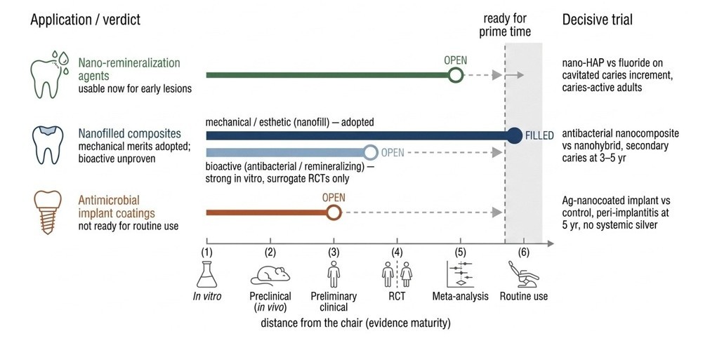
Figure 1. Three dental nanotechnologies, three distances from the chair.
|
Feature |
Nanofilled Composites |
Antimicrobial Implant Coatings |
Nano-Hydroxyapatite |
|
Current Evidence |
Proven mechanical and esthetic performance in clinical practice; bioactive and antibacterial claims untested in patients |
In vitro bactericidal efficacy; one combined laboratory and clinical study; no randomized trials with clinical endpoints |
Systematic review and meta-analysis of RCTs; most robust evidence of the three applications |
|
Clinical Readiness |
Established for restorations; experimental for bioactive functions |
Investigational; not yet suitable for routine use |
Closest to routine use for early and white-spot lesions; complements fluoride |
|
Advantages |
Improved polishability, wear resistance, and esthetic blend; widely adopted in clinical practice |
Strong in vitro bactericidal activity; directly addresses peri-implantitis risk |
Mimics enamel apatite crystal size; remineralizes early lesions; well-tolerated in trials |
|
Limitations |
Bioactive claims unproven in vivo; short follow-up; antibacterial agents may compromise mechanical performance |
Narrow therapeutic window; unknown long-term tissue fate of silver; no RCTs |
Few trials, short duration; no proven advantage over fluoride on definitive endpoints; surrogate endpoints predominate |
Across all three domains, standardized nanoparticle characterization and reporting would improve the comparability of future studies. The safety profile of nanoparticle exposure during routine clinical procedures also warrants dedicated investigation, including long-term surveillance of systemic nanoparticle accumulation. Until such evidence is available, clinicians should apply these materials within their current evidence base: nano-hydroxyapatite for early enamel lesions as a complement to fluoride, nanofilled composites for their proven mechanical properties, and antimicrobial implant coatings only within investigational protocols.
References
2. Weir MD, Chow LC, Xu HH. Remineralization of demineralized enamel via calcium phosphate nanocomposite. J Dent Res. 2012 Oct;91(10):979–84.
3. Haugen HJ, Makhtari S, Ahmadi S, Hussain B. The Antibacterial and Cytotoxic Effects of Silver Nanoparticles Coated Titanium Implants: A Narrative Review. Materials (Basel). 2022 Jul 19;15(14):5025.
4. Choi S, Jo YH, Han JS, Yoon HI, Lee JH, Yeo IL. Antibacterial activity and biocompatibility of silver coating via aerosol deposition on titanium and zirconia surfaces. Int J Implant Dent. 2023 Sep 4;9(1):24.
5. Karunakaran J, Jayakumar S, Rajamani S, Shri BS, Nandhini G, Srinivasan V. Revolutionizing dental restorations with nanoparticle-incorporated composites. J Conserv Dent Endod. 2025 Jun;28(6):498–504.
6. Mitra SB, Wu D, Holmes BN. An application of nanotechnology in advanced dental materials. J Am Dent Assoc. 2003 Oct;134(10):1382–90.
7. Lin Q, Chen H, Zheng S, Lin L, Zhu S. Effect of antibacterial agents on dentin bonding system: A systematic review and meta-analysis. J Prosthet Dent. 2025 Dec;134(6):2117–32.
8. Amir N, Mansoor A, Noor N, Hussain K. Clinical Efficacy of a Novel Titania Nanoparticle-Reinforced Bonding Agent in Reducing Post-Restorative Sensitivity: A randomized clinical trial. Pak J Med Sci. 2024 Aug;40(7):1332–37.
9. Derks J, Tomasi C. Peri-implant health and disease. A systematic review of current epidemiology. J Clin Periodontol. 2015 Apr;42 Suppl 16:S158–71.
10. Odatsu T, Kuroshima S, Sato M, Takase K, Valanezhad A, Naito M, et al. Antibacterial Properties of Nano-Ag Coating on Healing Abutment: An In Vitro and Clinical Study. Antibiotics (Basel). 2020 Jun 19;9(6):347.
11. Wierichs RJ, Wolf TG, Campus G, Carvalho TS. Efficacy of nano-hydroxyapatite on caries prevention-a systematic review and meta-analysis. Clin Oral Investig. 2022 Apr;26(4):3373–81.
12. Paszynska E, Pawinska M, Gawriolek M, Kaminska I, Otulakowska-Skrzynska J, Marczuk-Kolada G, et al. Impact of a toothpaste with microcrystalline hydroxyapatite on the occurrence of early childhood caries: a 1-year randomized clinical trial. Sci Rep. 2021 Jan 29;11(1):2650.
13. Grocholewicz K, Matkowska-Cichocka G, Makowiecki P, Droździk A, Ey-Chmielewska H, Dziewulska A, et al. Effect of nano-hydroxyapatite and ozone on approximal initial caries: a randomized clinical trial. Sci Rep. 2020 Jul 7;10(1):11192.
14. Karunakaran H, Krithikadatta J, Doble M. Local and systemic adverse effects of nanoparticles incorporated in dental materials- a critical review. Saudi Dent J. 2024 Jan;36(1):158–67.
15. Tafti F, Savant S, Saraf T, Pinge S, Thorat R, Sharma V. Hazards Associated With Nanotechnology in Clinical Dentistry. Cureus. 2023 Oct 13;15(10):e46978.