

Abstract
Pancreaticobiliary maljunction, an abnormal junction between pancreatic and Common Bile Duct (CBD) which forms before the opening in duodenum to form a long common channel, with or without biliary dilatation is a rare cause of recurrent pancreatitis in children. The anomalous junction results in reflux of pancreatic juices into the biliary tract leading to injury. This report describes a pediatric patient presenting with recurrent acute pancreatitis, suspected to have long common pancreaticobiliary maljunction based on ultrasound describing dilated common bile duct. The role of Magnetic Resonance Cholangiopancreatography (MRCP) in diagnosis and the role of endoscopy in management of the patient has been described.
Keywords
Long common channel, Pancreaticobiliary maljunction, Magnetic Resonance Cholangiopancreatography (MRCP), Endoscopic Retrograde Cholangiopancreatography (ERCP)
Background
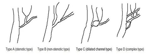
Pancreaticobiliary maljunction, also called anomalous pancreaticobiliary junction, resulting in reflux of pancreatic juices into bile duct and bile juice into pancreatic duct. It is more common in Asian population with an incidence of 1.5 to 3.2% and female preponderance (3 times) [1]. It can be associated with choledochal cyst [2], bile duct strictures, pancreatic anomalies [3], biliary cancers [4]. The Japanese study group on pancreaticobiliary maljunction classifies it into four types, A–D (Figure 1). A- Stenotic type with stenotic segment of Common Bile Duct (CBD) joining common channel. B- non stenotic distal CBD joins the common channel. C- narrow CBD joins dilated common channel. D- complex maljunction associated with complex duct systems, example, annular pancreas [5]. Approximately 80% cases are associated with biliary dilatation. Patients typically present with episodes of acute pancreatitis, vomiting, fever, and severe abdominal pain. The origin of long common channel may lie in the embryological adhesion of ventral pancreatic duct and terminal part of Bile Duct (CBD) [3]. Typically, amylase and lipase levels in serum and intrabiliary structures are elevated owing to destruction of CBD by pancreatic juices. Ultrasound has a limited role in diagnosis, but maybe a useful noninvasive screening modality depending on the experience of radiologist. Dilated CBD may be the first clue towards pancreaticobiliary maljunction. However, it is nonspecific, CT or MRI used for measurement of length of CBD, pancreatic duct and common channel are useful for diagnosis. Common channel length of ≥8 mm is diagnostic, though some authors use the cutoff as 15 mm [5]. Magnetic Resonance Cholangiopancreatography (MRCP) is the gold standard for diagnosis and is also superior to Endoscopic Retrograde Cholangiopancreatography (ERCP) to elicit the anatomy.
Case
A middle childhood (8 yrs) female presented to our side with complaints of fever, vomiting, and periumbilical abdominal pain for 3 days. The abdominal pain was severe in intensity and associated with 3–4 episodes of vomiting and relived in sitting position. The patient also had similar episodes 2 months back, when patient was admitted in hospital for one week where diagnosis of acute pancreatitis was made based on elevated serum amylase and serum lipase and abdominal ultrasound.
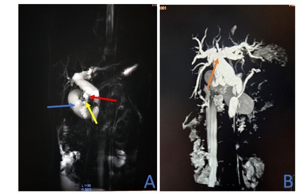
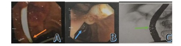
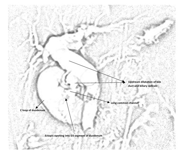
Owing to the recurrence of disease, further investigation was warranted. Screening ultrasound was done, suggesting dilated CBD, diffuse circumferential gall bladder wall edema and minimal peripancreatic free fluid raising suspicion of long common channel with dilated CBD. MRCP was planned which came to be suggested of abnormal pancreaticobiliary junction (long common channel with 29 mm length) with ectopic opening into D3 segment of duodenum, choledocholithiasis with upstream dilatation of CBD and mild bipolar intrahepatic biliary radical dilatation and acute cholecystitis confirming the diagnosis (Figure 2). On the basis of MRCP, a final diagnosis of type A pancreaticobiliary maljunction was made as per Japanese study group classification. ERCP was done with selective cannulation of CBD using papillotome, multiple balloon sweeping was done to retrieve sludge and 7Fr* 7 cm double pigtail stent was placed in CBD, draining bile freely (Figure 3). A diagrammatic representation of long common channel is shown in Figure 4. Over the course of hospital stay, abdominal pain and fever subsided.
Investigations
Abnormal pancreaticoduodenal junction, being a rare diagnosis, is not the upfront differential. A screening ultrasound may give some clue regarding diagnosis suggested by dilatation of CBD. An experienced radiologist can also talk about the length of CBD and pancreatic duct with opening in duodenum, but it is difficult. Ultrasound can help in suspicion but is not diagnostic. MRCP remains the gold standard for diagnosis as it can depict the anatomy in the best possible way.
Complete blood count of patient showed hemoglobin of 12.7 g/dL, total leucocyte count-4800 cells/mm3, N63.6%L2.8%E1.6%M7.4%B0.6%, platelet count-2.05 lac/mm3. Liver function test suggested total serum bilirubin of 0.60 mg/dl, SGOT- 20.54 IU/L, SGPT- 20.19 IU/L, serum alkaline phosphatase-619.18 IU/L. Serum amylase of 1448.45 IU/L and serum lipase of 185.64 IU/L suggestive of inflammation of gastrointestinal tract. A screening ultrasound was done suggesting dilated CBD, diffuse circumferential gall bladder wall edema and minimal peripancreatic free fluid. Dilated CBD raising suspicion of obstruction or stenosis; hence MRCP was performed which was suggestive of abnormal pancreaticobiliary junction (long common channel with 29 mm length) with ectopic opening into D3 segment of duodenum, choledocholithiasis with upstream dilatation of CBD and mild bipolar intrahepatic biliary radical dilatation and acute cholecystitis.
Differential Diagnosis
Owing to the rarity of abnormality, it is not an upfront differential in the physician’s mind. Other causes, such as trauma, genetic causes, metabolic disorders and certain medications can cause pancreatitis and needs to be ruled out based on history and investigations. Trauma being a common cause of pancreatitis in children can be ruled out based on history. Family history of pancreatitis may suggest genetic association. Hypercalcemia or hyperlipidemia may cause pancreatitis, so, other associations of such metabolic disorders are to be looked for. Medications including anti-seizure drugs and certain chemotherapeutic agents can cause pancreatitis, so drug history needs to be looked at.
Treatment
Once the diagnosis is confirmed, ERCP is the modality for treatment. In an asymptomatic patient, risk reduction prophylactic surgical correction is recommended to reduce the risk of cancers. For patients without biliary dilatation, prophylactic cholecystectomy is recommended owing to the high risk of gallbladder carcinoma. In symptomatic patients, ERCP is done after initial stabilization of patient followed by cholecystectomy.
In the above-mentioned patient, surgical correction (CBD was selectively cannulated using papillotome. Multiple balloon sweeps were done and sludge was retrieved. 7Fr * 7 cm double pigtail CBD stent was placed, draining bile freely.) through ERCP was done after the acute episode of acute pancreatitis subsided.
Outcome and Follow Up
Follow up of these patients is important even years after prophylactic or therapeutic surgery to look for complications like chronic pancreatitis, cholangitis, or biliary tract carcinomas, including gall bladder carcinoma. Screening ultrasound and any fresh complaints experienced by patients may be helpful in pointing towards residual disease or complications after the procedure which should be carefully looked for.
There were no procedural complications during ERCP and patient had no episode of abdominal pain since then. The patient is still under follow up and doing well.
Discussion
Pancreaticobiliary maljunction, also known as the long common channel, is a congenital anomaly where junction of pancreatic and bile ducts is located outside the duodenal wall allowing regurgitation between pancreatic duct and CBD and is no longer under the control of sphincter of Oddi. As the pressure in pancreatic duct is higher than CBD, pancreatic juice refluxes into the CBD, causing complications. Free communication is maintained between pancreatic and bile duct, even when sphincter of Oddi is contracted and can be diagnosed via MRCP or ERCP.
The activation of proteolytic pancreatic enzymes in the bile duct and its stagnation leads to injury to the epithelium, which over time can cause hyperplasia and metaplasia, which can progress to cancer. Commonly it leads to development of stones in gall bladder, bile duct and pancreatic duct.
No genetic association of pancreaticobiliary anomaly are yet known, but long common channel may be associated with other congenital anomalies, like, choledochal cyst, annular pancreas, pancreatic divisum etc. In our case no other anomaly was present. A single cell RNA based sequencing in patients with pancreaticobiliary anomaly reveals significant difference in cellular composition characterized by high proportion of endothelial cells and fibroblasts, whereas B and T lymphocytes were less abundant. Heightened activity of WNT and TWEAK signaling pathway was found. Also, there was increased ligand receptor interaction between fibroblasts and other cells [6].
It usually manifests as severe abdominal pain, vomiting and fever owing to the event of acute pancreatitis. It is an uncommon cause of recurrent pancreatitis in children and needs to be ruled out due to the risks of various carcinomas, gall bladder carcinoma being the most common one. Prophylactic cholecystectomy is recommended in cases of dilated bile duct.
In our case patient presented with recurrent episodes of acute pancreatitis with 2 episodes occurring within two months. Screening ultrasonography was done which was suggestive of dilated CBD, raising suspicion of long common channel. Careful examination with ultrasound can also tell about the length of bile and pancreatic duct. MRCP was further planned to confirm the diagnosis, once the patient was stabilized, confirming the disease. On the basis of MRCP, a diagnosis of type A pancreaticobiliary maljunction was made. Amylase and lipase levels in serum and bile duct are elevated, supporting the diagnosis.
Early diagnosis and timely intervention remain mainstay in management to prevent life threatening complications like carcinoma and also prevent absentees from work. Endoscopic duct reconstruction and removal of sludge is mostly used followed by cholecystectomy.
Owing to the rarity of disease, it may be missed. But high risk of suspicion and careful history taking and examination may prevent debilitating effects.
Learning Points
- Owing to the rarity of the disease, high risk suspicion helps in timely intervention and prevention of complications.
- Abdominal pain in children, which is a common illness, should not be ignored based on ultrasound and should be investigated properly in case of persistent complaint.
- Follow up of the patient after proper intervention is as important as initial management, owing to risk of further morbidities which can happen.
- Prophylactic cholecystectomy may be required in most of the patients which can prevent complications related to primary disease and sequel
Patient’s Perspective
With the severe pain which I had in abdomen and multiple episodes of vomiting, it was leading to loss of my studies with a week of hospital stay two months back and two weeks of hospital stay now. With that severe pain, I was not able to focus on my studies, which was thought to be tantrum initially. But with timely diagnosis and surgery, I will be able to focus on my studies and achieve my goals. Thanks to the managing team.
References
2. Urushihara N, Hamada Y, Kamisawa T, Fujii H, Koshinaga T, Morotomi Y, et al. Classification of pancreaticobiliary maljunction and clinical features in children. J Hepatobiliary Pancreat Sci. 2017 Aug;24(8):449–55.
3. Kamisawa T, Egawa N, Nakajima H, Tsuruta K, Okamoto A, Matsukawa M. Origin of the long common channel based on pancreatographic findings in pancreaticobiliary maljunction. Dig Liver Dis. 2005 May;37(5):363–7.
4. Ragot E, Mabrut JY, Ouaïssi M, Sauvanet A, Dokmak S, Nuzzo G, et al. Pancreaticobiliary Maljunctions in European Patients with Bile Duct Cysts: Results of the Multicenter Study of the French Surgical Association (AFC). World J Surg. 2017 Feb;41(2):538–45.
5. Ono A, Arizono S, Isoda H, Togashi K. Imaging of Pancreaticobiliary Maljunction. Radiographics. 2020 Mar-Apr;40(2):378–92.
6. Mao HM, Guo WL, Shi SL. Diversity and heterogeneity in human pancreaticobiliary maljunction revealed by single-cell RNA sequencing. Pediatr Surg Int. 2025 Mar 21;41(1):98.