Abstract
Purpose: The aim of this study was to investigate the efficacy of FAR 222 nm UV light in the treatment of apparently infected corneal ulcers.
Design: Patients with apparently infected corneal ulcers were offered the IRB approved protocol of FAR UV 222 nm UV light, in addition to standard antimicrobial intervention.
Methods: A total of 62 eyes of 61 patients referred for both acute and chronic corneal ulcers were cultured by direct culture media plating or E-Swabs sent to a local microbiology laboratory. Following IRB approved informed consent patients were treated with FAR UV 222 nm UV light. Fifty-eight patients underwent 60 seconds of treatment for presumed bacterial or fungal keratitis and 3 patients underwent 15 minutes of treatment for clinically consistent Acanthamoeba corneal ulcers. One of these Acanthamoeba patients had bilateral infections and both eyes were treated. After treatment the corneal ulcers were again cultured and the specimens sent to the same reference laboratory.
Results: Twenty-one patients were culture positive before FAR UV intervention and were culture negative after therapy. Thirty patient eyes were culture negative before treatment and remained culture negative after FAR UV. Seven ulcers were culture positive both before and after therapy. Two patients were culture negative before treatment and had positive cultures after treatment.
Conclusions: FAR UV 222 nm treatment of corneal ulcers is efficacious in eliminating many bacterial and fungal culture proven corneal ulcers. Although the presenting ulcer may be sterilized by FAR UV treatment, recrudescent infection from residual organisms in the conjunctival fornices may ensure and topical medications should be continued until inflammation subsides.
Keywords
Corneal ulcers, Bacterial keratitis, Acanthamoeba, Fungal infections
Introduction
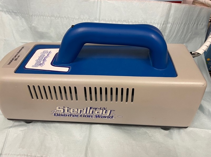
Microbial infections of the cornea are increasingly resistant to antibiotics, antifungals and antivirals [1–10]. We have investigated the use of FAR 222 nm UV light as adjunctive treatment in the treatment of acute and chronic corneal ulcers, many of which persist in the face of extant antimicrobial therapy. The SterilrayT provided by HEO3 was considered a good candidate for sterilizing corneal ulcers as it kills known common corneal pathogens, including Staphylococcus, Streptococcus, Pseudomonas, Serratia, E. coli, Candida, and Fusarium with under 20 seconds of application [11–18]. Acanthamoeba trophozoites and cysts can be killed in 7 minutes of application.
Materials and Methods
All study patients and protocols were obtained by HIPPA regulations. The corneal ulcer protocol was approved by the Western Institutional Review Board Study No : 1171772. In this study we recruited Florida patients that were resistant to current therapeutic interventions, had undergone unsuccessful therapy outside of our Institute, or were referred for acute corneal ulcers. All patients underwent a complete eye examination including visual acuity (VA), external exam, intraocular pressure (IOP) if no perforation was imminent, slit lamp exam, and fundus evaluation when visualization through the ulcer was possible.
Patients were precluded from this study in the following categories:
- Corneal perforation requiring immediate corneal transplantation
- Patients unable to undergo a complete eye exam.
- Inability to understand the significance of corneal infection and potential loss of vision without proper intervention.
- Life expectancy under one month.
Results
Seventy-nine patients have been evaluated and treated with the protocol outlined. Twenty-one patients were culture positive before FAR UV intervention and were considered a success, being culture negative immediately after therapy. A broad spectrum of culture positive organisms were seen (Table 1).
|
Organism |
Culture Positive Before FAR UV – Negative after UV |
Culture Positive Before and after Far UV |
Culture Negative Before FAR UV And Positive After |
|
Staphylococcus epidermidis |
6 |
|
|
|
Staphylococcus aureus |
2 |
3 |
|
|
Streptococcus |
2 |
1 |
|
|
Pseudomonas |
2 |
|
1 |
|
Serratia |
2 |
|
|
|
Propionibacterium acnes |
1 |
|
|
|
Bacillus |
1 |
|
|
|
Klebsiella |
1 |
|
|
|
Candida |
2 |
|
|
|
Aspergillus |
1 |
|
|
|
Fusarium |
1 |
3 |
|
|
Enterobacter |
|
|
1 |
|
Total |
21 |
7 |
2 |
Thirty-one patient eyes were culture negative before treatment and remained culture negative after FAR UV. Seven ulcers were culture positive both before and after therapy. Two patients were culture negative before treatment and had positive cultures after treatment.
Discussion
Corneal ulcers continue to occur, primarily from contact lens use [18,19], foreign bodies [20,21], and increasingly from nosocomial exposure [22]. The cultured ophthalmic organisms are increasingly resistant to current antibiotics or combinations thereof. Ultraviolet light has been utilized for sterilization for years for drinking water [23], air handling systems [24], operating rooms [25], and surgical instruments [26]. The shorter the UV wavelength the greater the killing power of UV light [27]. Therefore, the 222 Far UV light is a potential advance for superficial infections [28]. 222 Far UV provides excellent antimicrobial efficacy without the harmful skin effects of longer UV wavelengths [29]. The 222 wavelengths do not penetrate the stratum corneum of the skin and would be blocked by an intact epithelial layer of the cornea [30]. However, this epithelial layer is frequently absent in the presence of infectious corneal ulcers, allowing 222 UV penetration to superficial infections. Our unpublished SEM safety data demonstrated no endothelial toxicity in eye bank eyes from application of the 222 UV wavelength to the external cornea. We have, therefore, utilized this shorter wavelength UV for corneal ulcers with IRB approval. Rose Bengal photodynamic antimicrobial therapy has been utilized successfully for Pseudomonas keratitis isolates [31], Acanthamoeba Keratitis [32], Nocardia keratitis [33], and fungal keratitis [33] requiring 15–45 minutes of application and treatment time. We feel the 60 second killing power of the 222 wavelengths may be an auspicious advance for bacterial and fungal keratitis.
We noted that many of the ulcers had been treated for 1-90 days before referral, and this confounding variable may have accounted for the 31 negative culture results before any UV therapeutic intervention. The ulcers were not healing but had been adequately treated for infection before referral and the attendant ulcer tissue damage precluded epithelialization of the corneal surface. We are not able clinically to differentiate those corneal ulcers that remain infected at presentation and those which are already sterilized. We felt that delay of UV treatment for one to two weeks for initial culture results while routine ulcer treatment continued would not be safe or diagnostic.
Seven patients were culture positive before Far UV treatment and remained positive after UV treatment. Three of these ulcers were Fusarium. One additional patient with a Fusarium ulcer was culture negative after UV treatment. In addition, we have noted that some patients cannot easily tolerate the lid speculum and their eye movement during the two minutes of cultures, UV treatment and subsequent repeat cultures may allow reinoculation of the surface from untreated fornix organisms. Fornix organisms are not diminished or effaced by corneal treatment alone. In addition, in preclinical safety eye bank eye testing we found that Far UV light does not penetrate the cornea to the endothelium and this superficial level of treatment (250 microns) penetration may allow organisms to remain viable below the treated corneal surface. A corneal opacity may further block transmission of the FAR UV light to deeper corneal infections.
Two eyes had negative cultures before UV treatment but grew out organisms after the UV treatment: Pseudomonas (1), Enterobacter cloacae (1). This result may also be related to the culturing algorithm unavoidable eye movement.
The 222 Far UV light eliminated culture positivity in 21 of 28 infected eyes while 7 remained positive even with treatment. Many patients noted prompt relief of their ocular ulcer pain within hours of intervention. However, all patients should remain on standard ulcer therapy regiments until stromal inflammation subsides as fornix organisms will not be mitigated by corneal UV treatment alone. Adjacent skin flora may extend over the face with recontamination of the eye even as the ulcer heals. Therefore, prophylactic treatment for several weeks is appropriate even though ulcer treatment appears satisfactory. Longer UV wavelengths with deeper corneal penetration are anticipated.
Enhanced Disclosure
Funding support
The 222 instrument was supplied by HEO3, Inc.
High Energy Ozone LLC, 30 Centre Road, Suite 6, Somersworth, NH 03878.
Financial disclosure
S. Edward Neister, John Neister, Steve Hudson are all owners of HEO3. No other author has any financial interest in the Far 222 UV device or any financial disclosures.
Acknowledgements
No other Statisticians, Medical Writers or Expert Contributors have been utilized.
References
2. Sharma N, Goel M, Bansal S, Agarwal P, Titiyal JS, Upadhyaya AD, et al. Evaluation of moxifloxacin 0.5% in treatment of nonperforated bacterial corneal ulcers: a randomized controlled trial. Ophthalmology. 2013 Jun;120(6):1173–8.
3. Alfonso SA, Fawley JD, Alexa Lu X. Conjunctivitis. Prim Care. 2015 Sep;42(3):325–45.
4. Wang N, Yang Q, Tan Y, Lin L, Huang Q, Wu K. Bacterial Spectrum and Antibiotic Resistance Patterns of Ocular Infection: Differences between External and Intraocular Diseases. J Ophthalmol. 2015;2015:813979.
5. Asbell PA, Sanfilippo CM, Pillar CM, DeCory HH, Sahm DF, Morris TW. Antibiotic Resistance Among Ocular Pathogens in the United States: Five-Year Results From the Antibiotic Resistance Monitoring in Ocular Microorganisms (ARMOR) Surveillance Study. JAMA Ophthalmol. 2015 Dec;133(12):1445–54.
6. Watanabe K, Zhu H, Willcox M. Susceptibility of Stenotrophomonas maltophilia clinical isolates to antibiotics and contact lens multipurpose disinfecting solutions. Invest Ophthalmol Vis Sci. 2014 Dec 2;55(12):8475–9.
7. Sanfilippo CM, Morrissey I, Janes R, Morris TW. Surveillance of the Activity of Aminoglycosides and Fluoroquinolones Against Ophthalmic Pathogens from Europe in 2010-2011. Curr Eye Res. 2016 May;41(5):581–9.
8. Miller D, Chang JS, Flynn HW, Alfonso EC. Comparative in vitro susceptibility of besifloxacin and seven comparators against ciprofloxacin- and methicillin-susceptible/nonsusceptible staphylococci. J Ocul Pharmacol Ther. 2013 Apr;29(3):339–44.
9. Chang VS, Dhaliwal DK, Raju L, Kowalski RP. Antibiotic Resistance in the Treatment of Staphylococcus aureus Keratitis: a 20-Year Review. Cornea. 2015 Jun;34(6):698–703.
10. Rahimi F, Hashemian SM, Tafti MF, Mehjerdi MZ, Safizadeh MS, Pour EK, et al. Chlorhexidine Monotherapy with Adjunctive Topical Corticosteroids for Acanthamoeba Keratitis. J Ophthalmic Vis Res. 2015 Apr-Jun;10(2):106–11.
11. Yen S, Sokolenko S, Manocha B, Blondeel EJ, Aucoin MG, Patras A, et al. Treating cell culture media with UV irradiation against adventitious agents: minimal impact on CHO performance. Biotechnol Prog. 2014 Sep-Oct;30(5):1190-5.
12. Dai T, Kharkwal GB, Zhao J, St Denis TG, Wu Q, Xia Y, et al. Ultraviolet-C light for treatment of Candida albicans burn infection in mice. Photochem Photobiol. 2011 Mar-Apr;87(2):342–9.
13. Metzger Z, Better H, Abramovitz I. Immediate root canal disinfection with ultraviolet light: an ex vivo feasibility study. Oral Surg Oral Med Oral Pathol Oral Radiol Endod. 2007 Sep;104(3):425–33.
14. Neister E, Neister J, Hudson S. Comparison of UV Sources. Healthy Environment Innovations, LLC, 273 Locust Street, Suite B., Dover, NH 03802.
15. Clauß M. Higher effectiveness of photoinactivation of bacterial spores, UV resistant vegetative bacteria and mold spores with 222 nm compared to 254 nm wavelength. Acta hydrochimica et hydrobiologica. 2006 Dec;34(6):525–32.
16. Clauß M, Mannesmann R, Kolch A. Photoreactivation of Escherichia coli and Yersinia enterolytica after irradiation with a 222 nm excimer lamp compared to a 254 nm low‐pressure mercury lamp. Acta hydrochimica et hydrobiologica. 2005 Dec;33(6):579–84.
17. Neister E, Neister J, Hudson S. Virucidal Efficacy of 222 nm Far-UV light against Norovirus Feline calicivirus, Microtest. Healthy Environment Innovations, LLC, 273 Locust Street, Suite B., Dover, NH 03802.
18. Jeang L, Tuli SS. Therapy for contact lens-related ulcers. Curr Opin Ophthalmol. 2022 Jul 1;33(4):282–9.
19. Maier P, Betancor PK, Reinhard T. Contact Lens–Associated Keratitis—an Often-Underestimated Risk. Dtsch Arztebl Int. 2022 Oct 7;119(40):669–74.
20. Ahmed F, House RJ, Feldman BH. Corneal Abrasions and Corneal Foreign Bodies. Prim Care. 2015 Sep;42(3):363–75.
21. Lim Wen Siang J, Wu Zhuan O, Yi Chen N, Sok Lin N. Profile of Microbial Keratitis. Cureus. 2021 Dec 24;13(12):e20663.
22. Das A, Dey AK, Agarwal PK, Majumdar AK, Majumdar S, Chatterjee SS. Nosocomial ocular infection--a prospective study. J Indian Med Assoc. 2003 Aug;101(8):490–2.
23. Kim HJ, Yoon HW, Lee MA, Kim YH, Lee CJ. Impact of UV-C Irradiation on Bacterial Disinfection in a Drinking Water Purification System. J Microbiol Biotechnol. 2023 Jan 28;33(1):106–13.
24. Levetin E, Shaughnessy R, Rogers CA, Scheir R. Effectiveness of germicidal UV radiation for reducing fungal contamination within air-handling units. Appl Environ Microbiol. 2001 Aug;67(8):3712–5.
25. El Haddad L, Ghantoji SS, Stibich M, Fleming JB, Segal C, Ware KM, et al. Evaluation of a pulsed xenon ultraviolet disinfection system to decrease bacterial contamination in operating rooms. BMC Infect Dis. 2017 Oct 10;17(1):672.
26. Narain AS, Hijji FY, Yom KH, Kudaravalli KT, Haws BE, Singh K. Radiation exposure and reduction in the operating room: Perspectives and future directions in spine surgery. World J Orthop. 2017 Jul 18;8(7):524–30.
27. Hessling M, Haag R, Sieber N, Vatter P. The impact of far-UVC radiation (200-230 nm) on pathogens, cells, skin, and eyes - a collection and analysis of a hundred years of data. GMS Hyg Infect Control. 2021 Feb 16;16:Doc07.
28. Song BM, Lee GH, Han HJ, Yang JH, Lee EG, Gu H, et al. Ultraviolet-C light at 222 nm has a high disinfecting spectrum in environments contaminated by infectious pathogens, including SARS-CoV-2. PLoS One. 2023 Nov 28;18(11):e0294427.
29. Panzures A. 222-nm UVC light as a skin-safe solution to antimicrobial resistance in acute hospital settings with a particular focus on methicillin-resistant Staphylococcus aureus and surgical site infections: a review. J Appl Microbiol. 2023 Mar 1;134(3):lxad046.
30. Buonanno M, Ponnaiya B, Welch D, Stanislauskas M, Randers-Pehrson G, Smilenov L, et al. Germicidal Efficacy and Mammalian Skin Safety of 222-nm UV Light. Radiat Res. 2017 Apr;187(4):483–91.
31. Durkee H, Arboleda A, Aguilar MC, Martinez JD, Alawa KA, Relhan N, et al. Rose bengal photodynamic antimicrobial therapy to inhibit Pseudomonas aeruginosa keratitis isolates. Lasers Med Sci. 2020 Jun;35(4):861–6.
32. Prajna NV, Lalitha P, Sharma S, de Freitas D, Höfling-Lima A, Varnado N, et al. A double-masked, sham-controlled trial of rose bengal photodynamic therapy for the treatment of fungal and acanthamoeba keratitis: Rose Bengal Electromagnetic Activation with Green Light for Infection Reduction (REAGIR) study. Trials. 2024 Aug 28;25(1):566.
33. Adre E, Durkee H, Arboleda A, Alawa K, Maestre J, Mintz KJ, et al. Rose Bengal and Riboflavin Mediated Photodynamic Antimicrobial Therapy Against Selected South Florida Nocardia Keratitis Isolates. Transl Vis Sci Technol. 2022 Jan 3;11(1):29.