

Abstract
It has been almost four decades since CD28 was identified as a major player (co-stimulator) of T cell activation and has been recognized as one of the major milestones in the T cell field. However, the exact underlying mechanism involved in the activation of CD28 still remains poorly understood. Most importantly, the depletion of CD28 in mice did not lead to a major defect in T cell activation response, even when combined with the deletion of ICOS, a homolog of CD28. Here, we propose that CD28 is just a survival receptor, which works together with CD45 to maintain T cell survival. It does not participate in the activation of the T-cell activation process by antigen-presenting cells (APCs) in vivo. Furthermore, we propose that CD28 acts like CD4/CD8 to recruit LCK to activate the PI3 kinase pathway.
Keywords
CD28, Anti-CD28, CD3, Anti-CD3, LCK, CD45, PI3K, Galectin
Introduction
The anti-CD28 antibody (Mab 9.3) [1] was found to synergistically enhance T cell response when T cells were activated by anti-CD3 antibodies [2,3]. Furthermore, the anti-CD28 antibody combined with either PHA (Phytohemagglutinin) [1] or PMA (Phorbol Ester) [4,5] could bypass anti-CD3 to activate T cells. A series of research about the function of CD28 led to the assigned functional role of CD28 as the second major signal stimulator for T cell activation in the immunology field. However, the exact underlying mechanism by which CD28 promotes T cell activity remains unresolved, which causes a lot of confusion about how to design effective chimeric receptor-containing CAR-T cells to battle leukemia and other cancers.
The Underlying Mechanism of Artificial T cell Activation In Vitro
A groundbreaking discovery by two groups showed that the addition of an anti-CD3 antibody and an anti-CD28 antibody would trigger the activation and proliferation of T cells in vitro [2,3]. Early on, people found that plant lectin proteins were also able to activate T cells [6,7]. The exact underlying mechanism(s) of T cell activation by these artificial proteins remains mysterious. We recently proposed a novel T cell activation model to interpret how these artificial proteins work in vitro, which brings the Src kinase family (SKF) member LCK (through CD28) into proximity with the intracellular portions of the TCR; the latter is phosphorylated by LCK to trigger the downstream signal cascade (Zhang, unpublished). At the same time, in that model, we also proposed that CD28 is not required for T cell activation in vivo since CD4 and CD8 are capable of recruiting LCK to the proximity of the intracellular portions of TCR subunits. However, we did not provide strong evidence to show why CD28 is not required for T cell activation in vivo. Here, we are trying to find evidence to prove this hypothesis.
T cell In Vitro Activation without CD28
In our T cell activation model, we reason that CD4 and CD8 have the ability to replace CD28 to recruit LCK to activate T cells in vivo (Zhang, unpublished). This suggests that an anti-CD4 antibody or an anti-CD8 antibody could replace an anti-CD28 antibody to activate CD4 T cells or CD8 T cells in vitro, respectively. To our surprise, there is an early report showing that the addition of anti-CD4 antibody and anti-CD3 antibody did trigger the activation and proliferation of CD4 T cells, and it was also true for CD8 T cells, though anti-CD4 antibody alone led to the elimination of CD4 T cells [8]. In this regard, the anti-CD4 antibody and the anti-CD8 antibody could bypass CD28 to activate T cells in vitro.
CD28 is Not Required for T cell Activation In Vivo
Based on our current understanding of the conventional TCR activation pathway, APCs (antigen-presenting cells) will present antigens to T cells through their MHC II (MHC I) molecules with the help of CD4 (CD8) to activate the TCR pathway. At the same time, APCs will also contribute CD28 ligands such as CD80 and CD86 to activate CD28. The complete activation of T cells needs the two individual pathways [9]. It was reported that there is a PI3K-binding signature motif (YXXM) within the C-terminus of CD28 [9]. It is also reported that there is an LCK-binding motif at the tip of the C-terminus of CD28 [9], suggesting that LCK could be recruited by activated CD28 and phosphorylate the YXXM motif to recruit PI3K. Interestingly, the LAT (Linker for activation of T cells) adaptor protein not only recruits PI3K but also a large group of other adaptor proteins and enzymes [10]. It suggests that there are other candidates to act as CD28 to recruit essential components for a regular T cell activation process. Furthermore, it was reported that with the knockout of CD28 in mice, T cells still could be activated, though other defects did appear, such as the general survival of T cells without CD28 [11]. Interestingly, from the same report, cytotoxic T cells function normally without CD28 [11]. Another report showed that deletion of CD28 did affect the activation and proliferation of T cells but was just slightly compromised [12]. More impressively, even T cells with a double knockout of CD28 and ICOS were still properly primed, though showing decreased proliferation and reduced IL-4 and IL-10 production as well as compromised antibody class-switching when challenged by pathogens [13]. Surprisingly, a group of memory T cells lost their expression of CD28 on their surface but did not affect their re-activation later [14]. More and more reports showed that there exist T cell populations without CD28 [15]. These results suggest that CD28 may not be required for T cell activation in vivo.
The Exact Function of CD28
LCK, which associates with CD4 and CD8 in T cells, is one of the earliest characterized tyrosine kinases among eight SFK tyrosine kinase family members [16]. Knockout of LCK led to the complete disappearance of T cells [17,18], suggesting it is also essential for T cells to survive, besides the well-known role required for T cell activation (Zhang, unpublished) [19]. It is reported that LCK is constitutively active within T cells [20], which is licensed by CD45 rough removing the phosphor-group on Tyr505 [21] put on by CSK kinase [22–24] (Figure 1), while ZAP70 is only active when T cells are activated [25], supporting the previous discovery that LCK not only is required for T cell activation but also essential for T cells to survive at inactive status. CD45 is expressed on all nucleated hematopoietic cells [26]. Knockout of CD45 in mice led to almost complete loss of mature T cells [27], a similar phenotype to that of double knockout of Lck and Fyn [17] or Lck null alone (a few T cells could survive through a redundant role of Fyn) [18]. Interestingly, LCK is inactive in T cells with CD45 knockout (a few T cells could survive through a redundant phosphatase, CD148) [28], suggesting that CD45 is essential to keep LCK constitutively active. Based on the above analysis, it is likely that CD28 constantly recruits LCK (or is constitutively associated with LCK), which is activated by CD45 via removing the phosphate group on Tyr505 (Figure 1). Active LCK then further phosphorylates CD28 to recruit PI3K, which is essential for mature T cells to survive during the inactive status (Figure 1A). A question here is how LCK is brought into proximity to CD45 in vivo. One possible scenario is that a rich glycan-binding protein family in the microenvironment (blood circulation), the Galectin protein family, may bring both CD45 and CD28 together (Figure 1A). It was well established that Galectin proteins highly associate with CD45 [29–31], while the direct binding between Galectin proteins and CD28 or other family members started to emerge [32,33].
The Underlying Mechanism of CAR-T cells
Based on the understanding of T cell signaling, Weiss’ group pioneered the design of chimeric antigen receptors and introduced them into T cells (CAR-T) to treat cancer, first containing only CD3ζ [34], then adding the ITAM motif of CD28 or 4-1BB [35], which were approved by the FDA [36]. An interesting thing is that people do not know why the chimeric receptor with both ITAMs of CD3ζ and CD28 is not only much easier to grow (proliferation) but also more potent than that of both ITAMs of CD3ζ and 4-1BB. One possible interpretation is that ITAMs of CD28 constantly bring LCK (constitutively associated with LCK), which not only activates the PI3K pathway but also phosphorylates CD3ζ to activate the ZAP70 pathway (the regular T cell activation pathway). In this regard, this unique construction of the chimeric receptor (with CD3ζ and CD28) could keep the engineered T cells constitutively active with the help of CD45 (Figure 1B). However, for any differentiated cells, there is a Hayflick limit for proliferation (~50 limited divisions). To increase the efficacy or longevity of these CAR-T cells, there is a need to convert them into memory-like T cells, which have no limitation on cell division.
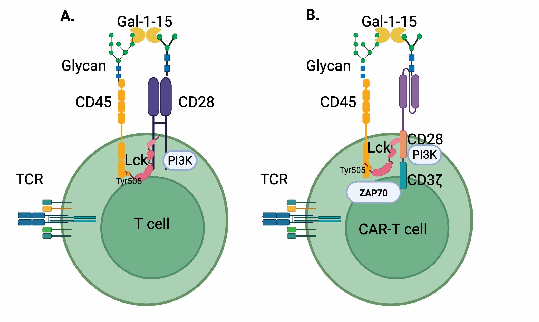
Figure 1. Comparison of a native T cell and a CAR-T cell. A. Native T cell: Constitutive signaling through Lck and PI3K is supported by CD45, while galectins bring CD45 and CD28 into proximity. B. CAR-T cell: The CAR contains ITAMs associated with CD28 and CD3ζ, which can promote constitutive signaling and proliferation, but may be limited by the Hayflick limit.
Conclusion
Based on the above analysis, it should be safe to claim that CD28 is not required for T-cell activation when T cells are activated by antigen-presenting cells via the TCR-MHC I/II pathways. Instead, CD28 and CD45 work together to keep T cells surviving in normal conditions through the LCK recruitment ability of CD28 in vivo, which is constitutively associated with inactive pTyr505 LCK, and could be activated by CD45 (via removing the phosphate group on Tyr505) when they are brought together by Galectin proteins in the circulation.
Acknowledgments
We thank members of National Jewish Health for their suggestions and support. We thank Dr. Arthur Weiss for his input and suggestions. The work is partially supported by an NIH grant GM135421 (G.Z.) and funds from NB Life Laboratory LLC, specifically private financial support from Cheng-Yuan Zhang, Peng Sun, Yun-Xia Jiang, and Yongmei Jiang. Figure 1 is generated by BioRender.
Contributions
GZ conceived the concepts, data analysis, and writing up the manuscript.
Conflicts of Interest
GZ holds equity in NB Life Laboratory LLC.
References
2. Martin PJ, Ledbetter JA, Morishita Y, June CH, Beatty PG, Hansen JA. A 44 kilodalton cell surface homodimer regulates interleukin 2 production by activated human T lymphocytes. J Immunol. 1986 May 1;136(9):3282–7.
3. Weiss A, Manger B, Imboden J. Synergy between the T3/antigen receptor complex and Tp44 in the activation of human T cells. J Immunol. 1986 Aug 1;137(3):819–25.
4. Hara T, Fu SM, Hansen JA. Human T cell activation. II. A new activation pathway used by a major T cell population via a disulfide-bonded dimer of a 44 kilodalton polypeptide (9.3 antigen). J Exp Med. 1985 Jun 1;161(6):1513–24.
5. Moretta A, Pantaleo G, Lopez-Botet M, Moretta L. Involvement of T44 molecules in an antigen-independent pathway of T cell activation. Analysis of the correlations to the T cell antigen-receptor complex. J Exp Med. 1985 Sep 1;162(3):823–38.
6. NOWELL PC. Phytohemagglutinin: an initiator of mitosis in cultures of normal human leukocytes. Cancer Res. 1960 May;20(4):462–6.
7. Dwyer JM, Johnson C. The use of concanavalin A to study the immunoregulation of human T cells. Clin Exp Immunol. 1981 Nov;46(2):237–49.
8. Jamali I, Field EH, Fleming A, Cowdery JS. Kinetics of anti-CD4-induced T helper cell depletion and inhibition of function. Activation of T cells by the CD3 pathway inhibits anti-CD4-mediated T cell elimination and down-regulation of cell surface CD4. J Immunol. 1992 Mar 15;148(6):1613–9.
9. Esensten JH, Helou YA, Chopra G, Weiss A, Bluestone JA. CD28 Costimulation: From Mechanism to Therapy. Immunity. 2016 May 17;44(5):973–88.
10. Zhang W, Sloan-Lancaster J, Kitchen J, Trible RP, Samelson LE. LAT: the ZAP-70 tyrosine kinase substrate that links T cell receptor to cellular activation. Cell. 1998 Jan 9;92(1):83–92.
11. Shahinian A, Pfeffer K, Lee KP, Kündig TM, Kishihara K, Wakeham A, et al. Differential T cell costimulatory requirements in CD28-deficient mice. Science. 1993 Jul 30;261(5121):609–12.
12. Green JM, Noel PJ, Sperling AI, Walunas TL, Gray GS, Bluestone JA, et al. Absence of B7-dependent responses in CD28-deficient mice. Immunity. 1994 Sep;1(6):501–8.
13. Suh WK, Tafuri A, Berg-Brown NN, Shahinian A, Plyte S, Duncan GS, et al. The inducible costimulator plays the major costimulatory role in humoral immune responses in the absence of CD28. J Immunol. 2004 May 15;172(10):5917–23.
14. Strioga M, Pasukoniene V, Characiejus D. CD8+ CD28- and CD8+ CD57+ T cells and their role in health and disease. Immunology. 2011 Sep;134(1):17–32.
15. Mou D, Espinosa J, Lo DJ, Kirk AD. CD28 negative T cells: is their loss our gain? Am J Transplant. 2014 Nov;14(11):2460–6.
16. Barber EK, Dasgupta JD, Schlossman SF, Trevillyan JM, Rudd CE. The CD4 and CD8 antigens are coupled to a protein-tyrosine kinase (p56lck) that phosphorylates the CD3 complex. Proc Natl Acad Sci U S A. 1989 May;86(9):3277–81.
17. van Oers NS, Lowin-Kropf B, Finlay D, Connolly K, Weiss A. alpha beta T cell development is abolished in mice lacking both Lck and Fyn protein tyrosine kinases. Immunity. 1996 Nov;5(5):429–36.
18. Lui VG, Hoenig M, Cabrera-Martinez B, Baxter RM, Garcia-Perez JE, Bailey O, et al. A partial human LCK defect causes a T cell immunodeficiency with intestinal inflammation. J Exp Med. 2024 Jan 1;221(1):e20230927.
19. Rudd CE. How the Discovery of the CD4/CD8-p56lck Complexes Changed Immunology and Immunotherapy. Front Cell Dev Biol. 2021 Mar 15;9:626095.
20. Nika K, Soldani C, Salek M, Paster W, Gray A, Etzensperger R, et al. Constitutively active Lck kinase in T cells drives antigen receptor signal transduction. Immunity. 2010 Jun 25;32(6):766–77.
21. Furlan G, Minowa T, Hanagata N, Kataoka-Hamai C, Kaizuka Y. Phosphatase CD45 both positively and negatively regulates T cell receptor phosphorylation in reconstituted membrane protein clusters. J Biol Chem. 2014 Oct 10;289(41):28514–25.
22. Bergman M, Mustelin T, Oetken C, Partanen J, Flint NA, Amrein KE, et al. The human p50csk tyrosine kinase phosphorylates p56lck at Tyr-505 and down regulates its catalytic activity. EMBO J. 1992 Aug;11(8):2919–24.
23. Nada S, Okada M, MacAuley A, Cooper JA, Nakagawa H. Cloning of a complementary DNA for a protein-tyrosine kinase that specifically phosphorylates a negative regulatory site of p60c-src. Nature. 1991 May 2;351(6321):69–72.
24. Nada S, Yagi T, Takeda H, Tokunaga T, Nakagawa H, Ikawa Y, et al. Constitutive activation of Src family kinases in mouse embryos that lack Csk. Cell. 1993 Jun 18;73(6):1125–35.
25. Chan AC, Iwashima M, Turck CW, Weiss A. ZAP-70: a 70 kd protein-tyrosine kinase that associates with the TCR zeta chain. Cell. 1992 Nov 13;71(4):649–62.
26. Holmes N. CD45: all is not yet crystal clear. Immunology. 2006 Feb;117(2):145–55.
27. Kishihara K, Penninger J, Wallace VA, Kündig TM, Kawai K, Wakeham A, et al. Normal B lymphocyte development but impaired T cell maturation in CD45-exon6 protein tyrosine phosphatase-deficient mice. Cell. 1993 Jul 16;74(1):143–56.
28. Stone JD, Conroy LA, Byth KF, Hederer RA, Howlett S, Takemoto Y, et al. Aberrant TCR-mediated signaling in CD45-null thymocytes involves dysfunctional regulation of Lck, Fyn, TCR-zeta, and ZAP-70. J Immunol. 1997 Jun 15;158(12):5773–82.
29. Earl LA, Bi S, Baum LG. N- and O-glycans modulate galectin-1 binding, CD45 signaling, and T cell death. J Biol Chem. 2010 Jan 22;285(4):2232–44.
30. Clark MC, Pang M, Hsu DK, Liu FT, de Vos S, Gascoyne RD, et al. Galectin-3 binds to CD45 on diffuse large B-cell lymphoma cells to regulate susceptibility to cell death. Blood. 2012 Nov 29;120(23):4635–44.
31. Gourdine JF, Nava P, Noll AJ, Duong DM, Seyfried NT, Cummings RD. CD45 and Basigin (CD147) Are Functional Ligands for Galectin-8 on Human Leukocytes. Biomolecules. 2025 Aug 27;15(9):1243.
32. Wang RZ, Yang F, Sui J. B7-H4 Binds Galectin-9 Glycosylation-Dependently and Attenuates Galectin-9-Mediated CD28/AKT Activation and T Cell Death. bioRxiv. 2025 Aug 1:2025–07.
33. Yang R, Sun L, Li CF, Wang YH, Yao J, Li H, et al. Galectin-9 interacts with PD-1 and TIM-3 to regulate T cell death and is a target for cancer immunotherapy. Nat Commun. 2021 Feb 5;12(1):832.
34. Irving BA, Weiss A. The cytoplasmic domain of the T cell receptor zeta chain is sufficient to couple to receptor-associated signal transduction pathways. Cell. 1991 Mar 8;64(5):891–901.
35. Sadelain M, Rivière I, Brentjens R. Targeting tumours with genetically enhanced T lymphocytes. Nat Rev Cancer. 2003 Jan;3(1):35–45.
36. Braendstrup P, Levine BL, Ruella M. The long road to the first FDA-approved gene therapy: chimeric antigen receptor T cells targeting CD19. Cytotherapy. 2020 Feb;22(2):57–69.