

Abstract
Biological evidence recovered from crime scenes is often subject to environmental conditions or suffers significant degradation, which can limit the accuracy of routine forensic genetic analyses. With their high stability and tissue-specific expression profiles, microRNAs (miRNAs) are emerging as promising forensic biomarkers for overcoming these limitations. This review focuses on the roles of miRNAs in the identification of body fluids, such as blood, semen, saliva, and vaginal secretions, as well as their current applications in postmortem interval (PMI) estimation, age determination, differentiation of monozygotic twins and forensic toxicology. While literature findings demonstrate that miRNAs provide robust results even in degraded samples, they also highlight significant methodological differences between the panels, reference genes, and analytical platforms employed.
Ultimately, miRNAs have significant potential as biomarkers in forensic genetics due to their high stability, tissue specificity, and wide range of applications. Nevertheless, due to shortcomings in the selection of reference genes, normalization strategies, and standardization of analytical workflows, no miRNA-based forensic test has yet been adopted for routine use. In the foreseeable future, the accuracy and clinical applicability of miRNA-based analyses are expected to improve through the integration of next-generation sequencing technologies, multi-biomarker panels, exosomal miRNA analyses, and artificial intelligence-supported data evaluation approaches into forensic applications. Consequently, the conduct of large-scale validation studies and the development of standardized analytical protocols to ensure inter-laboratory reproducibility are of critical importance from a forensic perspective.
Keywords
miRNA, Forensic genetics, Body fluid identification, Postmortem interval, Age estimation, Monozygotic twins
Introduction
In forensic genetic investigations, the accurate identification of biological evidence recovered from the crime scene is a critical step in identifying perpetrators and solving the crime. However, this evidence is often subject to environmental conditions at the crime scene or to cleaning or destruction by perpetrators seeking to tamper with the evidence [1]. Even though traditional forensic DNA profiling is regarded as the gold standard for identifying individuals based on the analysis of short tandem repeats (STRs) and single nucleotide polymorphisms (SNPs), it may prove inadequate for advanced forensic issues such as determining the type of biological fluid, age estimation, estimating the time of death, and distinguishing between monozygotic twins. Furthermore, as it is not always possible to obtain sufficient quantities of undegraded DNA, various studies in literature point to the development of new methods [2,3].
Although mRNA-based analyses have been widely employed in recent studies aimed at identifying biomarkers resistant to environmental conditions, their application to degraded forensic samples remains limited due to the relatively large size of the amplification products (approximately 200–300 nt) and the susceptibility of mRNA molecules to environmental factors, including heat, ultraviolet (UV) radiation, humidity, and ribonuclease (RNase) activity [4].
MicroRNAs (miRNAs) small non-coding RNA (ncRNA) molecules of approximately 18–24 nucleotides in length that play a role in post-transcriptional gene regulation—are highlighted in recent literature as promising biomarkers for forensic genetics [5–7]. They were first discovered in 1993 with the identification of the lin-4 gene in the Caenorhabditis elegans model [8]. It was established that miRNAs constitute an important class of gene-regulatory RNAs upon the realization that the second miRNA identified, let-7, is conserved across many species [9,10].
Because of their low molecular weights and high stability, miRNAs are resistant to degradation even under harsh conditions such as temperature and pH changes and chemical treatments. As they are present in blood, saliva, and other bodily fluids, they exhibit tissue-specific expression and offer significant advantages in the identification of biological samples in forensic genetics. Furthermore, as they can be analyzed with high sensitivity using methods such as qRT-PCR, microarrays, and NGS, they have become a powerful forensic biomarker [11,12].
This review comprehensively addresses the most up-to-date applications of miRNAs in forensic genetics, which are characterized by their high stability and tissue specificity. Within the scope of this study, the applications of miRNA expression profiles in important forensic cases such as the identification of body fluids and bloodstains, the estimation of postmortem interval (PMI), the estimation of biological age, the differentiation of monozygotic twins, and the determination of dependency profiles have been examined, and the analytical challenges in the field and future application perspectives have been discussed.
Identification of Body Fluids
Body fluids such as blood, semen, and saliva are frequently encountered at crime scenes. Accurate identification of the cellular origin of these biological samples represents one of the most fundamental steps in the investigation and resolution of forensic cases. It is necessary to know from which fluid a stain originates to prove the sequence of events (such as allegations of sexual assault or physical violence). Nevertheless, the washing away of evidence at the crime scene or the passage of a significant amount of time can reduce the sensitivity of traditional detection methods and lead to false-negative results [13,14].
Although mRNA-based applications are considered for identifying bodily fluids due to tissue-specific expression profiles, their greater susceptibility to degradation under environmental conditions compared to miRNAs has limited their applicability [15]. Additionally, the ability to simultaneously extract miRNA and genomic DNA from the same sample provides significant practicality and sample conservation in forensic analysis workflows [16].
miR-451 and miR-16, which are highly expressed during erythrocyte development, are widely recognized as the most reliable biomarkers for the identification of peripheral blood [17]. For semen, miR-135b and miR-10b have been reported as the most informative markers, whereas miR-658 and miR-205 are associated with saliva, and miR-124a and miR-372 with vaginal secretions [4]. Wang et al. (2013) identified miR-16 and miR-486 for venous blood, miR-888 and miR-891a for semen, miR-214 for menstrual blood, miR-124a for vaginal secretions, and miR-138-2 for saliva as potential markers [18]. Another important issue in forensic investigations is the discrimination between peripheral blood and menstrual blood. In this regard, miR-141-3p, miR-373-3p, miR-497-5p, miR-143-5p, and miR-136-5p have been identified as menstrual blood-specific biomarkers [19]. Moreover, RT-qPCR-based miRNA expression ratio analyses are being successfully applied to definitively distinguish between peripheral blood and menstrual blood. More specifically, the detection of body fluids via miRNA analysis has been achieved using the miR-451a/miR-21-5p ratio, even with trace amounts of RNA as little as 0.2 ng obtained from crime scenes [20]. Owing to their small molecular size and remarkable resistance to enzymatic degradation, miRNAs exhibit excellent stability in forensic samples. For example, miR-126 and miR-16 remained detectable in blood for up to two weeks, while miR-135b and miR-10b were successfully detected in semen stains after six months [21]. Chen et al. (2023) developed a combined detection strategy integrating the high tissue specificity of mRNA with miRNA analysis. This approach achieved reliable identification in diluted samples, mixed biological samples, and simulated crime scene evidence, with successful detection in blood at concentrations as low as 0.1 pg [22]. Furthermore, stability studies demonstrated that miRNA biomarkers are more resistant to environmental degradation than mRNA markers. Consequently, multiplex approaches combining the superior tissue specificity of mRNA with the exceptional stability of miRNAs have been proposed as a promising strategy for improving the analysis of forensic biological samples [23].
The detection of semen is particularly important in the investigation of sexual assault cases. Among the currently available biomarkers, miR-888-5p and miR-891a-5p are the most consistently validated semen-specific miRNAs [11,18,24,25]. In particular, hsa-miR-891a-5p has repeatedly been demonstrated as a highly reliable semen-specific biomarker, allowing semen to be distinguished from blood with high accuracy, even in mixed biological samples [26].
Studies focusing on the identification of saliva and vaginal secretions have identified miR-223 and miR-145 as saliva-specific biomarkers, whereas miR-1260b and miR-654-5p have been associated with vaginal secretions [27]. In addition, in stability analyses conducted under various environmental conditions, miR-205 is a promising candidate biomarker for saliva detection despite environmental stress factors [28]. In complex forensic cases, the combination of hsa-miR-203a-3p and hsa-miR-124-3p has successfully distinguished saliva from vaginal secretion samples, which share similar cellular origins. However, distinguishing between these fluids in mixed samples remains challenging [26]. Nevertheless, it has been determined that miR-203a-3p and miR-223-3p are expressed at higher levels in saliva and miR-1260b in vaginal secretions, compared to other body fluids [25].
Overall, miRNA-based analysis represents a highly promising approach for the identification of forensic body fluids because of the exceptional stability of miRNAs, their tissue-specific expression profiles, and their ability to provide reliable results even from minute amounts of biological material. Although there are areas requiring further development regarding the analysis of mixed samples and the differentiation of certain body fluids from one another, current studies indicate that the use of miRNA markers, either alone or in combination with other molecular markers, will make a significant contribution to the more accurate interpretation of crime scene evidence and the clarification of forensic investigations.
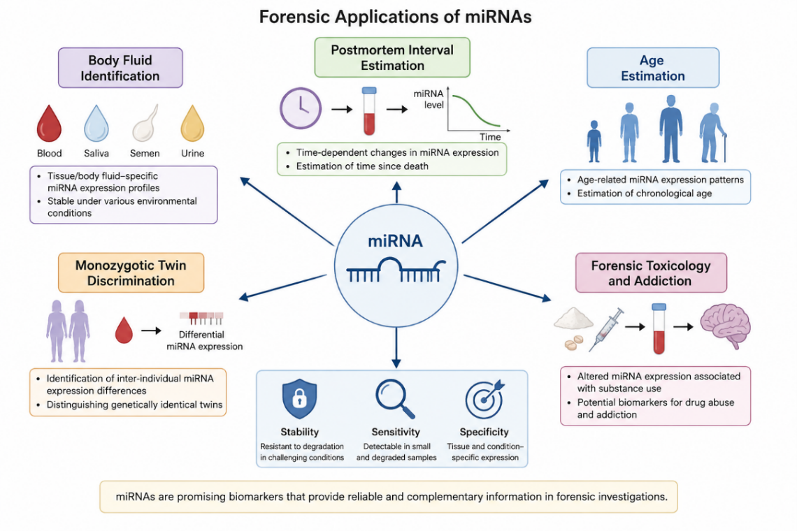
Figure 1. Overview of forensic applications of miRNAs.
Mixed Biological Samples
Biological evidence collected from a crime scene is often not found in its pure form in forensic cases but consists of a mixture of multiple biological sources. This is particularly evident in incidents such as sexual assault, physical alterations, and murder; mixed biological samples containing various combinations of biological fluids, such as semen, vaginal secretions, blood, and saliva, are frequently recovered from crime scenes [29]. The combined application of multiplex miRNA panels with high-throughput analytical techniques, such as reverse transcription quantitative polymerase chain reaction (RT-qPCR) and next-generation sequencing (NGS), enables simultaneous identification of multiple body fluids within the same biological sample [30]. A multiclass support vector machine (MSVM) model was evaluated using 17 mixed biological samples obtained from actual forensic casework. The model showed promising performance, particularly in the identification of semen–vaginal secretion mixtures from sexual assault cases; however, as vaginal secretions and saliva share similar epithelial cell characteristics, these two bodily fluids could not be accurately distinguished in some samples [31].
Analysis of Aged Blood Stains
As bloodstains are the most commonly encountered bodily fluid at a crime scene, they play a crucial role in solving criminal cases. Once blood has left the body, it is subject to various physical, chemical, and biological factors and is at risk of degradation. Traditional DNA profiling may prove inadequate in samples that have been exposed to environmental conditions, have deteriorated, or contain only trace amounts of material.
One of the comprehensive studies evaluating miRNA stability in bloodstains was carried out by Zhao et al. (2021). In this study, blood samples were exposed to different temperatures, humidity levels, UV light intensities, and natural conditions, and two miRNA markers (hsa-miR-16-5p, hsa-miR-451a) and a reference gene (U6 snRNA) were analyzed via RT-qPCR. As a result, miR-451a demonstrated high stability, with detection possible for up to 6 years under laboratory conditions and for 6 months at high temperatures. Additionally, rain accelerated miRNA degradation, and it was determined that the commonly used reference gene U6 was affected more rapidly by environmental conditions than the miRNAs. This situation raises questions regarding the reliability of the control genes used in miRNA analyses. However, the short nucleotide sequences make them more resistant to enzymatic and environmental effects, suggesting that they can be used as reliable biomarkers under challenging crime scene conditions [32].
Dalkiran et al. (2024), on the other hand, demonstrated that hsa-miR-451a is a robust miRNA biomarker that can be used to identify bloodstains. However, environmental conditions significantly impact the stability of this biomarker. In particular, while miRNA remains detectable for an extended period under appropriate storage conditions (–20°C, +4°C, and in airtight bags), environmental factors such as soil, water, bleach, and washing severely damage the miRNA, thereby reducing the success of the analysis [33].
In recent years, multiplex analytical approaches integrating miRNAs with other highly stable RNA molecules, such as circular RNAs (circRNAs), have increasingly been explored. For example, Chen et al. (2024) investigated the stability of miRNA and circRNA biomarkers under challenging crime scene conditions. Blood samples were exposed to high temperatures, ultraviolet radiation, cold environments, and disinfectants. Despite these challenging conditions, the target RNAs were successfully detected using RT-qPCR. In particular, circ0000095 demonstrated high stability and tissue specificity, and miRNAs and circRNAs can be used as reliable molecular markers in degraded biological samples [34].
Traditionally, the reverse transcription quantitative polymerase chain reaction (RT-qPCR) has been the most widely used method for miRNA analysis and stability assessment. However, recent advances in next-generation sequencing (NGS) and massively parallel sequencing (MPS) technologies have made substantial contributions to miRNA research in forensic molecular biology. In one of the pioneering studies in this field, Seashols-Williams et al. (2016) analyzed eight different body fluids, including blood, saliva, urine, feces, semen, vaginal fluid, menstrual blood, and sweat, using high-throughput sequencing on the Illumina platform [35]. Similarly, Wang et al. (2016) sequenced small RNAs isolated from blood and saliva samples using the Ion PGM platform. In addition to validating previously reported biomarkers, the authors identified one novel miRNA biomarker for blood and 15 novel miRNA biomarkers for saliva [36]. Comprehensive small RNA sequencing performed on six different body fluids and tissue samples identified 1,394 miRNAs, including 236 previously unreported miRNAs, along with numerous piRNAs, snoRNAs, and snRNAs. This analysis also revealed several potential biomarkers specific to different body fluids [37]. More recently, the integration of miRNA sequencing (miRNA-seq) data with machine learning algorithms has enabled the identification of the biological origin of body fluids and the more accurate and specific prediction of forensic parameters, such as the time since deposition (TsD) and postmortem interval (PMI) [38].
Estimation of the Postmortem Interval (PMI)
Postmortem interval (PMI) estimation is one of the most challenging issues in forensic medicine because it refers to the time elapsed between death and the examination of the body, and its accurate determination is complicated by numerous influencing factors. Reliable estimation of the PMI is essential in criminal investigations, as it contributes to crime scene reconstruction, verification of witness statements, and placement of potential suspects within the relevant time frame.
Traditional PMI estimation methods rely on macroscopic findings such as algor mortis, rigor mortis, livor mortis, and entomological variables; however, these parameters are highly susceptible to environmental conditions [39].
Molecular studies investigating the degradation of DNA, RNA, and proteins have also been employed to estimate the PMI. These studies have demonstrated that nucleic acid degradation is influenced by ribonuclease activity, bacterial decomposition, environmental factors such as temperature, humidity, and sunlight exposure, as well as the circumstances surrounding death. Furthermore, RNA stability varies considerably among tissues, with relatively higher stability observed in the brain, heart, and skeletal muscle, whereas lower stability has been reported in the pancreas and liver [11].
Recent studies have shown that miRNAs offer advantages in estimating the time of death [15,40,41]. Due to their small size and association with protective proteins, miRNAs are more resistant to postmortem degradation. With their high stability, miRNAs can be used as more consistent biomarkers for estimating the time of death [6].
In some studies, cardiac tissue has been used as a target to assess miRNA expression. Research has shown that the miRNAs miR-122, miR-133a, and miR-1 remain stable for an extended period during the postmortem process. For this reason, these miRNAs are suitable for use as reference genes in heart tissue. Lv Y et al. (2017) reported that miR-1 and miR-133a exhibited high and stable expression in the heart for up to 144 hours after death [42]. Likewise, Tu C et al. (2018) determined that miR-133a remains stable for up to 8 days post-mortem in both cardiac and skeletal muscle tissue [43].
Singh et al. (2023) examined heart tissue samples obtained from traffic accident cases for up to 196 hours. While miRNA-1 maintained its stability as a reference gene, miRNA-195, miRNA-206, and miRNA-378 exhibited a significant degradation profile proportional to the time elapsed since death [40]. Singh et al. (2025) used miRNAs in combination with the cardiac troponin I (cTnI) protein marker to estimate PMI. In particular, it has been determined that miRNAs undergo degradation depending on the cause of death in cases such as burns and electric shock. The findings demonstrated that miRNA-195 is a reliable biomarker for early-stage PMI prediction, whereas miRNA-378, due to its high stability, is a reliable biomarker for determining mid- and late-stage PMI [44].
miRNAs found within exosomes exhibit high stability during the postmortem process. These lipid-enveloped structures protect miRNAs from degradation and maintain their integrity for a longer period than free-circulating miRNAs. Kanno et al. (2023) found that exosomal structures and the miRNAs they contain successfully maintain their integrity for at least 3 days at storage temperatures such as 4°C and 20°C. Because of these properties, exosomal miRNAs can be used as reliable molecular markers for estimating the time of death [45].
Corradini et al. (2015) demonstrated that certain miRNAs are expressed at different levels depending on the time window in which death occurs. Statistically significant circadian rhythm differences were detected in the expression profiles of miR-106b and miR-96 in vitreous humor, and of miR-142-5p and miR-219 in blood. SNORD95 was the most stable reference gene in both blood and vitreous humor samples [46]. Further, Lazzari et al. (2026), in their study on vitreous humor samples, emphasized that hsa-miR-96-5p exhibits high stability and could serve as a reliable endogenous control in PMI analyses [41].
Biological Age Estimation
The ability to estimate an individual's age from biological samples recovered at a crime scene is an important area of research in forensic genetics. Age estimation can help narrow the profile of a suspect or victim and provides valuable information, particularly in cases where legal responsibility depends on age [47]. Traditional methods of age estimation are generally based on the examination of skeletal and dental development; however, where these tissues are unavailable, molecular biomarkers are required. Age can be estimated molecularly using telomerase length, mitochondrial DNA mutations and DNA methylation markers [48].
miRNAs not only provide tissue specificity but also exhibit differences in expression levels during the ageing process. Their high stability and resistance to environmental degradation further support their application as biomarkers for forensic age estimation. The first evidence that the expression of many miRNAs decreases with advancing age was reported by Hooten et al. (2010), who profiled more than 800 miRNAs in peripheral blood mononuclear cells from young and elderly individuals using real-time RT-PCR [49]. Supporting these findings, another study examined serum miRNA profiles in individuals aged 40–70 years and found that the expression of miR-29b and miR-106b decreased with age, whereas miR-92a, miR-222, and miR-375 showed increased expression [50]. More recent studies have focused on integrating miRNAs with other molecular datasets to reduce the limitations and potential sources of error associated with single-biomarker systems. Wang et al. (2022) analyzed 11 age-associated miRNAs and 4 circular RNAs (circRNAs) via RT-qPCR in blood samples collected from 200 individuals aged between 20 and 80 years. The Random Forest Regression model demonstrated the highest performance on the test set, with an average absolute error (MAE) of 6.84 years [51]. The inclusion of DNA methylation in miRNA analyses, which enhances the power of age prediction and resilience to environmental conditions [52,53], has improved prediction accuracy. Gao et al. (2025) combined the analysis of DNA methylation in the KLF14 gene region with miR-106b-5p expression in a single study and established a 4-plex droplet digital PCR (ddPCR) platform capable of simultaneous measurement alongside two reference genes, carrying out the analysis on 132 blood samples. Using their Random Forest model, they achieved high prediction accuracy with an average absolute deviation (MAD) of 3.51 years [54].
Distinguishing Monozygotic Twins
Monozygotic (MZ) twins share a genetically nearly identical genome, rendering traditional DNA identification methods based on STR profiling and SNPs inadequate for distinguishing between these individuals. The inability to distinguish monozygotic twins in forensic genetic applications creates challenges in investigative processes [55]. In monozygotic twins, the effects of environmental factors accumulate over the course of a person’s life, leading to age-related differences in DNA methylation patterns and miRNA expression profiles. Consequently, higher levels of expression differences are observed, particularly in older twins [56]. This suggests that the sensitivity of miRNA-based discrimination methods may increase over time and that age is a significant factor in the success of the analysis.
In recent years, studies have been conducted using miRNAs to distinguish monozygotic twins. Fang et al. (2019) identified the markers miR-151a-3p, miR-1-3p, and miR-451a—which exhibited the highest expression levels among six miRNAs expressed at different levels in MZ twins—using MPS and qRT-PCR methods [57]. Similarly, Xiao et al. (2019) identified 10 potential miRNA markers with high discriminatory power in monozygotic twins using the qRT-PCR method [58]. Studies have been conducted combining next-generation sequencing (miRNA-seq) and “droplet digital PCR” (ddPCR) methods to enhance analytical power. As a result, results with higher sensitivity compared to qPCR were obtained using the ddPCR technology. Five specific miRNA markers (hsa-miR-1273h-5p, hsa-miR-3192-5p, hsa-miR-188-5p, hsa-miR-206, and hsa-miR-6775-3p) were validated using this method. Additionally, these miRNAs remained stable without degradation in blood spots stored for 180 days and after 10 freeze-thaw cycles [59].
Forensic Toxicology and Addiction
Drug addiction is a significant problem from both a legal and a public health perspective, characterized by an individual’s inability to control their substance use, the development of a strong craving for the substance, and the emergence of withdrawal symptoms upon cessation of use. Clinically classified as “substance use disorder (SUD),” this condition can range in severity from mild use to severe addiction [60]. In the detection of substance use, drug substances and their metabolites are traditionally analyzed in blood samples; however, the levels of these compounds in the bloodstream decrease over time, limiting their detection [61]. Consequently, there is a need to develop stable biomarkers that can indicate drug use more accurately and reliably; miRNAs have emerged as biomarkers of interest in forensic toxicology due to their high stability and ability to regulate gene expression.
Cocaine use leads to persistent epigenetic changes in the brain associated with the reward mechanism. These changes can be investigated through miRNA analyses.
In studies conducted on animal models, following chronic cocaine use, levels of miR-124 and let-7d were reduced in the prefrontal cortex, ventral tegmental area, and caudate putamen, while miR-181a levels were increased in several regions. Furthermore, extracellular vesicles containing miR-124 can suppress cocaine-induced inflammation, highlighting the role of this miRNA in neuroinflammation [62]. Analyses of peripheral blood samples have also revealed elevated levels of miR-124 and miR-181 in cocaine users [63]. The effects of cocaine use on the vascular system have also been linked to miRNA alterations, and it is thought that miR-30c-5p, in particular, may play a role in the processes of atherosclerosis and oxidative stress [64].
Methamphetamine (METH) is an amphetamine-derived stimulant that affects the central nervous system and has a high potential for addiction. This substance exerts its effects by increasing levels of dopamine, noradrenaline and serotonin. With long-term use, it can cause psychosis-like symptoms, mood disorders and serious behavioral changes [65–67]. Studies conducted on plasma samples have found that levels of miR-181a, miR-15b, let-7e and let-7d are lower in methamphetamine users compared to healthy individuals. Furthermore, it has been noted that this reduction is associated with duration of use and severity of addiction [68]. In post-mortem examinations, let-7b-3p levels were found to be significantly elevated in the ventral tegmental area and nucleus accumbens in deaths attributed to methamphetamine toxicity. This suggests that the relevant miRNA could serve as a potential biomarker in forensic investigations [69].
Plasma levels of hsa-miR-592, hsa-miR-9-3p, hsa-miR-206, and hsa-let-7b-3p were significantly higher in methamphetamine-dependent individuals compared to the control group. In particular, while hsa-miR-9-3p alone demonstrated high discriminatory power with an AUC value of 0.756, when combined with hsa-miR-592, this value increased to 0.871, achieving a sensitivity of 82.7% and a specificity of 78.9% [70].
Limitations
While studies on miRNAs have demonstrated that they have broad potential for application in forensic genetics, there are still significant methodological barriers to the adoption of these markers for routine laboratory use. For the routine use of miRNAs, a miRNA panel must be validated across all laboratories. Independent studies have not yet demonstrated high reproducibility [71]. Although numerous reference genes have been investigated for normalization, a definitive reference gene that can be reliably used has not yet been identified [11].
Another significant limitation is that miRNA expression levels vary according to biological and environmental factors. Age, gender, ethnicity, hormonal status, infections, inflammatory processes, chronic diseases, medication use, and lifestyle factors can all cause changes in miRNA expression levels.
Even though miRNA-based analyses offer the advantages of high stability and sensitivity, full standardization across studies has not been achieved due to differences in extraction protocols, normalization strategies and the selection of reference genes. For this reason, large-scale validation studies are required for routine integration into forensic practice.
Conclusion
The reliable analysis of biological evidence in forensic science is of great importance for elucidating events. The studies reviewed in this article indicate that miRNAs have considerable potential as forensic biomarkers because of their stability and tissue-specific expression patterns. To date, encouraging findings have been reported in several forensic applications, including body fluid identification, age estimation, postmortem interval (PMI) estimation, the discrimination of monozygotic twins, and the assessment of substance use. Furthermore, the use of miRNAs along with other molecular markers, such as DNA methylation and circRNAs, has enhanced the accuracy of analyses.
For miRNA-based methods to be widely used in routine forensic practice, validation studies on larger sample groups, standardized analytical protocols, and reliable normalization strategies are required. With technological advancements, the widespread adoption of next-generation sequencing methods, and progress in AI-supported analytical approaches, miRNAs are expected to play a significant role in forensic science in the future and contribute to the resolution of complex cases.
References
2. Karadayı S, Yılmaz İ, Özbek T, Karadayı B. Transfer and persistence of microbiota markers from the human hand to the knife: A preliminary study. J Forensic Leg Med. 2024 Oct;107:102757.
3. Butler JM. Recent advances in forensic biology and forensic DNA typing: INTERPOL review 2019-2022. Forensic Sci Int Synerg. 2022 Dec 27;6:100311.
4. Hanson EK, Lubenow H, Ballantyne J. Identification of forensically relevant body fluids using a panel of differentially expressed microRNAs. Anal Biochem. 2009 Apr 15;387(2):303–14.
5. Courts C, Madea B. Micro-RNA - A potential for forensic science? Forensic Sci Int. 2010 Dec 15;203(1-3):106–11.
6. Pasaribu RS, Auerkari EI, Suhartono AW, Auerkari P. A small RNA, microRNA as a potential biomolecular marker to estimate post mortem interval in forensic science: a systematic review. Int J Legal Med. 2023 Sep;137(5):1313–25.
7. Wiraagni IA, Pidada IB, Heriyanto DS, Wiguna GN, Azhari MD, Amalia S, et al. The role of microRNA in forensic science: a bibliometric study of research trends and applications. Egyptian Journal of Medical Human Genetics. 2025 Dec 11;26(1):185.
8. Lee RC, Feinbaum RL, Ambros V. The C. elegans heterochronic gene lin-4 encodes small RNAs with antisense complementarity to lin-14. Cell. 1993 Dec 3;75(5):843–54.
9. Reinhart BJ, Slack FJ, Basson M, Pasquinelli AE, Bettinger JC, Rougvie AE, et al. The 21-nucleotide let-7 RNA regulates developmental timing in Caenorhabditis elegans. Nature. 2000 Feb 24;403(6772):901–6.
10. Pasquinelli AE, Reinhart BJ, Slack F, Martindale MQ, Kuroda MI, Maller B, et al. Conservation of the sequence and temporal expression of let-7 heterochronic regulatory RNA. Nature. 2000 Nov 2;408(6808):86–9.
11. Rocchi A, Chiti E, Maiese A, Turillazzi E, Spinetti I. MicroRNAs: An Update of Applications in Forensic Science. Diagnostics (Basel). 2020 Dec 26;11(1):32.
12. Silva SS, Lopes C, Teixeira AL, Carneiro de Sousa MJ, Medeiros R. Forensic miRNA: potential biomarker for body fluids? Forensic Sci Int Genet. 2015 Jan;14:1–10.
13. Glynn CL. Potential applications of microRNA profiling to forensic investigations. RNA. 2020 Jan;26(1):1–9.
14. Karadayı Ş, Karadayı B, Öner D, Çetin G. Evaluation of the relationship between the detectability of seminal stains on laundered fabric and stain age. Med Sci Law. 2021 Jul;61(3):198–207.
15. Montanari E, Giorgetti R, Busardò FP, Giorgetti A, Tagliabracci A, Alessandrini F. Suitability of miRNA assessment in postmortem interval estimation. Eur Rev Med Pharmacol Sci. 2021 Feb;25(4):1774–87.
16. Li Y, Zhang J, Wei W, Wang Z, Prinz M, Hou Y. A strategy for co-analysis of microRNAs and DNA. Forensic Sci Int Genet. 2014 Sep;12:24–9.
17. Bruchova H, Yoon D, Agarwal AM, Mendell J, Prchal JT. Regulated expression of microRNAs in normal and polycythemia vera erythropoiesis. Exp Hematol. 2007 Nov;35(11):1657–67.
18. Wang Z, Zhang J, Luo H, Ye Y, Yan J, Hou Y. Screening and confirmation of microRNA markers for forensic body fluid identification. Forensic Sci Int Genet. 2013 Jan;7(1):116–23.
19. Li Z, Bai P, Peng D, Wang H, Guo Y, Jiang Y, et al. Screening and confirmation of microRNA markers for distinguishing between menstrual and peripheral blood. Forensic Sci Int Genet. 2017 Sep;30:24–33.
20. Wang G, Wang Z, Wei S, Wang D, Ji A, Zhang W, et al. A new strategy for distinguishing menstrual blood from peripheral blood by the miR-451a/miR-21-5p ratio. Forensic Sci Int Genet. 2022 Mar;57:102654.
21. Al-Mawlah YH, Asma’a HM, Abd-Alameer AM, Hadi AM, Abdulabbas HS, Shaheed SH, et al. Assessment of the specificity and stability of Micro-RNAs as a forensic gene marker. Biomedical and Biotechnology Research Journal (BBRJ). 2023 Oct 1;7(4):569–76.
22. Chen X, Xu H, Zhu B. Forensic validation of a combined analysis of mRNA and miRNA markers for precise tissue origin inferences of five kinds of body fluids by RT-qPCR. Electrophoresis. 2023 Nov;44(21-22):1714–24.
23. Bamberg M, Bruder M, Kunz SN, Wöhrle T, Wiegand P. Really the best of both? Application of an mRNA/miRNA multiplex assay to casework samples, animal samples, and a storage study. Forensic Sci Int Genet. 2025 Jan;74:103129.
24. Liu Y, He H, Xiao ZX, Ji A, Ye J, Sun Q, et al. A systematic analysis of miRNA markers and classification algorithms for forensic body fluid identification. Brief Bioinform. 2021 Jul 20;22(4):bbaa324.
25. Fujimoto S, Manabe S, Morimoto C, Ozeki M, Hamano Y, Hirai E, et al. Distinct spectrum of microRNA expression in forensically relevant body fluids and probabilistic discriminant approach. Sci Rep. 2019 Oct 4;9(1):14332.
26. Sauer E, Reinke AK, Courts C. Differentiation of five body fluids from forensic samples by expression analysis of four microRNAs using quantitative PCR. Forensic Sci Int Genet. 2016 May;22:89–99.
27. Park JL, Park SM, Kwon OH, Lee HC, Kim JY, Seok HH, et al. Microarray screening and qRT-PCR evaluation of microRNA markers for forensic body fluid identification. Electrophoresis. 2014 Nov;35(21-22):3062–8.
28. Iroanya OO, Olutunde OT, Egwuatu TF, Igbokwe C. Stability of selected microRNAs in human blood, semen and saliva samples exposed to different environmental conditions. Forensic Sci Int. 2022 Jul;336:111338.
29. Arat Ö, Kaya-akyüzlü D. Vücut Sıvılarının Kimliklendirilmesinde miRNA Çalışmalarının Potansiyel Önemi: Güncel Bir Bakış. Adli Tıp Dergisi. 2024 Apr 4;38(1):80–92.
30. Uchimoto ML, Beasley E, Coult N, Omelia EJ, World D, Williams G. Considering the effect of stem-loop reverse transcription and real-time PCR analysis of blood and saliva specific microRNA markers upon mixed body fluid stains. Forensic Sci Int Genet. 2013 Jul;7(4):418–21.
31. Li S, Liu J, Xu W, Zhang S, Zhao M, Miao L, et al. A multi-class support vector machine classification model based on 14 microRNAs for forensic body fluid identification. Forensic Sci Int Genet. 2025 Feb;75:103180.
32. Zhao C, Zhao M, Zhu Y, Zhang L, Zheng Z, Wang Q, et al. The persistence and stability of miRNA in bloodstained samples under different environmental conditions. Forensic Sci Int. 2021 Jan;318:110594.
33. Dalkiran NL, Ercan B, Karatas O, Holumen N, Yukseloglu EH. Determination of blood stains exposed to environmental conditions using miRNA biomarkers. Medicine Science. 2024 Jun 1;13(2):499.
34. Chen X, Xu H, Lin Y, Zhu B. Forensic stability evaluation of selected miRNA and circRNA markers in human bloodstained samples exposed to different environmental conditions. Forensic Sci Int. 2024 Sep;362:112148.
35. Seashols-Williams S, Lewis C, Calloway C, Peace N, Harrison A, Hayes-Nash C, et al. High-throughput miRNA sequencing and identification of biomarkers for forensically relevant biological fluids. Electrophoresis. 2016 Oct;37(21):2780–8.
36. Wang Z, Zhou D, Cao Y, Hu Z, Zhang S, Bian Y, et al. Characterization of microRNA expression profiles in blood and saliva using the Ion Personal Genome Machine(®) System (Ion PGM™ System). Forensic Sci Int Genet. 2016 Jan;20:140–6.
37. Liu Z, Wang Q, Wang N, Zang Y, Wu R, Sun H. A Comprehensive Characterization of Small RNA Profiles by Massively Parallel Sequencing in Six Forensic Body Fluids/Tissue. Genes (Basel). 2022 Aug 25;13(9):1530.
38. Cai M, Lan Q, Xie T, Liu Q, Zhao M, Wu X, et al. Decoding temporal miRNA signatures of semen under in vitro exposure for forensic time since deposition estimation using machine learning‐driven modeling. Interdisciplinary Medicine. 2026 May;4(3):e70093.
39. Franceschetti L, Amadasi A, Bugelli V, Bolsi G, Tsokos M. Estimation of Late Postmortem Interval: Where Do We Stand? A Literature Review. Biology (Basel). 2023 May 28;12(6):783.
40. Singh P, Ali W, Sandhu S, Mishra S, Singh US, Verma AK, et al. Post-mortem interval estimation using miRNAs of road traffic accident cases: A forensic molecular approach. Sci Justice. 2023 Jul;63(4):485–92.
41. Lazzari J, Scatena A, Di Paolo M, Rocchi A. Selection and Validation of Endogenous Reference microRNAs for Post-Mortem Interval Estimation in Vitreous Humor: A Preliminary Study. Int J Mol Sci. 2026 Feb 24;27(5):2102.
42. Lv YH, Ma JL, Pan H, Zeng Y, Tao L, Zhang H, et al. Estimation of the human postmortem interval using an established rat mathematical model and multi-RNA markers. Forensic Sci Med Pathol. 2017 Mar;13(1):20–7.
43. Tu C, Du T, Shao C, Liu Z, Li L, Shen Y. Evaluating the potential of housekeeping genes, rRNAs, snRNAs, microRNAs and circRNAs as reference genes for the estimation of PMI. Forensic Sci Med Pathol. 2018 Jun;14(2):194–201.
44. Singh P, Ali W, Sandhu S, Wamique M, Kushwaha R, Verma AK, et al. Exploring the paradigm of miRNA and cardiac troponin I expression in prolonged post-mortem interval. Egyptian Journal of Forensic Sciences. 2025 May 15;15(1):33.
45. Kanno S, Sakamoto T, Fukuta M, Kato H, Aoki Y. Stability of exosomes in the postmortem serum and preliminary study on exosomal miRNA expression profiling in serum from myocardial infarction cadavers. Int J Legal Med. 2023 May;137(3):825–34.
46. Corradini B, Alu M, Radheshi E, Gabbolini V, Ferrari F, Santunione AL, et al. Estimation of the time of death through the analysis of clock miRNAs expression in blood and vitreous humor. Forensic Science International: Genetics Supplement Series. 2015 Dec 1;5:e204–6.
47. Eipel M, Mayer F, Arent T, Ferreira MR, Birkhofer C, Gerstenmaier U, et al. Epigenetic age predictions based on buccal swabs are more precise in combination with cell type-specific DNA methylation signatures. Aging (Albany NY). 2016 May;8(5):1034–48.
48. Correia Dias H, Cunha E, Corte Real F, Manco L. Age prediction in living: Forensic epigenetic age estimation based on blood samples. Leg Med (Tokyo). 2020 Nov;47:101763.
49. Noren Hooten N, Abdelmohsen K, Gorospe M, Ejiogu N, Zonderman AB, Evans MK. microRNA expression patterns reveal differential expression of target genes with age. PLoS One. 2010 May 20;5(5):e10724.
50. Zhang H, Yang H, Zhang C, Jing Y, Wang C, Liu C, et al. Investigation of microRNA expression in human serum during the aging process. J Gerontol A Biol Sci Med Sci. 2015 Jan;70(1):102–9.
51. Wang J, Zhang H, Wang C, Fu L, Wang Q, Li S, et al. Forensic age estimation from human blood using age-related microRNAs and circular RNAs markers. Front Genet. 2022 Nov 22;13:1031806.
52. Yang F, Qian J, Qu H, Ji Z, Li J, Hu W, et al. DNA methylation-based age prediction with bloodstains using pyrosequencing and random forest regression. Electrophoresis. 2023 May;44(9-10):835–44.
53. Er S, Abik Z, Ersoy G, Filoglu G, Ozkara H, Bulbul O. A semen-specific deoxyribonucleic acid methylation model for epigenetic age estimation and its robustness under environmental challenges. Electrophoresis. 2024 Oct;45(19-20):1820–33.
54. Gao N, Li J, Yang F, Yu D, Huo Y, Liu X, et al. Forensic Age Estimation From Blood Samples by Combining DNA Methylation and MicroRNA Markers Using Droplet Digital PCR. Electrophoresis. 2025 Apr;46(7-8):424–32.
55. Liu MH, Xia X, Wang YL, Wang DY, Wang SW, Chen YZ, et al. Current progress and future perspectives in personal identification of monozygotic twins in forensic medicine. Forensic Sci Int Genet. 2025 Mar;76:103231.
56. Bell JT, Spector TD. A twin approach to unraveling epigenetics. Trends Genet. 2011 Mar;27(3):116–25.
57. Fang C, Zhao J, Liu X, Zhang J, Cao Y, Yang Y, et al. MicroRNA profile analysis for discrimination of monozygotic twins using massively parallel sequencing and real-time PCR. Forensic Sci Int Genet. 2019 Jan;38:23–31.
58. Xiao C, Pan C, Liu E, He H, Liu C, Huang Y, et al. Differences of microRNA expression profiles between monozygotic twins' blood samples. Forensic Sci Int Genet. 2019 Jul;41:152–8.
59. Wang DY, Tian MH, Chen YZ, Wang SW, Xing XY, Sun ML, et al. Profiling microRNA expression differentiates monozygotic twins in peripherical blood by droplet digital PCR. Forensic Sci Int Genet. 2025 Mar;76:103230.
60. Occhipinti C, La Russa R, Iacoponi N, Lazzari J, Costantino A, Di Fazio N, et al. miRNAs and Substances Abuse: Clinical and Forensic Pathological Implications: A Systematic Review. Int J Mol Sci. 2023 Dec 4;24(23):17122.
61. Mancinelli R, Guiducci MS. Aspetti procedurali e problemi d'interpretazione nell'analisi di sostanze d'abuso [Procedural aspects and interpretation problems in the analysis of drugs of abuse]. Ann Ist Super Sanita. 2002;38(3):305–13.
62. Chandrasekar V, Dreyer JL. microRNAs miR-124, let-7d and miR-181a regulate cocaine-induced plasticity. Mol Cell Neurosci. 2009 Dec;42(4):350–62.
63. Viola TW, Heberle BA, Zaparte A, Sanvicente-Vieira B, Wainer LM, Fries GR, et al. Peripheral blood microRNA levels in females with cocaine use disorder. J Psychiatr Res. 2019 Jul;114:48–54.
64. Zhu W, Wang H, Wei J, Sartor GC, Bao MM, Pierce CT, et al. Cocaine Exposure Increases Blood Pressure and Aortic Stiffness via the miR-30c-5p-Malic Enzyme 1-Reactive Oxygen Species Pathway. Hypertension. 2018 Apr;71(4):752–60.
65. Pantoni MM, Kim JL, Van Alstyne KR, Anagnostaras SG. Correction to: MDMA and memory, addiction, and depression: dose-effect analysis. Psychopharmacology (Berl). 2022 Jul;239(7):2369.
66. Li MJ, Shoptaw SJ. Clinical management of psychostimulant withdrawal: review of the evidence. Addiction. 2023 Apr;118(4):750–62.
67. Alanazi AS, Alahmad KM, Almutairi SH, Alanazi BF, Bahkali KJ, Alkhamis AN, et al. Methamphetamine Use: Neurochemical Effects Health Outcomes and Addiction Trajectories. Journal of Medical and Life Science. 2026 Mar 1;8(1):156–71.
68. Zhao Y, Zhang K, Jiang H, Du J, Na Z, Hao W, et al. Decreased Expression of Plasma MicroRNA in Patients with Methamphetamine (MA) Use Disorder. J Neuroimmune Pharmacol. 2016 Sep;11(3):542–8.
69. Demirel G, Tanoglu EG, Aslıyuksek H. Evaluation of microRNA let-7b-3p expression levels in methamphetamine abuse. Rev Assoc Med Bras (1992). 2023 Apr 17;69(4):e20221391.
70. Li W, Liu D, Liu X, Lu Y, Zhang L, Yu F, et al. Combined Diagnostic Value of Hsa-miR-592 and Hsa-miR-9-3p in Plasma for Methamphetamine Addicts. Int J Mol Sci. 2024 Aug 16;25(16):8952.
71. Zendjabil M. Preanalytical, analytical and postanalytical considerations in circulating microRNAs measurement. Biochem Med (Zagreb). 2024 Jun 15;34(2):020501.