Loading
Archives of Clinical Ophthalmology
ISSN: 2771-7925
Editors Choice Articles
Comment on “Retinitis Pigmentosa and Molar Tooth Sign Caused by Novel AHI1 Compound Heterozygote Pathogenic Variants: A Case Report”
Qing Lv, Ailian Du
Joubert syndrome (JS) is a rare congenital neurodevelopmental disease which is basically a primary Ciliopathy. It’s characteristic manifestation on imaging is so called ‘molar tooth sign’ in the brainstem and cerebellum. JS can involve multiple organs, mainly including retina, kidney, bone and liver. Clinical signs of early onset JS include hypotonia, developmental delay, breathing abnormalities, and ocular motor apraxia.
Arch Clin Ophthalmol, 2022, Volume 2, Issue 1, p1-2 | DOI: 10.33696/Ophthalmology.2.005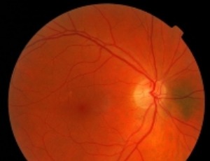
Repeatability of Scheimpflug Corneal Tomography in Patients with Keratoconus and Different Body Mass Indices
James S Lewis, Lize Angelo, Akilesh Gokul, Charles NJ McGhee, Mohammed Ziaei
To evaluate the repeatability of corneal tomographic parameters in keratoconus patients across different body mass index (BMI) categories. This prospective study was conducted at the University of Auckland, New Zealand, from June 2021 to June 2022. A total of 243 eyes from keratoconus patients aged 18-45 years were categorized into normal (BMI ≤24.9; n=55), overweight (BMI 25.0-29.9; n=58), and obese (BMI ≥30.0; n=130) groups. Patients underwent three consecutive scans using the Pentacam AXL.
Arch Clin Ophthalmol, 2025, Volume 4, Issue 1, p1-11 | DOI: 10.33696/Ophthalmology.4.015
Stroke and Visual Loss in a Young Girl with Dengue Fever – Report of a Case and a Mini Review
Sweety Trivedi, Ambar Chakravarty
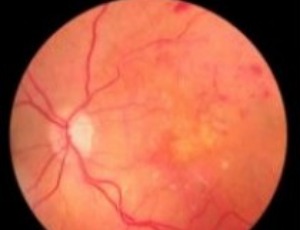
The case of a young girl with Dengue fever presenting with seizures and bilateral visual loss is presented. At the time of presentation, she had right hemiplegia and dysarthria but was not dysphasic. Fundoscopy revealed presence of macular and disc oedema in the right eye and vitreous haemorrhage in the left eye.
Arch Clin Ophthalmol, 2022, Volume 2, Issue 1, p3-8 | DOI: 10.33696/Ophthalmology.2.006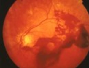
Focal Aggregates of Normal or Near Normal Uveal Melanocytes (FANNUMs) in the Choroid. A Practical Clinical Category of Small Ophthalmoscopically Evident Discrete Melanocytic Choroidal Lesions
James J. Augsburger
Focal aggregate of normal or near normal uveal melanocytes (FANNUM) of the choroid is a term the author has proposed to categorize small melanocytic choroidal lesions that are not detectably thicker than surrounding normal choroid by B-scan ocular ultrasonography. In this article, the author describes the clinical features of small melanotic choroidal lesions he categorizes clinically as FANNUMs and discusses the presumed compositional spectrum of such lesions.
Arch Clin Ophthalmol, 2021, Volume 1, Issue 1, p8-19 | DOI: 10.33696/Ophthalmology.1.002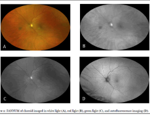
Multidisciplinary Acute Care of Central Retinal Artery Occlusion with a Stroke Paradigm: A Call to Action
Stacey Q. Wolfe, Stephanie A. Coffman, Mark Perez, Katriel Lee, Bartlett H.Hayes, Tamra Ranasinghe, Patrick A. Brown, Kyle M. Fargen
Central retinal artery occlusion (CRAO) is an ophthalmologic emergency that can result in permanent vision loss. Over 25% of CRAO are associated with acute cerebral ischemia, and there are many parallels between CRAO and acute ischemic stroke. There are no definitive treatment algorithms for CRAO, however there may be opportunities to treat CRAO as an “eye stroke”. Given the similarities to acute ischemic stroke, multidisciplinary involvement and stroke algorithms should be considered and tested for this disease.
Arch Clin Ophthalmol, 2021, Volume 1, Issue 1, p20-26 | DOI: 10.33696/Ophthalmology.1.004