

Abstract
Introduction: Chronic non-specific lower back pain (LBP) is a common musculoskeletal condition that may be associated with biomechanical alterations in the lower extremities. Flatfoot, characterized by a reduction in the medial longitudinal arch, may influence lower-limb alignment and spinal mechanics, potentially contributing to changes in lumbar lordosis and low back pain. Customized total contact insoles (TCIs) may help address these biomechanical alterations by providing appropriate plantar support. However, evidence regarding their effects on lumbar lordosis, low back pain-related disability, and foot alignment in individuals with flatfoot remains limited.
Methodology: A prospective single-arm pre-post interventional study was conducted involving 15 participants with chronic non-specific LBP and flatfoot. Customized TCIs were provided for eight weeks. Lumbar lordosis angle was measured using standardized lateral lumbar spine radiographs, while low back pain-related disability was assessed using the Modified Oswestry Disability Questionnaire (MODQ). Foot alignment was evaluated by measuring Meary’s angle on lateral foot radiographs. All three parameters were assessed before and after 8 weeks of the intervention.
Results and conclusion: Pre- and post-intervention comparisons with and without orthosis of the lumbar lordosis angle and Meary’s angle were statistically significant (p < 0.001), and MODQ scores showed p = 0.001. a significant negative correlation was also observed between changes in foot alignment and lumbar lordosis (p = 0.013). These findings indicate that customized TCIs were associated with significant changes in lumbar curvature, low back pain-related disability, and radiographic foot alignment over eight weeks. Customized TCIs may represent a useful conservative approach for individuals with flatfoot and chronic non-specific LBP.
Keywords
Flatfoot, Chronic non-specific low back pain, Total contact insoles, Lumbar lordosis, Meary’s angle
Introduction
Lower back pain (LBP) is a prominent cause of disability worldwide, and its multiple etiologies make it difficult to treat [1]. Global Burden of Disease research ranks LBP as one of the top 10 diseases and injuries that individuals face on a daily basis. In developed countries, the prevalence of LBP varies from 60–70%, with 15–45% of persons developing LBP each year. This frequency is particularly high among people between 35 and 55 years old, and it will significantly increase with age due to trauma, stress, or intervertebral disc disorders [2]. A 2025 retrospective cross-sectional study reported considerable variation in LBP cases across the 2019–2023 period, with estimates ranging from 42% to 83% and the highest proportion of cases observed in 2023 (22.63%) among the analyzed patient records. These findings highlight the substantial clinical burden of LBP and the need for effective approaches to its management in the Indian population [3].
LBP is characterized as either structural (specific/non-mechanical) or non-structural (mechanical/ nonspecific), with mechanical LBP accounting for over 90% of cases. The term "non-specific low back pain" (LBP) refers to discomfort that arises from the facet joints, ligaments, vertebrae, or intervertebral disks but has no identifiable underlying cause. In contrast, recognizable illnesses such as infection, cancer, osteoporosis, spinal fractures, structural deformities, inflammatory disorders, radicular syndrome, or cauda equina syndrome are linked to structural or specific LBP. Flatfoot has been associated with mechanical low back pain, potentially due to alterations in foot biomechanics and lower-limb alignment. Lulupoy et al. investigated this relationship using Clarke’s angle to assess foot posture in individuals with mechanical low back pain. These findings suggest that flatfoot may contribute to altered biomechanical loading and postural adaptations, although its direct association with non-specific low back pain requires further investigation [4,5]. Therefore, researchers have made efforts to understand the mechanism of non-specific LBP by examining the lower limb distal problems, such as deviations of the foot and ankle. Faulty foot biomechanics can have a negative impact on all supporting joints above the foot/ankle complex, including LBP. Previous investigations have proven a relationship between nonspecific low back pain and podiatric abnormalities such as flatfoot. Therefore, early detection of foot posture changes becomes necessary in them to help identify the presence of low back pain, as emerging research suggests that foot posture abnormalities, particularly flatfoot, may contribute to such non-specific/mechanical LBP [6].
Flatfoot syndrome, also known as pes planus, is characterized by various static and dynamic abnormalities as well as partial or total collapse of the medial longitudinal arch (MLA). Furthermore, it is characterized by the abduction of the forefoot, internal rotation, plantar flexion of the talus, and calcaneal eversion, which induce compensatory changes in the lower extremities, pelvis, and lumbar spine, potentially changing spinal biomechanics and increasing susceptibility to pain and injury [5,6]. Flatfoot causes hyper-pronation of the subtalar joint, which is accompanied by talus adduction and plantarflexion, as well as calcaneal eversion. In closed-chain kinematics, foot hyper-pronation produces internal rotation of the tibia and femur. Internal rotation of the femur causes the head of the femur to migrate posteriorly and the pelvis to shift posteriorly [7]. In order to achieve postural balance, the trunk shifts more anteriorly, causing the center of mass of the body to shift in the same direction. This forces the pelvis to also tilt anteriorly, thereby increasing the lumbar lordosis arch. This increased lumbar lordosis in individuals with flatfoot is assumed to be the cause of non-specific low back pain. Therefore, it has been assumed that by maintaining the foot arches and hindfoot in neutral conservatively using total contact insoles, we can treat lumbar lordosis and its indirect linkage with non-specific low back pain in patients with flatfoot [8–10].
Emerging researchers were able to find the link between non-specific low back pain and flatfoot. This linkage shows that patients with flatfoot who complain of low back pain can be treated conservatively by taking steps to correct the podiatric deviation, i.e., flatfoot. Previous researchers have identified the effect of custom foot orthosis as a conservative treatment in flat-footed individuals to improve low back pain [11,12] but were not able to evaluate its impact on lumbar lordosis previously, along with related linkage with lower back pain, to the extent of the authors’ knowledge. Therefore, this study was designed to help bridge the gap in knowledge and give information regarding the appropriate conservative treatment option.
Methods
Participants
A prospective single-arm pre-post interventional clinical trial was designed for this study, which was conducted at Artificial Limbs Centre (ALC) of Indian Railway Post Graduate Institute of Medical Sciences and Research (IRPGIMSR), New Delhi. A convenience sample of 15 participants consisted of 6 males and 9 females with a mean age of 38.34 ± 9.21 years was clinically identified for non-specific low back pain and flatfoot. Informed consent was obtained from the study participants. The sample size was calculated using G*Power software (version 3.1.9.4), where the Cohen’s d value was taken as 1.00 from the post-treatment Oswestry disability value obtained from Castro-Mendez et al. [12], a two-sided significance level of 0.05 and 95% power, with a statistical test taken between two dependent means. The total sample size obtained was 16. One participant failed to report in the middle of the trial; hence, was excluded from the study. The inclusion criteria were as follows: Bilateral flatfoot confirmed using static digital foot scanning, flexible flatfoot using Jack’s test [13]; age ranging from 18–50 years [2,12], Non-specific low back pain (> 3 months), and willingness to wear TCI daily and follow up. The exclusion criteria were as follows: history of spinal fractures, disc herniation, spondylolisthesis, or other spinal disorders; any neurological disorders affecting lower limb biomechanics and gait (e.g., CP, Muscular dystrophy), presence of recent/ acute lower limb injury or trauma (within 6 months); and pregnancy.
Procedure
Patients who visited the center with non-specific LBP underwent lower limb examination using digital foot scanning at static weight-bearing. Arch Index (AI), used as the recruiting parameter, was calculated using the digital foot scan shown in Figure 1, as it is defined as the ratio of the midfoot area (B) to the area of the entire foot excluding the toes (A+B+C), i.e., AI = (B)/ (A+B+C). An AI value greater than 0.26 is indicative of flatfoot [14]. Accordingly, subjects were proposed to participate in this study after verification of inclusion/exclusion criteria. At baseline, lumbar lordosis and flatfoot were assessed using a lateral (LAT) lumbar spine (L1–S1) and Bilateral LAT foot radiographs, respectively, obtained in the standing position without wearing total contact insoles (TCIs). Radiographic procedures were performed following standard safety protocols. The angles were measured using Kinovea software (version 2023.1.2). Three authors independently measured each angle while blinded to the other measurements, and the mean of the three readings was used for the final analysis. Low back pain (LBP)-related disability was evaluated using the Modified Oswestry Disability Index (ODI), which assesses the impact of LBP on daily activities across 10 sections.
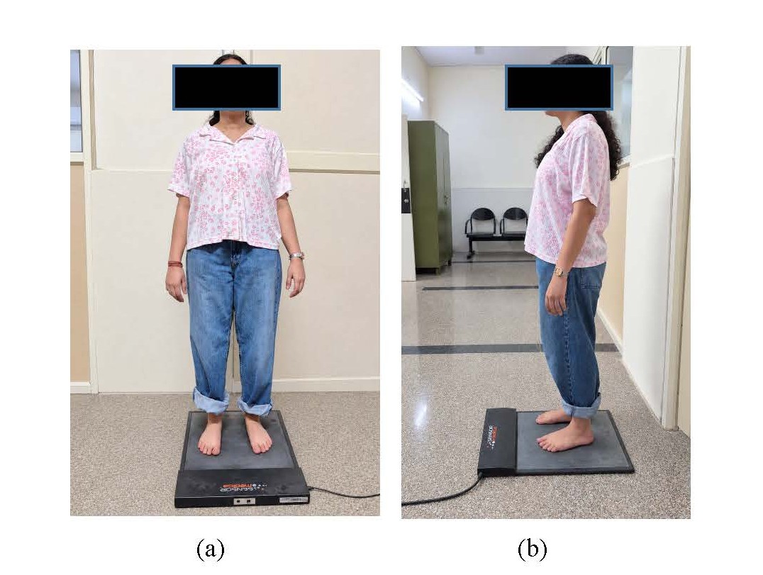
Figure 1. Performing digital foot scanning during static weight-bearing to calculate Arch Index (AI); a) Front view, b) Lateral view.
Following the baseline assessment, foot casts were taken on the first day to fabricate custom-made TCIs for each participant. The insoles were provided within 2–3 working days, along with instructions regarding their appropriate use. Participants were advised to wear the insoles for 8–10 hours daily over an eight-week intervention period. After eight weeks of TCI use, a follow-up assessment was conducted to evaluate changes in the study outcomes. Lumbar lordosis, Meary’s angle, and MODQ were reassessed while participants wore their custom-made TCIs after 8 weeks. Outcome measures taken into account for the study were as follows:
Primary outcome measures
1. Lumbar Lordosis angle: Lumbar lordosis is typically measured using the Cobb angle technique on a standing lateral lumbar radiograph, as it is still considered to be the gold standard tool for the assessment of lumbar lordosis. In this method, a line is drawn through the superior endplate of the first vertebra (L1), and a second line is drawn parallel to the superior endplate of the sacral base (S1). Then, perpendiculars are traced, and the angle at the intersection is precisely measured. Normal Cobb’s L1–S1 angle: Between 42° and 59° [15,16].
2. Modified Oswestry Disability Questionnaire (MODQ): MODQ score was used to evaluate the percentage of disability an individual encounters while performing everyday activities, where a greater percentage indicates more disability [8]. MODQ consists of ten items that assess the level of pain and interference with several physical activities: sleeping, self-care, social life, travelling, and homemaking. The validity and reliability of the English and another Indian language, including Hindi, have already been established [17]. Scoring instructions: For each section, the total possible score is 5: if the first statement is marked, the section score = 0; if the last statement is marked, it = 5. If all 10 sections are completed, the score is calculated as follows: Example: 16 (total scored) 50 (total possible score) x 100 = 32% [18].
Secondary outcome measure
1. Talo-first metatarsal or Meary’s angle: Meary’s angle (MA) measures the tilting of both the Talus and the first metatarsal bones [15]. The following axes were drawn on the weight-bearing lateral view: longitudinal axes of the talus (line bisecting the upper and lower edges) and longitudinal axes of Metatarsal 1 (line parallel to the upper edges) [19]. A normal Méary’s angle is 0 ± 4°. An angle greater than 4° with a superior apex indicates pes cavus, while an inferior apex indicates pes planus. Deformity severity is classified as mild (< 15°), moderate (15–30°), or severe (> 30°) [20].
Fabrication of the foot orthosis
Negative impressions of both feet were obtained in non-weight-bearing. During casting, the foot was positioned to achieve the subtalar joint in neutral and lock the hindfoot in neutral as well by applying subtle pressure over the sinus tarsi (ST) and peroneal groove. The natural elevation of the medial longitudinal arch (MLA) is obtained by first metatarsophalangeal joint dorsiflexion [21]. The negative cast is then filled with liquid plaster to obtain a positive mold of the foot. The mold rectification was done to support the MLA and the ST-peroneal groove; the rest of the mold was shaped to accommodate the plantar surface and to achieve total contact without excessive localized pressure. All the TCIs were made with polypropylene sheet of 3-mm thickness from the heel to just behind the metatarsal heads; the center of the heel area of 1 cm radius in polypropylene sheet was excavated and filled with 3-mm thick polyethylene foam sheet instead for added comfort. 4-mm thick polyethylene foam sheet is heated and vacuum molded and then adhered to the upper surface of the molded polyethylene covering the heel-to-toe region. Lastly, a 2-mm thick polyethylene sheet is heated and adhered onto the outer/lower surface of the molded orthosis from the rearfoot to surpassing the distal part of the toes (Figure 2).
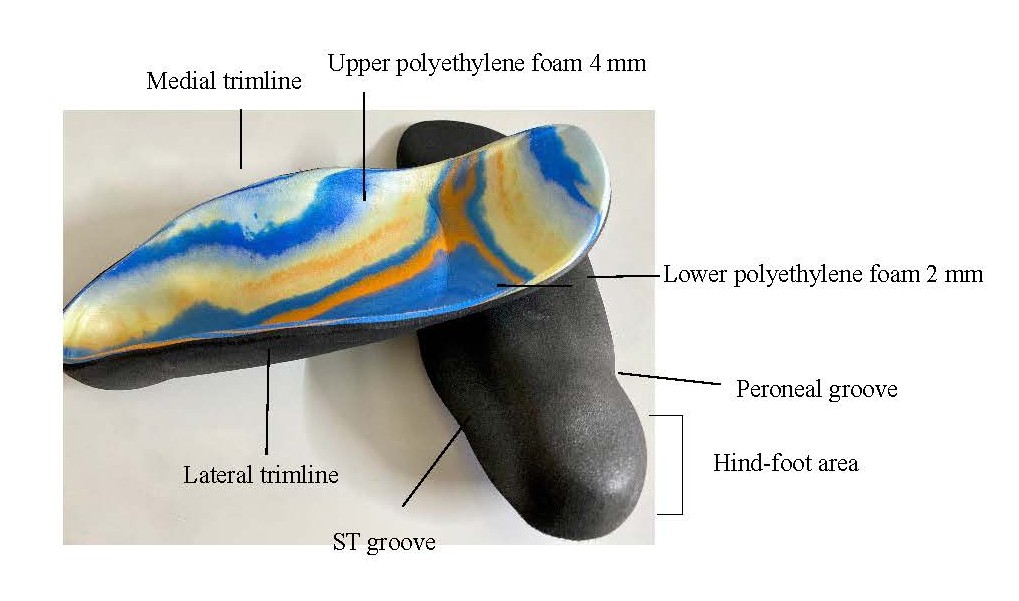
Figure 2. Customize Total Contact Insole (TCI) fabricated for each individual subjects.
Data analysis
The data were analyzed with the IBM SPSS software version 25.0. Descriptive data analysis for each of the dependent variables was assessed using the Shapiro-Wilk test and Kolmogorov-Smirnov test. The student t-test was used to compare pre-and post-intervention values of the lumbar Cobb angle and left and right Meary’s angle, provided the differences were approximately normally distributed. The Wilcoxon signed-rank test was used for MODQ scores when the assumptions for the paired t-test were not met. Correlation analysis was performed to examine the relationship between changes in foot alignment and lumbar lordosis following the intervention. Changes in left and right Meary’s angles were correlated separately with changes in the lumbar Cobb angle using Spearman’s correlation, as appropriate. A p-value of less than 0.05 was considered statistically significant.
Results
The experimental group consisted of 15 participants (6 males and 9 females) aged 18 to 50 years (mean 38.34 ± 9.21 years), with subsequent descriptive data of height, weight, Arch Index (AI), and BMI listed in Table 1. Table 2 presents the mean, standard deviation, 95% CI, median, and interquartile range of Lumbar Lordosis angle, Meary’s angle of the foot (right and left), and the MODQ questionnaire for the participants at the two moments of measurement, i.e., without TCI and after 8 weeks with TCI.
|
S. No |
Descriptive data |
Mean ± SD |
|
1 |
Age |
38.34 ± 9.21 |
|
2 |
Height |
161.4 ± 11.13 |
|
3 |
Weight |
75.85 ± 11.73 |
|
4 |
BMI |
29.10 ± 3.46 |
|
5 |
AI |
0.34 ± 0.06 |
|
*BMI: Body Mass Index; AI: Arch Index |
||
|
|
|
Mean ± SD |
95% CI lower limit |
95% CI Upper limit |
Median |
Interquartile range |
|
Cobb’s angle |
Pre Post |
46.79 ± 8.89 39.47 ± 7.02 |
41.86 35.58 |
51.71 43.36 |
46.20 39.0 |
14.2 10.4 |
|
Meary’s angle (Right foot) |
Pre Post |
-12.65 ± 6.6 -5.66 ± 3.5 |
-16.32 -7.60 |
-8.98 -3.71 |
-11.10 -5.0 |
7.4 4.6 |
|
Meary’s angle (Left foot) |
Pre Post |
-14.55 ± 7.35 -7.72 ± 3.53 |
-18.62 -9.67 |
-10.47 -5.76 |
-14.0 -8.1 |
9.5 5.3 |
|
MODQ |
Pre Post |
37.87± 17.49 22.33 ± 10.65 |
28.18 16.43 |
47.55 28.23 |
34.0 20.0 |
22 10 |
|
*CI: Confidence Interval; MODQ: Modified Oswestry Disability Questionnaire |
||||||
A paired-sample Student’s t-test was used to assess the pre- and post-intervention changes in Meary's angle for the right and left feet and cobb’s angle for lumbar lordosis. The results are presented in terms of mean difference and associated 95% CI, t-values, and p-values shown in Table 3.
|
Pre-Post of dependent variables |
|
Mean ± SD |
95% CI of the difference |
t-value |
p-value |
|
|
Lower |
Upper |
|||||
|
Meary’s angle |
Right foot |
-6.99 ± 5.27 |
-9.91 |
-4.07 |
-5.14 |
0.000* |
|
Meary’s angle |
Left foot |
-6.83 ± 4.61 |
-9.39 |
-4.27 |
-5.73 |
0.000* |
|
Cobb’s angle |
|
7.32 ± 2.13 |
6.13 |
8.50 |
13.25 |
0.000* |
For Meary’s angle, the mean pre-post difference was -6.99 (SD = 5.27; 95% CI: −9.91 to −4.07; t (14) = −5.14, p < 0.001) for the right foot and −6.83 (SD = 4.61; 95% CI: −9.39 to −4.27; t (14) = -5.73, p < 0.001) for the left foot. These findings indicate a significant shift in Meary’s angle toward more positive values, consistent with improved foot alignment toward a normal medial longitudinal arch. Cobb’s angle also showed a statistically significant pre–post difference (mean difference = 7.32, SD = 2.13; 95% CI: 6.13 to 8.50; t (14) = 13.25, p < 0.001). This reduction in Cobb’s angle indicates a decrease in lumbar lordosis following eight weeks of continuous insole use, suggesting a favorable change in lumbar alignment. The Modified Oswestry Disability Questionnaire (MODQ) scores decreased from 37.87 ± 17.49 at pre-intervention to 22.33 ± 10.65 after eight weeks of customized total contact insole use. The Wilcoxon signed-rank test demonstrated in Table 4 showed a statistically significant reduction in MODQ scores (Z = −3.413, p = 0.001), indicating an improvement in self-reported disability associated with low back pain following the intervention. All 15 participants showed a reduction in their MODQ scores, with no positive ranks observed. All pre-test post-test results for Meary’s angle, lumbar lordosis angle, and MODQ scores are graphically represented in Figures 3a–3c, respectively.
|
|
|
N |
Mean Rank |
Sum of ranks |
z-value |
p-value |
|
MODQ (pre-post) |
Negative rank Positive rank |
15 0 |
8.00 0.00 |
120.00 0.00 |
-3.413 |
0.001* |
|
*Significant at the 0.05 level (two-tailed). |
||||||
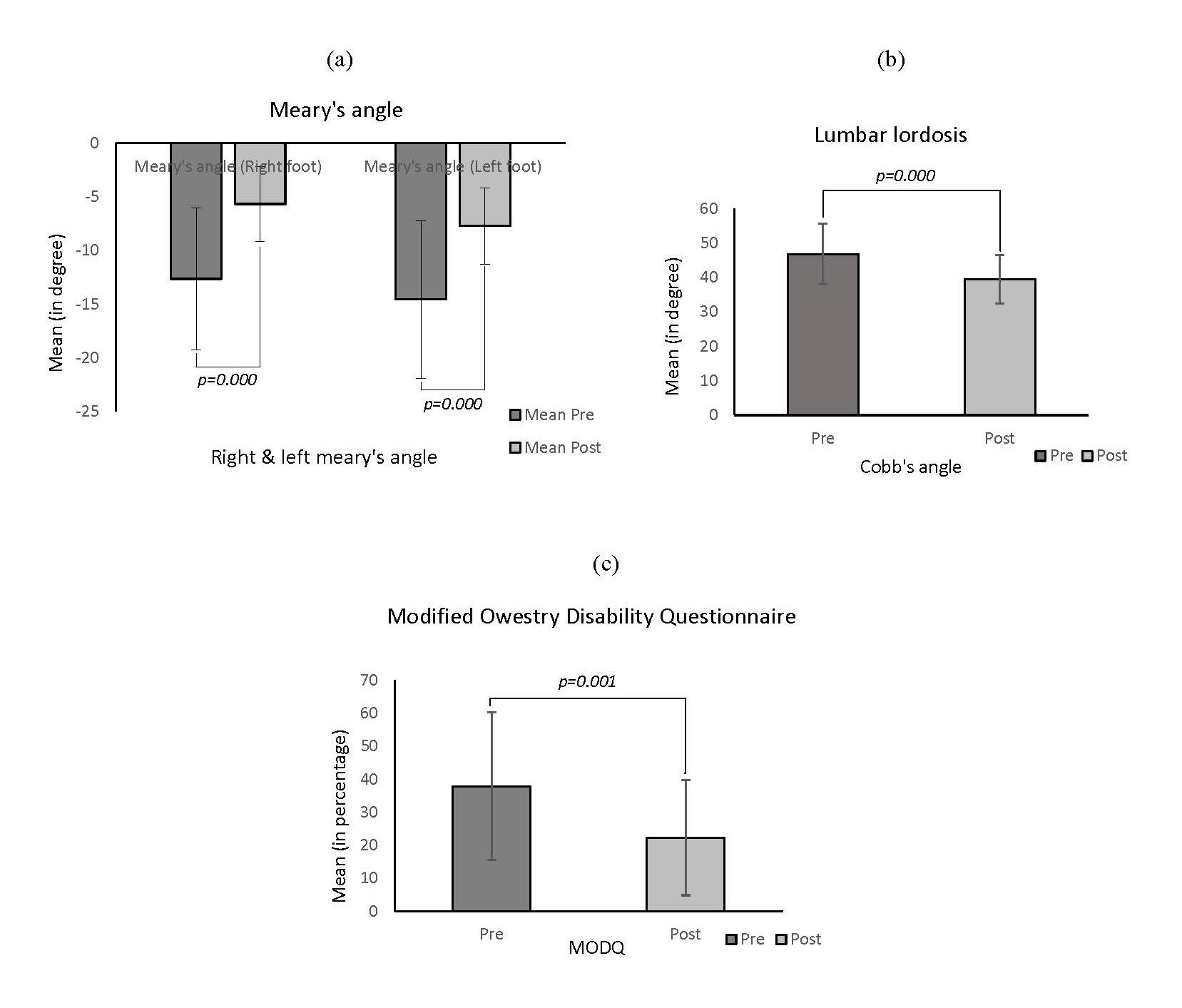
Figure 3. a) Pre- and post-test results of Meary’s angle-right and left foot, b) Pre- and post-results of Lumbar lordosis, c) Pre-post results of Modified Owestry Disability Questionnaire (MODQ).
Spearman’s rank correlation was used to examine the association between changes in bilateral foot alignment and changes in lumbar lordosis following eight weeks of customized total contact insole (TCI) use. To obtain an overall measure of foot alignment, the mean change in Meary’s angle from the right and left feet was calculated for each participant, rather than analyzing each foot separately. This mean bilateral change was then correlated with the change in lumbar Cobb’s angle, with both changes calculated as post-intervention minus pre-intervention measurements. The test revealed a statistically significant negative correlation between the change in lumbar Cobb’s angle and the mean bilateral change in Meary’s angle following eight weeks of customized total contact insole use [(rs = -0.626), (p = 0.013), (n = 15)] as shown in Table 5. The inverse association indicated that greater positive changes in Meary’s angle tended to be associated with greater reductions in lumbar lordosis. These findings suggest an association between changes in bilateral foot alignment and lumbar curvature when assessed while wearing the customized insoles.
|
Variables
|
Statistical test |
Correlation coefficient (rs) |
p-value |
N |
|
Mean B/L change in Meary’s angle Vs. Change in lumbar Cobb’s angle |
Spearman’s rank correlation |
-0.626 |
0.013* |
15 |
|
*Significant at the 0.05 level (two-tailed). Change scores were calculated as post-intervention minus pre-intervention values. The mean bilateral change in Meary’s angle was calculated by averaging the right and left foot changes for each participant. |
||||
Discussion
The present study examined changes in bilateral foot alignment, lumbar lordosis, and low-back-pain-related disability following eight weeks of customized total contact insole (TCI) use. The findings showed improvements in Meary’s angle in both feet, a reduction in lumbar lordosis, and lower MODQ scores after the intervention. A significant correlation between changes in bilateral foot alignment and lumbar curvature was also observed, suggesting a possible relationship between foot alignment and lumbo-pelvic posture.
The improvement in Meary’s angle indicates that the foot alignment shifted toward a more normal configuration while participants wore the insoles. Meary’s angle is an established radiographic measure for evaluating the relationship between the talus and first metatarsal and is commonly used in assessing medial longitudinal arch alignment [19,20]. Customized insoles may provide support to the medial longitudinal arch and redistribute plantar loading, potentially influencing lower-limb and lumbo-pelvic alignment. However, the observed changes should be interpreted in the context of the assessment conditions, as post-intervention radiographs were obtained while participants were wearing the insoles. The relationship between foot posture and lumbar alignment has been investigated in previous studies, although the evidence remains mixed. Dharmayat et al. [8] examined the association between foot posture and lumbar lordosis in teachers, while Borges et al. [10] explored the relationship between lumbar changes and plantar arch modifications in women with low back pain. The systematic review by O’Leary et al. [6] also highlighted the need for further evidence regarding the relationship between podiatric deviations and nonspecific chronic low back pain. These studies provide a basis for investigating whether changes in foot alignment are accompanied by changes in lumbar posture, although differences in populations, assessment methods, and study designs limit direct comparisons.
The significant negative correlation observed in the present study suggests that greater positive changes in mean bilateral Meary’s angle tended to accompany greater reductions in lumbar lordosis. This finding supports a possible association between changes in foot alignment and lumbar curvature. Nevertheless, the correlation does not establish a causal relationship, and the observed changes may also reflect other postural or biomechanical adaptations during insole use.
The reduction in MODQ scores indicates an improvement in self-reported disability associated with low back pain. This finding is broadly consistent with research investigating foot orthoses for individuals with low back pain. Castro-Méndez et al. [12] reported short-term effects of custom-made foot orthoses in individuals with excessive foot pronation and low back pain, while Cambron et al. [14] investigated shoe orthotics in a randomized controlled trial involving chronic low back pain. However, differences in orthotic design, treatment duration, and study populations should be considered when comparing these findings. Moreover, improvement in disability does not necessarily indicate that changes in lumbar curvature directly accounted for symptom improvement. This study has some limitations. The small sample size and absence of a control group limit the generalizability of the findings and prevent the observed changes from being attributed solely to the intervention; therefore, future studies involving larger samples, appropriate control groups, and assessments both with and without insoles would provide more clarity on the relationship between foot alignment, lumbar posture, and low-back-pain-related disability.
Conclusion
In conclusion, eight weeks of customized TCI use was associated with improvements in foot alignment, lumbar lordosis, and low-back-pain-related disability. The observed correlation between bilateral foot alignment and lumbar curvature highlights the potential relationship between foot mechanics and lumbo-pelvic posture. However, the findings do not establish lasting correction without the insoles or demonstrate that changes in lordosis caused the improvement in disability but instead helped in understanding a linkage of lumbar curvature with foot alignment and the subsequent effect of podiatric correction on the associated symptoms of LBP. Further controlled studies with larger samples are needed to confirm these findings.
Conflict of Interest
Nil.
Funding or Source of Support
None.
References
2. Almutairi AF, BaniMustafa A, Bin Saidan T, Alhizam S, Salam M. The Prevalence and Factors Associated with Low Back Pain Among People with Flat Feet. Int J Gen Med. 2021 Jul 20;14:3677–85.
3. Shetty SH, S AS, Karmakar A, Mitra M, Madkholkar N, Pawar R, et al. Evaluation of the Prevalence, Trends, and Correlates of Low Back Pain in India: A Retrospective Cross-Sectional Study. Cureus. 2025 May 5;17(5):e83518.
4. Cahya SA, Santoso WM, Husna M, Munir B, Kurniawan SN. Low back pain. Journal of Pain, Vertigo and Headache; 2021.1:13–7.
5. Lulupoy FA, Pradhana TM, Siahaan YM. Flat Foot and Its Association with Mechanical Low Back Pain: A Case-Control Study Utilizing Clarke’s Angle Measurement. World Journal of Clinical Medicine Research. 2025;5(1):34–9.
6. O'Leary CB, Cahill CR, Robinson AW, Barnes MJ, Hong J. A systematic review: the effects of podiatrical deviations on nonspecific chronic low back pain. J Back Musculoskelet Rehabil. 2013;26(2):117–23.
7. Levangie PK, Norkin CC. Joint Structure and Function: A Comprehensive Analysis. 4th ed. Philadelphia: F. A. Davis Company; 2005.
8. Dharmayat S, Thakkar MP, Kolmule S. Association of foot posture with lumbar lordosis angle in teachers. Indian journal of physical therapy and research. 2021 Jan 1;3(1):46–50.
9. Chun SW, Lim CY, Kim K, Hwang J, Chung SG. The relationships between low back pain and lumbar lordosis: a systematic review and meta-analysis. The Spine Journal. 2017 Aug 1;17(8):1180–91.
10. Borges CD, Fernandes LF, Bertoncello D. Relationship between lumbar changes and modifications in the plantar arch in women with low back pain. Acta ortopedica brasileira. 2013;21:135–8.
11. Cambron JA, Dexheimer JM, Duarte M, Freels S. Shoe orthotics for the treatment of chronic low back pain: a randomized controlled trial. Archives of Physical Medicine and Rehabilitation. 2017 Sep 1;98(9):1752–62.
12. Castro-Méndez A, Munuera PV, Albornoz-Cabello M. The short-term effect of custom-made foot orthoses in subjects with excessive foot pronation and lower back pain: a randomized, double-blinded, clinical trial. Prosthet Orthot Int. 2013 Oct;37(5):384–90.
13. Mosca VS. Flexible flatfoot in children and adolescents. J Child Orthop. 2010 Apr;4(2):107–21.
14. Liao HR, Yu HC, Li SJ. A deep learning-based plantar pressure measurement system for accurate foot arch index estimation. Applied Sciences. 2025 Sep 17;15(18):10156.
15. Eddine HK, Saleh S, Hajjar J, Harati H, Nasser Z, Desoutter A, et al. Evaluation of the accuracy of new modalities in the assessment and classification of lumbar lordosis: A comparison to Cobb's angle measurement. Heliyon. 2023 Aug 1;9(8):e18952.
16. Okpala FO. Comparison of four radiographic angular measures of lumbar lordosis. Journal of neurosciences in rural practice. 2018 Jul;9(3):298–304.
17. Chhabra HS, Kapoor KS. New modified english and hindi oswestry disability index in low back pain patients treated conservatively in Indian population. Asian spine journal. 2014 Oct 18;8(5):632–8.
18. Alcántara-Bumbiedro S, Flórez-García MT, Echávarri-Pérez C, García-Pérez F. Oswestry low back pain disability questionnaire. REHABILITACION-MADRID-. 2006;40(3):150–8.
19. Bourdet C, Seringe R, Adamsbaum C, Glorion C, Wicart P. Flatfoot in children and adolescents. Analysis of imaging findings and therapeutic implications. Orthopaedics & Traumatology: Surgery & Research. 2013 Feb 1;99(1):80–7.
20. Stein AM, Havasy J, Bitterman A, Herzenberg JE. The Méary angle: a historical perspective and contemporary review. Foot & Ankle Orthopaedics. 2025 Oct;10(4):24730114251386357.
21. Sichting F, Ebrecht F. The rise of the longitudinal arch when sitting, standing, and walking: Contributions of the windlass mechanism. PLoS One. 2021 Apr 8;16(4):e0249965.