Abstract
Normally, there are 23 pairs of chromosomes found in human beings. But in some conditions, a mother has a fetus with an additional chromosome at T21(21st position) which is said to be the fetus with Trisomy-21 (Down Syndrome). There are many factors of Down Syndrome - nasal bone absence, short nasal bone, thick amniotic fluid (greater than 3 mm), fetal heart rate (FHR), Crown Lump Length, short arm, and thighs’ bones; are screened during 10-14th (First Trimester) and 15-22nd week (Second Trimester) of pregnancy. These screenings are very crucial tasks as invasive methods, hence ultrasound imaging technique comes into the picture as a non-invasive methods. With this article, we put a glance at various reasons of occurrence of Down Syndrome and comment on some analysis of societal impact of Down Syndrome. Further, we should focus on how a fetus can be diagnosed with Down Syndrome during the early age of her mother’s pregnancy using Computerized techniques such as machine learning and deep learning approaches?
Keywords
Trisomy-21, Down Syndrome, Ultrasonography image, Ultrasound image, Deep Learning
Commentary
Down Syndrome is a genetic disorder caused by an additional copy of chromosome to the 21st chromosome during cell division in the fetus during the 10th-22nd week of pregnancy. During cell division (Meiosis-1 and Meiosis-2), sperm or egg may come with an error that is an extra copy of chromosome by which when the gamete fuses with a normal gamete, the zygote forms with three copies of chromosome 21, it is called nondisjunction [1-3].
Another reason is Robertsonian Translocation. In this scenario, 3-4% of cases of birth take place with trisomy-21 by genetic carriage, which means parents may be having the Trisomy-21. Actually, a part of chromosome 21 is sometimes associated with another chromosome as chromosome 14 usually. This translocation is inherited from parents who are balanced carriers [1-3].
The next reason is Mosaicism, in which some cells consist of three copies of chromosome 21, whereas others typically have two. This may lead to carrying milder features of Down Syndrome (Trisomy-21) as many proportion of cells with the extra chromosome. This can be seen in 1-2% of cases [1-3].
Last but not the least is chromosomal impact, in which the additional copy of chromosome 21 exhibits overexpression of genes joined to this chromosome. It can intervene with the normal regulatory process and lead to physiological and developmental abnormalities [1-3].
Down Syndrome cases have reached 1,579,784 (95% UI 1,251,955-1,962,089) in 2019 from 1,257,110 (95% UI 989,416-1,573,671) in 1990 [4].
An individual with Down syndrome has some characteristics by which we can identify visually as follows:
- Facial characteristics: Flattened face, slant-up eyes, protruding tongue, small ears, and short neck (Figure 1).
- Slow growth rate and shorter stature.
- Short fingers, wide gap between the first and second toes, deep palmar crease.
- Intellectual disabilities: They have mild to moderate intellectual disability with IQ ranges from 30 to 70.
- Slow cognitive development, slow in speech and motor skills. Therefore, they require some practicing of skills in routine to improve cognitive skills.
- Cardiac: Congenital defects in the heart for example, ventricular septal defects, and atrioventricular septal defects occur in approximately 50% of cases.
- Gastrointestinal problem: duodenal atresia and Hirschsprung’s disease.
- Immunologic: Higher susceptibility to infections because of immune system anomalies.
- Hematologic: Higher risk of leukemia, especially acute lymphoblastic leukemia (ALL).
- Endocrine: Hypothyroidism and thyroid dysfunctions.
- Neurological: Higher risk of early-onset Alzheimer’s disease and seizures.
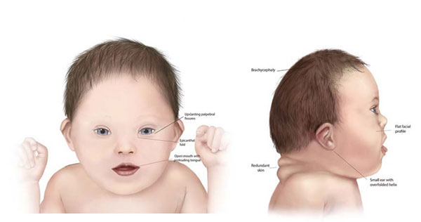
Figure 1: Physical characteristics of a child with Down Syndrome [25].
For assurance of a fetus with Down Syndrome, we prefer several screening tests (prenatal diagnosis) during the first trimester and second trimester- blood tests and ultrasound images to measure the thickness of amniotic fluid (Nuchal Translucency). NIPT (Non-invasive prenatal testing) provides cell-free fetal DNA analysis from maternal blood. Another test is a diagnostic test (part of prenatal diagnosis), i.e. CVS (Chorionic villus sampling). Amniocentesis gives a definitive diagnosis by an analysis of the fetus chromosome. As a postnatal diagnosis, we consider physical examination and karyotype analysis. Physical examination provides physical characteristics, and karyotype analysis makes assurance of the extra chromosome. An individual with Down syndrome needs extra care from the age of pregnancy to adulthood, and that care varies according to the age and the individual’s interests. Those cares include occupational therapy, physical therapy, and speech therapy to address delays in development. They also need specialized edutainment (education + entertainment) programs to enhance their skills [1-3].
They need regular medical check-ups to monitor their health-related issues like heart defects, vision and hearing problems, and thyroid dysfunction. For these issues, they are required to get medications like treatment for hypothyroidism and managing congenital heart anomalies. Apart from the mentioned care, they deserve family support, everyday counseling, programs for developing their social skill, and community involvement [1-3].
Research Scope
- Genetic Research- Gene expression study: Understanding the combination and expression of genes on the 21st chromosome and their impact on health and development.
- Genetic Research-Therapeutic Approaches: Research on highly efficient and capable gene therapies and other cures to reduce the effects of the additional chromosome.
- Biomedical Research- Neuro-development: Studies on brain growth in Trisomy-21 (Down Syndrome) to make interventions for cognitive development.
- Biomedical Research-Pharmacological Treatments: Drug trial for making improvements in cognitive functions and other symptoms of Down Syndrome.
In this study, we reviewed various methods for detecting the fetus or birth with Down Syndrome or Trisomy-21. We found that a blood test of maternal blood helps to find a fetus whether having any chromosomal abnormality or not. Blood tests in the first-trimester help to measure proteins made by PAPP-A, placenta beta, HCG [5], and blood tests in the second trimester help to triple and quadruple measures and AFP (Alpha-Fetoproteins) test. Another method for testing the risk of Down Syndrome is an Ultrasound image that is safe and non-invasive. In this review, we found-
- a technique i.e., Wavelet Analysis used to sharpen the edge of the boundary of nuchal translucency, and in this way, features are extracted from the image and give classification with 97.4% accuracy [6].
- Hierarchical structural models to detect nuchal translucency-used techniques are SVM classifier, Dynamic programming, and distance [7].
- Three-dimensional ultrasound measurement of nuchal translucency-used technique is a dynamic programming [8].
- Using ultrasound video stream to measure the nuchal translucency of the fetus-done by experts [9].
- Variation of nuchal translucency with increasing crown-lump length and gestational age in normal singleton pregnancies-used technique is linear regression [10].
- Nasal bone detection from ultrasound image [11].
- Nasal bone detection in first-trimester screening using an artificial neural network- used techniques are BPNN (back propagation neural network), DCT (Discrete cosine transform), and wavelet transform [12].
- Screening test to detect trisomy-21 [13].
- Nasal bone identification to detect Down Syndrome at an early stage- used technique is BPNN, DCT, wavelet transform, and Otsu thresholding [12].
- Map matching method for finding nasal bone [14].
- Fetal nasal bone length using Euclidean distance, morphological process, and Otsu thresholding [15].
- Cross-correlation techniques for nasal bone segmentation [16].
- Considering mid-second trimester for detection of hypoplasia [17]. Used techniques are mean shift analysis and canny operator [18].
- Classification of image features to detect Down Syndrome using Haralick features- used technique is GLCM (Gray-level co-occurrence matrix), Lee & frost filter, and SVM classifier. Obtained accuracy is 94.4% [19].
Extracting features from an ultrasound image of the fetus is a very crucial task as we see in the above list of works because it comes under a non-invasive technique for visualizing amniotic fluid thickness, i.e., nuchal translucency. We apply many deep learning approaches for classification, and before the classification, we need to provide segmented images in the deep learning algorithms [11,20-23]. It is expected to enhance the accuracy for getting better results and extraction of features like nuchal translucency, nasal bone, CRL, etc. In account of accuracy enhancement and false detection challenges in trisomy-21 diagnosis, it is suggested to recognize and exhibit all relevant screenings which have the FMF (Frontomaxillary facial), FHR (fetal heart rate), bowels shine, swelling, crow’s feet, an augmented nuchal translucency, dilated brain ventricles, little bit kidney damage or swelling.
We observed the highest accuracy of 97.4% for nuchal translucency [6], 97.34% for facial difference [24], and 96.26% for nasal bone [16].
References
2. Down Syndrome. Mayo Clinic. https://www.mayoclinic.org/diseases-conditions/down-syndrome/symptoms-causes/syc-20355977
3. Down Syndrome. MedlinePlus. https://medlineplus.gov/genetics/condition/down-syndrome/
4. Chen L, Wang L, Wang Y, Hu H, Zhan Y, Zeng Z, et al. Global, Regional, and National Burden and Trends of Down Syndrome From 1990 to 2019. Front Genet. 2022 Jul 15;13:908482.
5. Cicero S, Longo D, Rembouskos G, Sacchini C, Nicolaides KH. Absent nasal bone at 11-14 weeks of gestation and chromosomal defects. Ultrasound Obstet Gynecol. 2003 Jul;22(1):31-5.
6. Sciortino G, Tegolo D, Valenti C. A non-supervised approach to locate and to measure the nuchal translucency by means of wavelet analysis and neural networks. In2017 XXVI International Conference on Information, Communication and Automation Technologies (ICAT) 2017 Oct 26 (pp. 1-7). IEEE.
7. Deng Y, Wang Y, Chen P. Automated detection of fetal nuchal translucency based on hierarchical structural model. In2010 IEEE 23rd International Symposium on Computer-Based Medical Systems (CBMS) 2010 Oct 12 (pp. 78-84). IEEE.
8. Siqing Nie, Jinhua Yu, Ping Chen, Yuanyuan Wang, Yi Guo, Jian Qiu Zhang. Automatic measurement of fetal Nuchal translucency from three-dimensional ultrasound data. Annu Int Conf IEEE Eng Med Biol Soc. 2017 Jul;2017:3417-20.
9. Anzalone A, Fusco G, Isgro F, Orlandi E, Prevete R, Sciortino G, et al. A system for the automatic measurement of the nuchal translucency thickness from ultrasound video stream of the foetus. InProceedings of the 26th IEEE International Symposium on Computer-Based Medical Systems 2013 Jun 20 (pp. 239-44). IEEE.
10. Mahale N, Kumar A, Rayapureddi VC, Mahale A. Variaton of nuchal translucency with increasing crown rump length and gestational age in normal singleton pregnancies. IOSR Journal of Dental and Medical Sciences. 2013;6(3):16-9.
11. Rafeek T, Gunasundari A. Reliable Non invasive First Trimester Screening Test Using Image processing and Artificial Neural Network. International Journal of Engineering Research and Applications. 2013 May;3(3):662-8.
12. Sonia R, Shanthi V. Early detection of down syndrome marker using fetal nasal bone length during first and second trimester. Indian journal of applied research. 2015 Jul;5(7):152-6.
13. Maiz N, Valencia C, Kagan KO, Wright D, Nicolaides KH. Ductus venosus Doppler in screening for trisomies 21, 18 and 13 and Turner syndrome at 11-13 weeks of gestation. Ultrasound Obstet Gynecol. 2009 May;33(5):512-7.
14. Wee LK, Supriyanto E. Automatic detection of fetal nasal bone in 2 dimensional ultrasound image using map matching. In12th WSEAS International Conference on Automatic Control, Modeling & Simulation 2010 May 29 (pp. 305-09).
15. VK VD, Rajesh R. A study on Down syndrome detection based on Artificial Neural Network in Ultra sonogram images. In2016 International Conference on Data Mining and Advanced Computing (SAPIENCE) 2016 Mar 16 (pp. 204-9). IEEE.
16. Wee LK, Arooj A, Supriyanto E. Computerized automatic nasal bone detection based on ultrasound fetal images using cross correlation techniques. WSEAS Transactions on Information Science and Applications. 2010 Aug 1;7(8):1068-77.
17. Narayani BH, Radhakrishnan P. Mid-second Trimester Measurement of Nasal Bone Length in the Indian Population. J Obstet Gynaecol India. 2013 Aug;63(4):256-9.
18. Nirmala S, Palanisamy V. Clinical decision support system for early prediction of Down syndrome fetus using sonogram images. Signal, Image and Video Processing. 2011 Jun;5:245-55.
19. Sonia R, Shanthi V. Ultrasound image classification for Down syndrome during first trimester using Haralick features. Int J Eng Technol. 2014;6(2):781-8.
20. Kumar S, Selvakumar K. Various Methods for Computing Risk Factors of Down Syndrome in Fetus. Archives of Computational Methods in Engineering. 2024 Jun 24:1-4.
21. Thomas MC, Arjunan SP. Deep learning measurement model to segment the nuchal translucency region for the early identification of down syndrome. Measurement Science Review. 2022 Aug 1;22(4):187-92.
22. Nirmala S, Palanisamy V. Measurement of nuchal translucency thickness in first trimester ultrasound fetal images for detection of chromosomal abnormalities. In2009 International Conference on Control, Automation, Communication and Energy Conservation 2009 Jun 4 (pp. 1-5). IEEE.
23. Anjit TA, Rishidas S. Identification of nasal bone for the early detection of down syndrome using Back Propagation Neural Network. In2011 International conference on communications and signal processing 2011 Feb 10 (pp. 136-140). IEEE.
24. Saraydemir S, Taspinar N, Erogul O, Kayserili H, Dinckan N. Down syndrome diagnosis based on Gabor Wavelet Transform. J Med Syst. 2012 Oct;36(5):3205-13.
25. DynaFisio. https://dynafisio.com/service/down-syndrome/