Abstract
The reported incidences of 10.6 million tuberculosis cases worldwide with 1.6 million deaths in 2021 indicate that this disease, caused by Mycobacterium tuberculosis pathogen is difficult to treat and requires exploring newer possible therapeutic interventions. To identify novel drug targets, it is important to understand the basic physiological processes of each pathogen in detail. Cell division is the fundamental physiological process which maintains the replicative state of bacteria. This process requires remodelling of the cell wall, which is performed by two spatio-temporal organized complexes, elongasome and divisome. These two complexes, elongasome and divisome function in synthesis of peptidoglycan (PG) at poles or septum of the cell, respectively. This review article is focused on illustrating differential features and composition of mycobacterial elongasome complex. This sort of understanding would allow identification of new drug targets and design of Mycobacterium specific drugs.
Keywords
Cell Division, Drug target identification, Elongasome, Mycobacterium
Tuberculosis- A Threat to the Human Host
The transfer of tuberculosis disease from infectious to healthy individuals happens upon uptake of Mycobacterium tuberculosis (Mtb) containing aerosol droplets, where it reaches the lung alveoli and enters macrophage cell. In an immunocompromised state of an individual, bacilli replicate continuously by preventing the process of phagolysosomal fusion. As soon as this contained disease gets converted into an active form, the hematogenous spread of lesions starts from the pulmonary region to many vital organs like kidney, uterus, bones, eyes, brain (military tuberculosis) and causes detrimental forms of disease. The reported rise in tuberculosis cases worldwide in sequential years from 2019 to 2020 to 2021 indicate the intensity of this threat (Figure 1). Moreover, the data showing increase in rifampicin resistant TB cases from 2020 to 2021 in WHO tuberculosis report 2022 further suggest the need of intensified efforts to target physiological switches of causative organism, Mtb inside hosts.
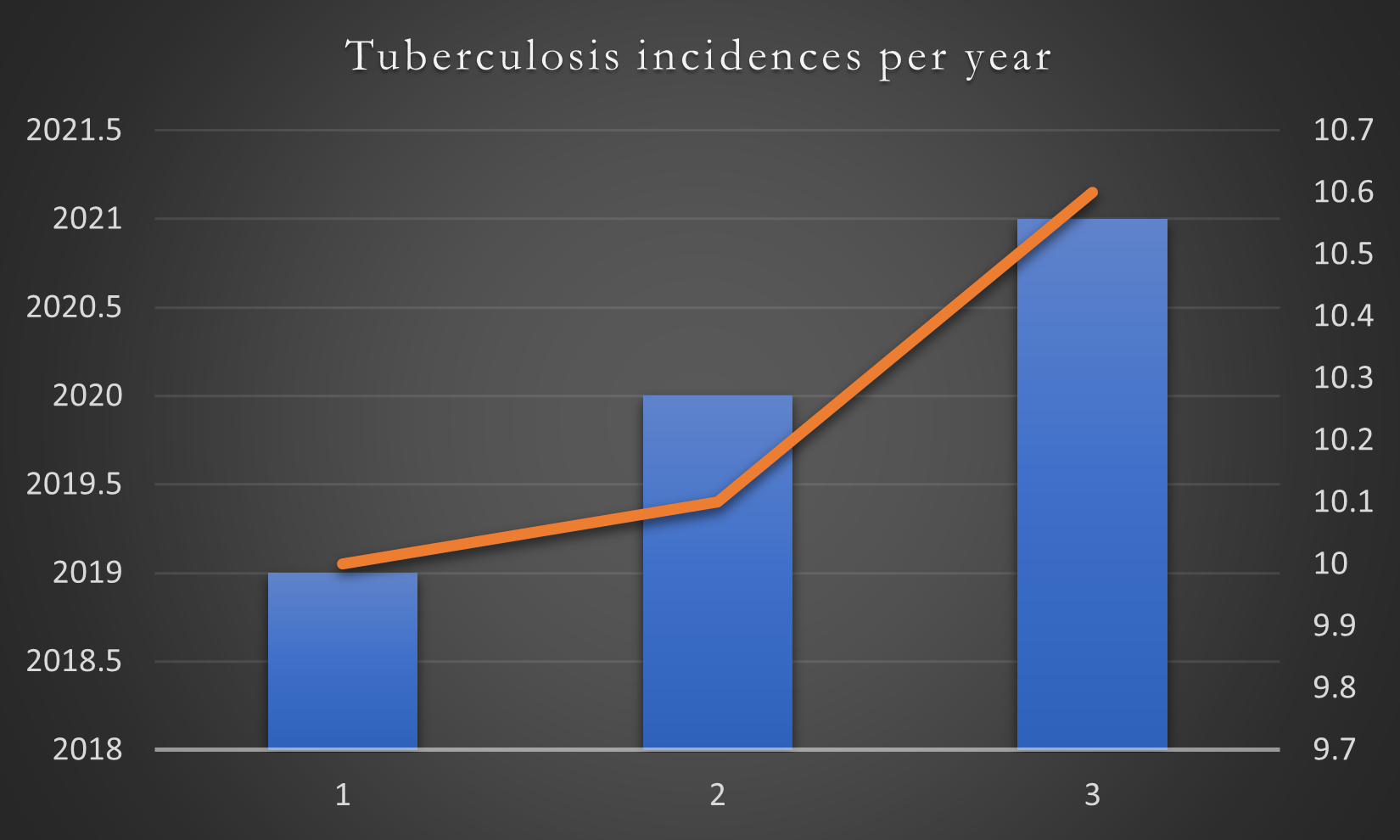
Figure 1 illustrates the rate of increase in tuberculosis incidences worldwide as the year progresses from 2019 to 2020 to 2021.
Cell Division- the Regulatory, Physiological Switch of Mycobacterium Pathogenesis
Pathogen, Mycobacterium is documented to exist in two states: replicative and dormant state. The switch between these two states is determined by factors such as socio- economic factors, host nutritional and immunity status. These determinants regulate this regulatory switch via modifying the functionalities of many cell division proteins; mainly categorised in two macromolecular complexes as elongasome and divisome. While the elongasome functions in mediating the peripheral peptidoglycan (PG) synthesis and duplicating the cell size, the function of the divisome is reported to carry out the septal PG synthesis and in dividing the cell mass. These two complexes are present in every bacterial species; however, their composition varies between species. One of the reasons for the presence of variable features in these two macromolecular complexes of mycobacterium could be their differential mode of growth and regulation. While most of the bacterial species incorporate PG along the lateral wall in patches, Mycobacterium grows asymmetrically from their poles and eventually generates a heterogeneous population upon cell division. This heterogeneous population exhibits differential antibiotic susceptibilities and disparate physiological niches in the human host [1].
Mutations in most of the cell division proteins of different bacterium species including Mycobacterium results in either loss of normal cell shape/size or gain of differential antibiotic susceptibilities [2]. The loss of normal cell shape/size happens because of failure in the mechanism, which ensures the occurrence of correct pattern of cell division within a given time frame. Among the different patterns of cell division, binary fission is the most common form adopted by bacterial kingdoms. The simple binary fission mode of reproduction involves doubling of cell mass, initiating, and terminating one round of chromosome replication, decatenate or segregating replicated chromosomes, localization of division machinery at mid cell and finally, cytokinesis. Completion of this complex proposition within a defined time requires presence of overlapping events, which are categorized in B, C, and D periods and assembly of elongasome and divisome at a precise position. B period is the phase between cell birth and DNA replication initiation, C, the time of chromosome replication and D, the phase between termination of replication and beginning of division [3].
Many of the cell division proteins, belonging to elongasome, divisome, and chromosome segregation units, are registered as potential drug targets. MreB protein, which is the part of the Rod system of the elongasome unit, is reported to be the target of A22 analogs in Escherichia coli (E. coli) [4]. Another MreB inhibitor, TXH11106 have been identified which is broad spectrum and reported to be bactericidal against many gram-negative pathogens, E coli, Klebsiella pneumoniae, Acinetobacter baumannii, and Pseudomonas aeruginosa [5]. Comparative analysis has demonstrated superiority of TXH11106 over A22 analogs [5]. Similarly, many of the penicillin binding proteins (PBPs) of the elongosome unit have been identified to be targeted by β lactam antibiotics [6]. Although these reports have suggested the elongasome unit of different bacterial species as potential drug targets, reports building the understanding of mycobacterial elongasome as the unit of potential drug targets is limited. Moreover, it is important to identify differential features of elongasome units, which can be readily targeted by drugs. This review article is focused on understanding the elongasome unit of Mycobacterium and presenting its comparative analysis with other bacterial species. This sort of analysis will allow the identification of newer Mycobacterium specific drug targets and design of the future therapeutic regimen.
Elongation Complex/Elongasome in the Bacterial Kingdom
The function of elongation complex during cell division is to double the size of a cell, which is considered a trigger for manifestation of chromosome segregation and division events [7]. This complex is known to localize and regulate incorporation of nascent cell wall components at a specific position of the cell. While in E. coli and Bacillus subtilis (B. subtilis), the PG precursors are integrated along the wall; it assembles at poles in Streptomyces coelicolor and Mycobacterium [8-11]. What determines the specific localization of this complex in different organisms is still a question, some insights have been brought into the role of cytoskeletal like proteins in this process. One of the known cytoskeletal proteins, MreBEc (E. coli)/Bs (B. subtilis)/Cc (Caulobacter crescentus) is an actin like protein, which tends to form filaments by continuously undergoing polymerization – depolymerization [12-14]. This continuous recycling of MreB protein is thought to be governed by the rate of PG synthesis. Depletion of specific elongation class B transpeptidase (PBP2) abolishes MreBEc mediated insertion of PG along the lateral cell wall in the absence of functional FtsZ [15]. Additionally, MreBEc is known to interact with conserved inner membrane proteins MreC, MreD, and RodZ (transmembrane protein) as well as lipid II synthesis enzymes indicating crucial role of MreBEc (actin like filaments) in organization of elongation complex [16-19]. Moreover, presence of three actin homologs (MreB, Mbl and MreBH) in B. subtilis and CreS in C. crescentus advocates the importance of cytoskeletal like proteins in maintaining cell shape [20-22]. The elongation complex is roughly composed of PG remodelling modules, carrying functions of its synthesis and hydrolysis. Though various synthetases and hydrolases are functionally redundant, some of them show a major impact over others in regulating the rate of PG turnover. Interestingly, their ability to switch between elongasome and divisome to provide harmonious coordination between events has been observed [23]. The main functional activities of PG synthetases are transglycosylation (TG) and transpeptidation (TP). Based on these activities, the synthetases are classified into three functional categories in E. coli. First category includes bifunctional class A PBPs-GTase–TPases (PBP1A, PBP1B and PBP1C) which harbors both transglycosylation and transpeptidation activities [24]. While transglycosylation activity is independent from transpeptidation, transpeptidation requires efficient ongoing transglycosylation activity indicating transpeptidation activity is required after transglycosylation within a cell [25,26]. Though one between PBP1A and PBP1B is required for viability [27], PBP1C is essential in the host [28]. The other categories include monofunctional transpeptidases PBP2 or PBP3 and monofunctional transglycosylase MgtA [29,30]. Furthermore, PBP1A and PBP1B are thought to participate in two different complexes of elongation and division by their ability to interact with monofunctional transpeptidases PBP2 and PBP3, respectively [31]. While precursors for transpeptidation and transglycosylation reactions i.e. lipid II are synthesized in the cytoplasmic region, its insertion to the PG sacculus occurs in the periplasmic region. Consecutive actions of various Mur ligases generate lipid II (UDP-GlcNAc-MurNAc-pentapeptide anchored to undecaprenyl phosphate carrying lipid membrane) in the cytoplasmic region, which is flipped outside by MurJ/RodA/FtsW and crosslinked with existing PG by PBPs [32]. Mur ligases are cytosolic and comprise an N-terminus UDP-MurNAc binding domain, connected to a C-terminus domain via central ATP binding domain. The function of C-terminus of Mur enzymes is to attach aminoacid to stem pentapeptide [33]. Enzymatic activities of MurA-MurF lead to the generation of UDP-MurNAc-pentapeptide, which is a substrate for MraY. MraY ligates it to an undecaprenyl phosphate carrying lipid generating membrane and results in membrane linked UDP-MurNAc pentapeptide (lipid I). Finally, MurG catalyzes transfer of GlcNAc molecules from UDP-GlcNAc to lipid I, generates lipid II, a substrate for action of RodA (flippase) and PBPs [34,35]. Importance of RodA and PBP2/PBP1A/PBP1B is demonstrated in E. coli when mutants lacking such genes have been found to grow as spherical cells [36]. Similarly, in B. subtilis, mutation in RodA or PBPs such as in PBP2a or PbpH leads to conversion of rod shape cells into spherical cells [37,38]. Mutants of Streptococcus thermophilus lacking homologues of PBP2 or RodA have been shown to adopt spherical morphology rather than ovococci [39]. Importance of PBP1BEc is demonstrated by its ability to interact with M1tA through MipA (outer membrane protein), indicating the existence of a proper channel to transfer signal from cytoplasm to periplasm [40]. Additional known elongasome component, RodZEc/Bs/Cc is known to communicate signals from cytoplasm to periplasmic region [41]. While N-terminus cytoplasmic region of RodZEc/Cc has been shown to interact with MreB, C-terminus periplasmic region interacts with MreC, which associates with MreD, Class A (PBP1A /PBP1B) and Class B PBPs (PBP2) indicating the role of RodZ as an adaptor molecule [41]. Additionally, MreBEc/Cc interaction with MurG or MraY is demonstrated, indicating the presence of complex morphogenetic apparatus, which establishes signal-transducing network to communicate signals between two distinct cytoplasmic and periplasmic components and targets PG intermediates to a specific position of a cell [42,43]. Another component of PG remodeler, coined as hydrolases, is abundant in number (13 in E. coli) and is functionally redundant [44]. Deletion of one or two hydrolases does not impair the growth and survival. The functionality of hydrolases is classified into glycosidases, amidases, lytic transglycosylases and endopeptidases. As a result of their actions, soluble fragments from the existing sacculus are generated, which is reused via an efficient peptidoglycan-recycling pathway. E. coli is known to have 5 amidases (AmiA, AmiB, AmiC, AmpD, MepA) and are redundant in function [45,46]. Two amidase activators EnvC and NlpD have been identified which causes conformational change upon interaction with amidases [47]. In B. subtilis, four cell wall amidases CwlD, LytC, CwlC, and CwlB have been identified and found to play a crucial role in the process of sporulation [48,49]. Once PG is synthesized, it undergoes minor changes for e.g., newly synthesized pentapeptide enriched PG in E. coli is characterized by the presence of glycan chains with an average of 50-60 disaccharide units. While DD-carboxypeptidases are known to convert pentapeptides into tetrapeptides enriched peptidoglycan, LD- carboxypeptidases trims tetrapeptides into tripeptides. In addition, lytic transglycosylases are known to modify glycan chains of peptidoglycan. There are 6 DD-carboxypeptidases reported in E. coli in which PBP5 (DacA) is highly active, supported by an observation of altered morphology and aberrant cell shape when PBP5 was removed with additional PBPs [50,51]. Increased expression of DD-carboxypeptidase PBP6 during the stationary phase has been found to result in shorter cell length [52]. On the other hand, in C. crescentus, the role of DD carboxypeptidase is very less because pentapeptide enriched PG is the primary component of sacculus growth [53]. Conclusively, the elongasome unit of a typical bacterial cell comprises cytoskeletal like proteins, PG synthetic, PG hydrolytic, and PG modifiers modules (Figure 2).
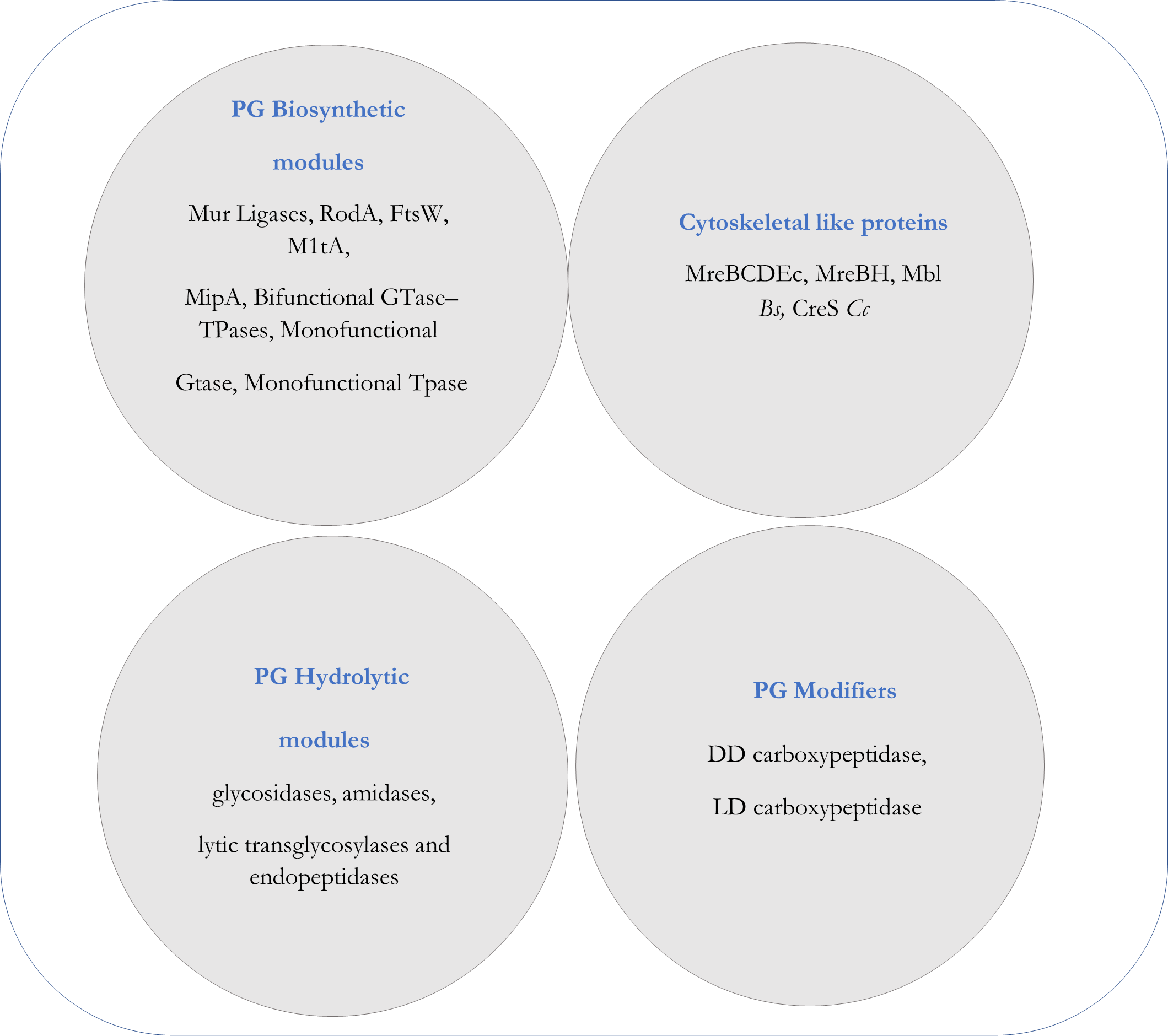
Figure 2 presents the components of the elongasome unit of a typical bacterial cell. The main components identified are cytoskeletal like proteins, PG biosynthetic, PG hydrolytic, and PG modifiers.
Elongation Complex/Elongasome in Mycobacterium
The functionality of the elongation complex in Mycobacterium is equivalent to other bacterial kingdoms. However, PG precursors are incorporated at subpolar regions. To achieve the aim of subpolar growth, which requires positioning of an elongation complex at poles, the elongation complex is equipped with a protein known as polar growth determinant (Wag31) [54]. The addition of polar growth determining protein is considered as one of the compensatory mechanisms for absence of cytoskeletal elements in Mycobacterium whose function is thought to be in localization of elongation complex in other bacterial species. Wag31 positioning at subpolar space requires recognition of concave membrane curvature, which is mediated through presence of hydrophobic and positively charged residues. Additionally, deletion or overexpression of Cell Wall Synthesis protein A (CwsA) has been shown to cause differential localization of Wag31, indicating accurate localization of Wag31 is dependent upon CwsA [55]. Another cell wall synthesis regulatory module, MviN-FhaA complex has been identified. MviN is an essential pseudokinase protein and known to recruit FhaA (forkhead associated) protein upon phosphorylation in pseudokinase domain. Depletion of MviN has been found to accumulate solvent extractable PG precursors, indicating its importance in precursor assimilation to existing sacculus [56]. Basic enzymes of PG remodeling, Mur complexes (MurA-MurG) have been found functionally conserved in Mycobacterium. These enzymes synthesize lipid II, which gets flipped in periplasm and gets cross-linked by conserved PBPs. While Mtb has two HMM class-A PBPs i.e., PonA1 and PonA2, Mycobacterium smegmatis (non-pathogenic mycobacterium, Msmeg) is characterized by the presence of three Class A penicillin binding proteins PonA1, PonA2, and PonA3 [57]. PonA1 interaction with Wag31 demonstrates its functionality during elongation [58]. Its localization at both septum and poles in Msmeg emphasizes its importance during both elongation and division phases of bacterial growth. Additionally, PonA1 has been demonstrated to decrease hydrolytic activity by binding with the C-terminus endopeptidase domain of RipA (hydrolase) [58]. PonA1 is important for maintenance of correct cell lengths in both Mtb / Msmeg and required for establishing successful infection in hosts. Mutants of PonA1 and PonA2 showed an unusual hypersensitivity towards β-lactam antibiotics. While PonA2 deletion impacts morphology and shows higher susceptibility towards antibiotics in the stationary phase, PonA3 deletion does not show such kind of defects under standard laboratory conditions. Moreover, PonA3 can partially substitute for PonA2 indicating the crucial role of these two proteins in stationary phase or non-replication stage [57]. Other than class A PBPs, Mtb has two HMM class-B PBPs PbpA/PBP3 and six PBPs in LMM class-C (carboxypeptidases and β-lactamases) category. While PbpA is a homolog of PBP2 (elongation complex component), it is known to be involved in division rather than elongation in Mycobacterium. Absence of PbpA at an elongation site is probably compensated by the presence of non-canonical transpeptidases (LdtA or LdtB) [59]. PBP3 is known to form ternary complexes with FtsZ and FtsW, which constitutes an important component of divisome. The roles of LMM class C proteins need further investigation to understand the complete mechanism of PG synthesis and regulation. RipA, RipB, and RipD, hydrolases in Mycobacterium are categorized into NlpC type PG hydrolases [60]. However, RipD presents an example of non-catalytic PG binding function [61]. These proteins are individually dispensable in Msmeg [62]. Interestingly, RipA is produced as a zymogen, which requires proteolytic processing for its activation and presents a wonderful example of self-inhibitory protein [63]. In vitro evidence nullifies the importance of the N-terminal region in this auto inhibition of RipA, as removal of N-terminus does not change the activity of either RipA or RipB [60]. Moreover, RipA is known to bind RpfB and RpfE resuscitation promoting factors, which is known to reactivate dormant bacteria. The observed colocalization of RipA with RpfB at septum indicates the concerted or coordinated action of hydrolases in cell division [65]. Mtb possesses five resuscitation promoting factors starting from RpfA to E with all are found to be dispensable for mycobacterial growth. The observed interaction between RipA and PonA1 indicates the coordinated action of synthases and hydrolases in PG remodeling [58]. Conclusively, the elongasome unit of Mycobacterium comprises polar elongation complex, PG biosynthetic and PG hydrolytic modules (Figure 3). Unlike other model bacterial species, the composition of PG biosynthetic and hydrolytic modules has been found different. The identification of PG modifiers in Mycobacterium is under investigation.
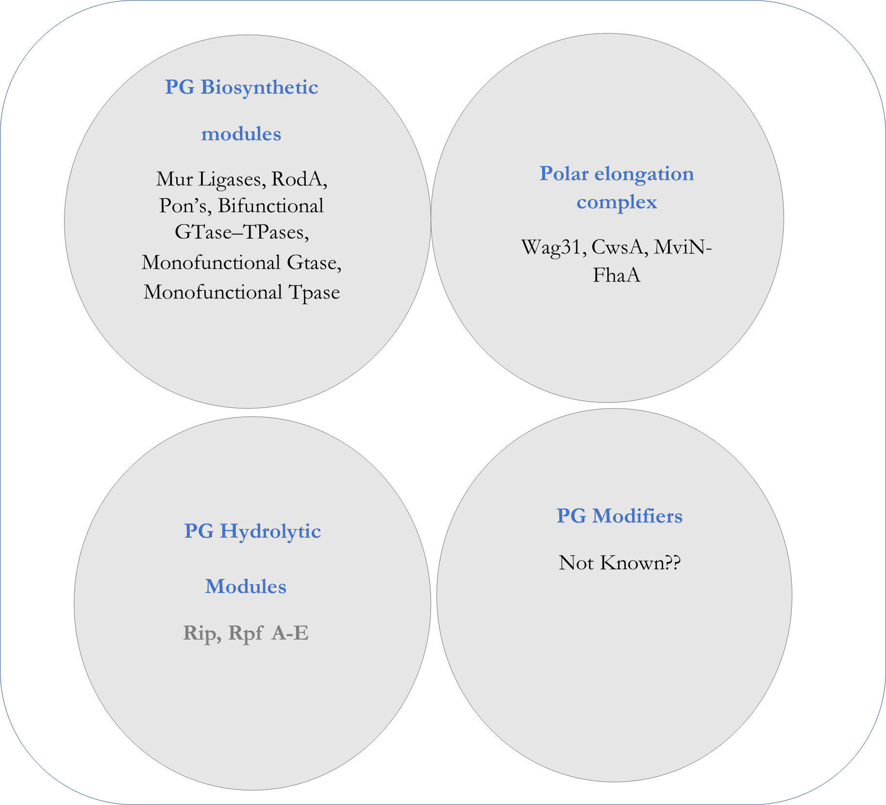
Figure 3 presents the components of the elongasome unit of a typical mycobacterial cell. The main components identified are polar elongation complex, PG biosynthetic, and PG hydrolytic modules.
Conclusions
Like every other bacterial species, the function of Mycobacterium elongasome is reported to carry out the peripheral PG synthesis and double the cell size. However, its composition differs and is reported to possess differential proteins with unique properties. For e.g., the subcomplexes, Wag31-CwsA- CrgA and MviN-FhaA are registered as unique proteins in Mycobacterium elongasome and have a role to play in the polar growth of the cell. Few non canonical proteins i.e., Ldts, Rips, and Rpfs have been found present in mycobacteria, suggesting their potential as drug targets. The presence of these unique proteins could be either the compensation of the absence of similar functional proteins or evolve to perform some alternative function as per requirement of the cell. The absence of PG modifiers in mycobacterial elongasome indicates that the PG layer is synthesized as needed in this pathogen. However, we could not nullify the point that PG modifiers may remain undiscovered in Mycobacterium. In short, this review article has enlisted components of mycobacterial elongasome complex, some of which are either similar or dissimilar to other bacterial species.
Future Perspectives
The rise in tuberculosis cases drives the urgency to identify novel drug targets and drugs. Not only it is important to identify drug targets belonging to the basic physiological processes, but also there is an urge to identify multiple drug targets for a single drug. Identification of multiple targets for a single drug allows targeting multiple proteins of same or different macromolecular complexes at the same time. Thus, research regarding identification of those drug- drug targets pair is of special interest.
Identification of drug targets belonging to the cell division process is relatively difficult. Most of the cell division proteins do not show any activities which can be assayed in vitro. Moreover, most of the cell division proteins are essential. Thus, the only approach available to study the gene effect is to create conditional knockouts. Most of the approaches, which can be used for generating conditional knockouts in Mycobacterium, generate a loosely controlled system, which do not allow the study of complete effects of gene knockouts. Moreover, knockouts of cell division genes result in cells with disturbed size and shape, which is difficult to grow under standard laboratory conditions.
Despite these challenges, researchers are constantly engaged in discovering novel drug targets of macromolecular complexes of the cell division processes in mycobacterium. Elongasome and divisome, the two macromolecular complexes, are known to be therapeutically targeted. Based on reports, the elongasome macromolecular complex of Mycobacterium may have many potential drug candidates, which needs to be explored and investigated. This review article is a step towards achieving this aim and is focused on dissecting differential features of the elongasome complex of Mycobacterium. This study is expected to reveal the newer possible drug targets for future therapeutics. Moreover, the identified differential proteins of elongasome complexes can be investigated further for studying their functional domains.
Declaration of Competing Interest
The authors declare that they have no known competing financial interests or personal relationships that could have appeared to influence the work reported in this paper.
Data Availability
No data was used for the research described in the article.
References
2. Mahone CR, Goley ED. Bacterial cell division at a glance. Journal of Cell Science. 2020 Apr 1;133(7):jcs237057.
3. Chien AC, Hill NS, Levin PA. Cell size control in bacteria. Current biology. 2012 May 8;22(9):R340-9.
4. Buss JA, Baidin V, Welsh MA, Flores-Kim J, Cho H, Wood BM, et al. Pathway-Directed Screen for Inhibitors of the Bacterial Cell Elongation Machinery. Antimicrob Agents Chemother. 2018 Dec 21;63(1):e01530-18.
5. Bryan EJ, Sagong HY, Parhi AK, Grier MC, Roberge JY, LaVoie EJ, et al. TXH11106: A Third-Generation MreB Inhibitor with Enhanced Activity against a Broad Range of Gram-Negative Bacterial Pathogens. Antibiotics (Basel). 2022 May 20;11(5):693.
6. Beadle BM, Nicholas RA, Shoichet BK. Interaction energies between beta-lactam antibiotics and E. coli penicillin-binding protein 5 by reversible thermal denaturation. Protein Sci. 2001 Jun;10(6):1254-9.
7. Haeusser DP, Levin PA. The great divide: coordinating cell cycle events during bacterial growth and division. Curr Opin Microbiol. 2008 Apr;11(2):94-9.
8. de Pedro MA, Quintela JC, Höltje JV, Schwarz H. Murein segregation in Escherichia coli. J Bacteriol. 1997 May;179(9):2823-34.
9. Tiyanont K, Doan T, Lazarus MB, Fang X, Rudner DZ, Walker S. Imaging peptidoglycan biosynthesis in Bacillus subtilis with fluorescent antibiotics. Proc Natl Acad Sci U S A. 2006 Jul 18;103(29):11033-8.
10. Gray DI, Gooday GW, Prosser JI. Apical hyphal extension in Streptomyces coelicolor A3(2). J Gen Microbiol. 1990 Jun;136(6):1077-84.
11. Thanky NR, Young DB, Robertson BD. Unusual features of the cell cycle in mycobacteria: polar-restricted growth and the snapping-model of cell division. Tuberculosis (Edinb). 2007 May;87(3):231-6.
12. Figge RM, Gober JW. Cell shape, division and development: the 2002 American Society for Microbiology (ASM) conference on prokaryotic development. Mol Microbiol. 2003 Mar;47(5):1475-83.
13. Divakaruni AV, Baida C, White CL, Gober JW. The cell shape proteins MreB and MreC control cell morphogenesis by positioning cell wall synthetic complexes. Mol Microbiol. 2007 Oct;66(1):174-88.
14. Daniel RA, Errington J. Control of cell morphogenesis in bacteria: two distinct ways to make a rod-shaped cell. Cell. 2003 Jun 13;113(6):767-76.
15. Varma A, Young KD. In Escherichia coli, MreB and FtsZ direct the synthesis of lateral cell wall via independent pathways that require PBP 2. J Bacteriol. 2009 Jun;191(11):3526-33.
16. Alyahya SA, Alexander R, Costa T, Henriques AO, Emonet T, Jacobs-Wagner C. RodZ, a component of the bacterial core morphogenic apparatus. Proc Natl Acad Sci U S A. 2009 Jan 27;106(4):1239-44.
17. Kruse T, Bork-Jensen J, Gerdes K. The morphogenetic MreBCD proteins of Escherichia coli form an essential membrane-bound complex. Mol Microbiol. 2005 Jan;55(1):78-89.
18. Shiomi D, Sakai M, Niki H. Determination of bacterial rod shape by a novel cytoskeletal membrane protein. EMBO J. 2008 Dec 3;27(23):3081-91.
19. van den Ent F, Leaver M, Bendezu F, Errington J, de Boer P, Löwe J. Dimeric structure of the cell shape protein MreC and its functional implications. Mol Microbiol. 2006 Dec;62(6):1631-42.
20. Gitai Z, Dye NA, Reisenauer A, Wachi M, Shapiro L. MreB actin-mediated segregation of a specific region of a bacterial chromosome. Cell. 2005 Feb 11;120(3):329-41.
21. Soufo HJ, Graumann PL. Actin-like proteins MreB and Mbl from Bacillus subtilis are required for bipolar positioning of replication origins. Curr Biol. 2003 Oct 28;13(21):1916-20.
22. Ausmees N, Kuhn JR, Jacobs-Wagner C. The bacterial cytoskeleton: an intermediate filament-like function in cell shape. Cell. 2003 Dec 12;115(6):705-13.
23. Bertsche U, Kast T, Wolf B, Fraipont C, Aarsman ME, Kannenberg K, et al. Interaction between two murein (peptidoglycan) synthases, PBP3 and PBP1B, in Escherichia coli. Mol Microbiol. 2006 Aug;61(3):675-90.
24. Goffin C, Ghuysen JM. Multimodular penicillin-binding proteins: an enigmatic family of orthologs and paralogs. Microbiol Mol Biol Rev. 1998 Dec;62(4):1079-93.
25. Born P, Breukink E, Vollmer W. In vitro synthesis of cross-linked murein and its attachment to sacculi by PBP1A from Escherichia coli. J Biol Chem. 2006 Sep 15;281(37):26985-93.
26. Bertsche U, Breukink E, Kast T, Vollmer W. In vitro murein peptidoglycan synthesis by dimers of the bifunctional transglycosylase-transpeptidase PBP1B from Escherichia coli. J Biol Chem. 2005 Nov 11;280(45):38096-101.
27. Yousif SY, Broome-Smith JK, Spratt BG. Lysis of Escherichia coli by beta-lactam antibiotics: deletion analysis of the role of penicillin-binding proteins 1A and 1B. J Gen Microbiol. 1985 Oct;131(10):2839-45.
28. Budd A, Blandin S, Levashina EA, Gibson TJ. Bacterial alpha2-macroglobulins: colonization factors acquired by horizontal gene transfer from the metazoan genome? Genome Biol. 2004;5(6):R38.
29. Matsuhashi M, Wachi M, Ishino F. Machinery for cell growth and division: penicillin-binding proteins and other proteins. Res Microbiol. 1990 Jan;141(1):89-103.
30. Derouaux A, Wolf B, Fraipont C, Breukink E, Nguyen-Distèche M, Terrak M. The monofunctional glycosyltransferase of Escherichia coli localizes to the cell division site and interacts with penicillin-binding protein 3, FtsW, and FtsN. J Bacteriol. 2008 Mar;190(5):1831-4.
31. Banzhaf M, van den Berg van Saparoea B, Terrak M, Fraipont C, Egan A, Philippe J, et al. Cooperativity of peptidoglycan synthases active in bacterial cell elongation. Mol Microbiol. 2012 Jul;85(1):179-94.
32. Ruiz N. Lipid Flippases for Bacterial Peptidoglycan Biosynthesis. Lipid Insights. 2016 Jan 13;8(Suppl 1):21-31.
33. Barreteau H, Kovac A, Boniface A, Sova M, Gobec S, Blanot D. Cytoplasmic steps of peptidoglycan biosynthesis. FEMS Microbiol Rev. 2008 Mar;32(2):168-207.
34. Bouhss A, Trunkfield AE, Bugg TD, Mengin-Lecreulx D. The biosynthesis of peptidoglycan lipid-linked intermediates. FEMS Microbiol Rev. 2008 Mar;32(2):208-33.
35. Scheffers DJ, Pinho MG. Bacterial cell wall synthesis: new insights from localization studies. Microbiol Mol Biol Rev. 2005 Dec;69(4):585-607.
36. Pedro MA, Donachie WD, Höltje JV, Schwarz H. Constitutive septal murein synthesis in Escherichia coli with impaired activity of the morphogenetic proteins RodA and penicillin-binding protein 2. J Bacteriol. 2001 Jul;183(14):4115-26.
37. Henriques AO, Glaser P, Piggot PJ, Moran CP Jr. Control of cell shape and elongation by the rodA gene in Bacillus subtilis. Mol Microbiol. 1998 Apr;28(2):235-47.
38. Wei Y, Havasy T, McPherson DC, Popham DL. Rod shape determination by the Bacillus subtilis class B penicillin-binding proteins encoded by pbpA and pbpH. J Bacteriol. 2003 Aug;185(16):4717-26.
39. Thibessard A, Fernandez A, Gintz B, Leblond-Bourget N, Decaris B. Effects of rodA and pbp2b disruption on cell morphology and oxidative stress response of Streptococcus thermophilus CNRZ368. J Bacteriol. 2002 May;184(10):2821-6.
40. Vollmer W, von Rechenberg M, Höltje JV. Demonstration of molecular interactions between the murein polymerase PBP1B, the lytic transglycosylase MltA, and the scaffolding protein MipA of Escherichia coli. J Biol Chem. 1999 Mar 5;274(10):6726-34.
41. van den Ent F, Johnson CM, Persons L, de Boer P, Löwe J. Bacterial actin MreB assembles in complex with cell shape protein RodZ. EMBO J. 2010 Mar 17;29(6):1081-90.
42. Ha S, Walker D, Shi Y, Walker S. The 1.9 A crystal structure of Escherichia coli MurG, a membrane-associated glycosyltransferase involved in peptidoglycan biosynthesis. Protein Sci. 2000 Jun;9(6):1045-52.
43. White CL, Kitich A, Gober JW. Positioning cell wall synthetic complexes by the bacterial morphogenetic proteins MreB and MreD. Mol Microbiol. 2010 May;76(3):616-33.
44. Vollmer W, Joris B, Charlier P, Foster S. Bacterial peptidoglycan (murein) hydrolases. FEMS Microbiol Rev. 2008 Mar;32(2):259-86.
45. Heidrich C, Templin MF, Ursinus A, Merdanovic M, Berger J, Schwarz H, et al. Involvement of N-acetylmuramyl-L-alanine amidases in cell separation and antibiotic-induced autolysis of Escherichia coli. Mol Microbiol. 2001 Jul;41(1):167-78.
46. Jacobs C, Joris B, Jamin M, Klarsov K, Van Beeumen J, Mengin-Lecreulx D, et al. AmpD, essential for both beta-lactamase regulation and cell wall recycling, is a novel cytosolic N-acetylmuramyl-L-alanine amidase. Mol Microbiol. 1995 Feb;15(3):553-9.
47. Uehara T, Parzych KR, Dinh T, Bernhardt TG. Daughter cell separation is controlled by cytokinetic ring-activated cell wall hydrolysis. EMBO J. 2010 Apr 21;29(8):1412-22.
48. Smith TJ, Blackman SA, Foster SJ. Autolysins of Bacillus subtilis: multiple enzymes with multiple functions. Microbiology (Reading). 2000 Feb;146 ( Pt 2):249-262.
49. Senzani S, Li D, Bhaskar A, Ealand C, Chang J, Rimal B, et al. An Amidase_3 domain-containing N-acetylmuramyl-L-alanine amidase is required for mycobacterial cell division. Sci Rep. 2017 Apr 25;7(1):1140.
50. de Pedro MA, Young KD, Höltje JV, Schwarz H. Branching of Escherichia coli cells arises from multiple sites of inert peptidoglycan. J Bacteriol. 2003 Feb;185(4):1147-52.
51. Nelson DE, Young KD. Penicillin binding protein 5 affects cell diameter, contour, and morphology of Escherichia coli. J Bacteriol. 2000 Mar;182(6):1714-21.
52. Sarkar SK, Dutta M, Chowdhury C, Kumar A, Ghosh AS. PBP5, PBP6 and DacD play different roles in intrinsic β-lactam resistance of Escherichia coli. Microbiology (Reading). 2011 Sep;157(Pt 9):2702-2707.
53. Markiewicz Z, Glauner B, Schwarz U. Murein structure and lack of DD- and LD-carboxypeptidase activities in Caulobacter crescentus. J Bacteriol. 1983 Nov;156(2):649-55.
54. Kang CM, Nyayapathy S, Lee JY, Suh JW, Husson RN. Wag31, a homologue of the cell division protein DivIVA, regulates growth, morphology and polar cell wall synthesis in mycobacteria. Microbiology (Reading). 2008 Mar;154(Pt 3):725-735.
55. Plocinski P, Martinez L, Sarva K, Plocinska R, Madiraju M, Rajagopalan M. Mycobacterium tuberculosis CwsA overproduction modulates cell division and cell wall synthesis. Tuberculosis (Edinb). 2013 Dec;93 Suppl:S21-7.
56. Gee CL, Papavinasasundaram KG, Blair SR, Baer CE, Falick AM, King DS, et al. A phosphorylated pseudokinase complex controls cell wall synthesis in mycobacteria. Sci Signal. 2012 Jan 24;5(208):ra7.
57. Patru MM, Pavelka MS Jr. A role for the class A penicillin-binding protein PonA2 in the survival of Mycobacterium smegmatis under conditions of nonreplication. J Bacteriol. 2010 Jun;192(12):3043-54.
58. Hett EC, Chao MC, Rubin EJ. Interaction and modulation of two antagonistic cell wall enzymes of mycobacteria. PLoS Pathog. 2010 Jul 29;6(7):e1001020.
59. Schoonmaker MK, Bishai WR, Lamichhane G. Nonclassical transpeptidases of Mycobacterium tuberculosis alter cell size, morphology, the cytosolic matrix, protein localization, virulence, and resistance to β-lactams. J Bacteriol. 2014 Apr;196(7):1394-402.
60. Böth D, Schneider G, Schnell R. Peptidoglycan remodeling in Mycobacterium tuberculosis: comparison of structures and catalytic activities of RipA and RipB. J Mol Biol. 2011 Oct 14;413(1):247-60.
61. Böth D, Steiner EM, Izumi A, Schneider G, Schnell R. RipD (Rv1566c) from Mycobacterium tuberculosis: adaptation of an NlpC/p60 domain to a non-catalytic peptidoglycan-binding function. Biochem J. 2014 Jan 1;457(1):33-41.
62. Martinelli DJ, Pavelka MS Jr. The RipA and RipB Peptidoglycan Endopeptidases Are Individually Nonessential to Mycobacterium smegmatis. J Bacteriol. 2016 Apr 14;198(9):1464-75.
63. Ruggiero A, Marasco D, Squeglia F, Soldini S, Pedone E, Pedone C, et al. Structure and functional regulation of RipA, a mycobacterial enzyme essential for daughter cell separation. Structure. 2010 Sep 8;18(9):1184-90.
64. Hett EC, Chao MC, Steyn AJ, Fortune SM, Deng LL, Rubin EJ. A partner for the resuscitation-promoting factors of Mycobacterium tuberculosis. Mol Microbiol. 2007 Nov;66(3):658-68.
65. Kana BD, Mizrahi V. Resuscitation-promoting factors as lytic enzymes for bacterial growth and signaling. FEMS Immunol Med Microbiol. 2010 Feb;58(1):39-50.