

Keywords
Antibody Drug Conjugate Therapy, Cancer therapy, Ocular toxicity
Introduction
Antibody–drug conjugates (ADCs), which merge the specificity of monoclonal antibodies with potent cytotoxic payloads, are becoming more common as a therapeutic option for cancer patients with multiple approvals and many more in clinical development [1,2]. As of March 2026, 15 ADCs have been approved by the FDA for use in oncological indications (Table 1), and despite their targeted design, ocular adverse events (OAEs) have emerged as adverse events of special interest in patients across various tumor types [3,4]. OAEs can disrupt treatment and diminish patient quality of life. This review discusses underlying mechanisms, scientific gaps, and quantitative modeling approaches relevant to ADC-associated OAEs.
|
ADC |
Target |
Drug Conjugate |
Top 5 OAEs (EBGM >1) 1 |
Disease Indication 2 |
|
Belantamab mafodotin |
BCMA |
MMAF |
Keratopathy, night blindness, corneal epithelial microcysts, ocular toxicity NOS, punctate keratitis |
Multiple myeloma |
|
Brentuximab vedotin |
CD30 |
MMAE |
Ectropion, blindness, visual acuity reduced transiently, papilloedema |
Cutaneous mesenchymal large cell lymphoma |
|
Datopotamab deruxtecan |
TROP-2 |
DXd |
No OAE data available3 |
Breast cancer |
|
Enfortumab vedotin |
Nectin-4 |
MMAE |
Dry eye, ocular toxicity NOS, lacrimation increased |
Urothelial cancer |
|
Gemtuzumab ozogamicin |
CD33 |
Calicheamicin |
No significant OAEs |
Acute myelogenous leukemia |
|
Inotuzumab ozogamicin |
CD22 |
Calicheamicin |
No significant OAEs |
Acute lymphoblastic leukemia |
|
Loncastuximab tesirine |
CD19 |
SG3199 |
No significant OAEs |
B-cell lymphoma |
|
Mirvetuximab soravtansine |
FRα |
DM4 |
Keratitis, keratopathy, ocular toxicity NOS, cataract, dry eye |
Platinum-resistant epithelial ovarian cancer |
|
Moxetumomab pasudotox |
CD22 |
IT |
No significant OAEs |
Hairy cell leukemia |
|
Polatuzumab vedotin |
CD79 |
MMAE |
No significant OAEs |
Diffuse large B-cell lymphoma |
|
Sacituzumab govitecan |
Trop-2 |
SN-38 |
No significant OAEs |
Breast cancer |
|
Telisotuzumab vedotin |
c-Met |
MMAE |
No OAE data available3 |
Non-small cell lung cancer |
|
Tisotumab vedotin |
Tissue factor |
MMAE |
Ocular toxicity NOS, keratitis, ulcerative keratitis, punctate keratitis, dry eye |
Cervical cancer |
|
Trastuzumab deruxtecan |
HER2 |
DXd |
Ocular toxicity NOS, eye haematoma, keratitis, excessive eye blinking, dry eye |
Breast cancer |
|
Trastuzumab emtansine |
HER2 |
DM1 |
Asthenopia, scintillating scotoma, excessive eye blinking, corneal disorder, lacrimation increased |
Breast cancer |
|
1 Top 5 OAEs are derived from FAERS data, indicating all AEs with EBGM above 1. 2 Information retrieved from the database of antibody–drug conjugates (ADCdb; https://adcdb.idrblab.net) (33) 3 No FAERS data were available as these ADCs were approved after the Q4 2024 data cutoff and therefore were not represented in the pharmacovigilance dataset. |
||||
ADCs are comprised of three components: an antibody, a cytotoxic payload, and a chemical linker connecting the two. The antibody component confers target specificity (ideally recognizing antigens preferentially expressed on tumor cells) and prolonged systemic circulation which facilitates tumor accumulation of the cytotoxic payload [5–8]. In some cases, the Fc region of antibodies may also elicit an antitumor immune response via Fc-mediated effector functions such as antibody-dependent cell-mediated cytotoxicity (ADCC) [9]. When the antibody binds to its target antigen, the ADC-antigen complex is internalized, allowing release of the payload inside the tumor. Cytotoxic payloads are primarily categorized into three main classes: microtubule inhibitors, DNA-damaging agents, and topoisomerase inhibitors, all of which ultimately lead to tumor cell death (albeit through different mechanisms, [5]). The linker attaches the cytotoxic payload to the antibody, and is designed to remain stable in circulation while allowing efficient release of the payload following internalization into target cells [5]. There are several comprehensive reviews that give an in-depth overview of the intricacies of these components and the evolution of ADC design [5–8]. While the ADC modality is designed to selectively deliver a cytotoxic payload to antigen-expressing tumor cells through antibody binding, internalization, linker processing, and intracellular drug release, this multi-component mechanism has been shown to introduce unique safety considerations that can manifest as OAEs in some, but not all approved ADCs.
This review highlights some of the current insights into the complex and multifactorial nature of ADC-associated ocular toxicity, spanning the mechanisms that may drive these events, the translational challenges in linking preclinical findings with clinical ocular outcomes, and the application of quantitative approaches such as Markov modeling to better characterize and predict OAEs over time. Together, these perspectives support the development of improved strategies for ocular risk assessment, monitoring, and mitigation during ADC development and clinical use, with the goal of safeguarding vision while preserving the therapeutic potential of ADCs in cancer therapy.
Pharmacovigilance Data of OAEs in ADCs
As ADCs transition from controlled clinical trials into broader real-world use, post-marketing pharmacovigilance analyses provide an important opportunity to further characterize the frequency, spectrum, and clinical impact of OAEs across larger and more heterogeneous patient populations. To further investigate ADC-associated ocular toxicity in real-world settings, publicly available data from the FDA Adverse Event Reporting System (FAERS) database (Q1 2004 through Q4 2024) were retrieved and analyzed using the faers R package [10]. This dataset included 13 FDA-approved ADCs and identified 2,855 unique adverse event reports associated with ADC treatment (datopotamab deruxtecan and telisotuzumab vedotin were not included because they were approved after the FAERS data cutoff). The analysis showed that the severity and spectrum of OAEs varied across agents, ranging from mild conditions such as dry eye to more severe manifestations including keratopathy.
Disproportionality analyses identified ocular toxicity signals for several ADCs, defined as an empirical Bayes geometric mean (EBGM) > 1, indicating higher-than-expected reporting frequencies relative to the overall FAERS database. These included belantamab mafodotin, brentuximab vedotin, enfortumab vedotin, tisotumab vedotin, mirvetuximab soravtansine, trastuzumab deruxtecan, and trastuzumab emtansine (Figure 1). Of these, belantamab mafodotin, trastuzumab emtansine, and mirvetuximab soravtansine have linker-payload physicochemical properties that may increase susceptibility to ocular toxicity [11]. For example, in this analysis, a disproportionality signal for papilloedema was identified for brentuximab vedotin, suggesting the potential involvement of mechanisms extending beyond ocular surface toxicity alone (e.g., neuro-ophthalmologic or central nervous system-related processes). Because this finding is derived solely from pharmacovigilance analyses and has not been mechanistically validated, it should be interpreted with caution.
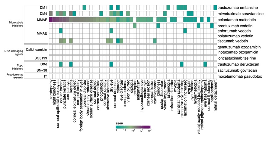
Although pharmacovigilance data are inherently limited by reporting bias, variable adverse event reporting practices, concomitant medications, indication-confounded denominators, and the inability to establish causality, these data remain valuable for identifying emerging toxicity patterns across larger and more heterogeneous patient populations than those typically captured in clinical trials.
Mechanistic Basis of ADC-Associated Ocular Toxicity
OAEs may occur due to on-target and off-target effects of ADCs and are generally related to the target expression, payload class, physicochemical properties, and/or linker stability.
To evaluate whether target expression patterns may inform potential risks associated with ADC-related ocular toxicities, the target expression for various approved ADCs was examined using publicly available Tabula Sapiens (V2; (12)) datasets. This dataset was used to enable a more granular evaluation of ADC target expression, which compiles single-cell gene expression data across multiple donors and 28 organs, including the eye. Analysis of this dataset highlighted pronounced cell type-specific expression patterns of ADC targets (Figure 2). Notably, conjunctival epithelial cells, corneal epithelial cells, and retinal pigment epithelial cells exhibited moderate to high expression of several ADC targets, including TACSTD2 (Trop-2), NECTIN4, MET, and F3 (tissue factor). In contrast, expression of targets of ADCs developed for hematologic malignancies was largely restricted to immune cell compartments, including CD79B, CD22, and TNFRSF17 (BCMA) within the B-cell lineage, with CD22 additionally detected in monocyte and basophil populations.
Target expression alone does not fully explain the occurrence of OAEs, as illustrated by ADCs targeting Trop-2. TACSTD2, the gene encoding Trop-2, is highly expressed in conjunctival and corneal epithelial cells (Figure 2), suggesting the potential for target-mediated ocular uptake. However, sacituzumab govitecan (SN-38 payload, TRODELVY®; approved in April 2020), did not demonstrate significant ocular disproportionality signals in the pharmacovigilance analyses, and its prescribing information does not include major warnings related to ocular toxicity [14]. In contrast, datopotamab deruxtecan (DXd payload, DATROWAY®), a second Trop-2-directed ADC approved in January 2025 (after the FAERS analysis cutoff), includes warnings for ocular adverse reactions such as dry eye, keratitis, and blepharitis in its prescribing information [15]. Consistent with this safety profile, ocular surface events of any grade were reported in approximately 40% of patients treated with datopotamab deruxtecan in the Phase 3 TROPION-Breast01 study [16]. The distinct ocular toxicity profiles of these two Trop-2-directed ADCs may reflect differences between the SN-38 and DXd payload platforms, although other properties such as linker stability, intracellular payload release, and catabolite permeability may also play a role in ADC-mediated ocular toxicity.
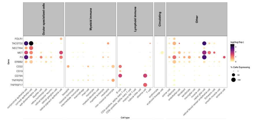
Figure 2. Single-cell gene expression profiles from the Tabula Sapiens (v2) dataset showing the expression patterns of target genes for 15 FDA-approved ADCs across cell types. Dot size represents the percentage of cells expressing the gene, and color indicates average expression (log-transformed). Cell types are grouped by ocular specialized cells, immune cells (myeloid and lymphoid lineages), circulating and other categories (such as stromal or supporting cell types).
Beyond the two topoisomerase I inhibitor payload examples discussed above, other payloads may also impact the ocular toxicity profile of ADCs. ADCs with the microtubule-disrupting payloads monomethyl auristatin F (MMAF), DM1 (a maytansinoid), or DM4 (a ravtansine) tend to have higher rates of ocular toxicity, even when their targets are not expressed in corneal tissue (e.g., folate receptor alpha [FRα; mirvetuximab soravtansine] and BCMA [belantamab mafodotin]). In these cases, toxicity is considered off-target at the tissue level; however, the underlying mechanisms may differ across ADCs. For belantamab mafodotin, soluble BCMA detected in lacrimal fluid has been proposed to form ADC-target complexes that may facilitate corneal epithelial cell uptake via non-specific processes such as pinocytosis [17]. In contrast, for DM- and potentially other microtubule-disrupting payload-containing ADCs, ocular toxicity is more commonly attributed to direct effects of the payload on proliferating corneal epithelial cells following non-specific uptake processes [18]. Monomethyl auristatin E (MMAE)-conjugated ADCs (vedotins), which also harbor microtubule-disrupting payloads, are generally not associated with clinically meaningful ocular toxicity unless the target is expressed in ocular tissues (e.g., Nectin-4 [enfortumab vedotin] and tissue factor [F3, tisotumab vedotin]).
A recent spatially resolved study suggests that ADC-associated ocular toxicity may result from a multi-compartmental cascade of injury rather than a single mechanism [19]. In a case-control, single-cell study in rats treated intravenously with mirvetuximab soravtansine (ELAHERE®; FRα-DM4 ADC), the authors describe a cascade involving potential on-target vascular damage leading to a pro-inflammatory microenvironment, which in turn increased drug penetration into corneal tissue, leading to differentiation blockade of corneal cells. Despite their relevance for improving our understanding of the mechanisms underlying ADC-induced ocular toxicity, such mechanistic and spatially resolved investigational studies remain scarce, limiting the ability to fully disentangle the relative contributions of target expression, payload-related effects, and tissue-specific susceptibility to ADC-associated OAEs.
Translational Gap between Preclinical Models and Clinical Ocular Toxicity
Based on published ADC nonclinical data, ocular toxicity signals in animals were relatively weak and largely underpredicted the clinical events seen in humans [20,21]. Belantamab mafodotin-associated ocular toxicity was not well predicted by nonclinical species. In patients, the hallmark finding was clinically significant microcystic keratopathy characterized by corneal pseudomicrocysts, intracytoplasmic inclusions, epithelial apoptosis, occasional inflammation, and visual symptoms. In contrast, rats and rabbits demonstrated corneal epithelial apoptosis and evidence of ADC exposure in ocular tissues, supporting a mechanistic link to MMAF-mediated epithelial injury, but neither species developed the characteristic pseudomicrocystic lesions observed clinically. Furthermore, rats exhibited increased basal epithelial mitoses and mitotic arrest consistent with microtubule inhibition, findings not identified in human corneal samples. Notably, cynomolgus monkeys showed no meaningful ophthalmologic or histopathologic corneal changes, indicating that the non-human primate model was largely non-predictive of the clinically relevant ocular toxicity observed in patients. Overall, animal studies provided mechanistic evidence of corneal epithelial injury but only partially translated to the unique and clinically significant corneal findings seen in humans.
In cynomolgus monkeys dosed with enfortumab vedotin (EV) or tisotumab vedotin (TV), the “eye findings” were mainly external, observation-level changes (e.g., sunken eyes with EV, reddened/partially closed eyes and conjunctivitis with TV that improved with antibiotic drops), and were not confirmed as corneal injury by more detailed ophthalmology exams or histopathology. In rats, an increased number of corneal mitotic figures was reported in histopathology assessment following treatment with EV (and in non-GLP work with a non-targeted vedotin control ADC), but the authors highlight that the dataset was too limited to determine the relevance of this observation to human corneal toxicity. In contrast, target-associated ocular toxicities observed in patients such as corneal keratopathies and conjunctival changes are observed for vedotin ADCs when the target is expressed in ocular tissues, underscoring a translational gap between standard animal studies and human risk. The overall poor predictivity likely reflects biology and exposure mismatches. Species differences in corneal epithelial turnover/repair, tear film dynamics, and ocular surface barrier properties can change both susceptibility and the phenotype of injury. Even when ocular target expression exists, toxicity depends on antigen accessibility/internalization and local payload delivery, which may not be replicated in animals under typical systemic dosing regimens. Standard toxicology endpoints may also be insensitive to early epithelial stress that later manifests clinically as symptomatic keratopathy, requiring more detailed evaluations such as the quantification of individual mitotic figures in the corneal epithelium.
New approach methodologies (NAMs) may offer an opportunity to address some of these translational limitations by incorporating human-relevant ocular biology into preclinical assessment frameworks [22–24]. Human ocular surface models, including corneal epithelial organoids, induced pluripotent stem cell (iPSC)-derived ocular systems, and eye-on-chip microphysiological platforms incorporating dynamic tear-flow conditions, can be used to evaluate human target biology, characterize barrier function, monitor stress and repair responses, and investigate relationships between ADC or catabolite exposure and cellular injury pathways. In addition, these systems can generate quantitative mechanistic endpoints, including transepithelial electrical resistance (TEER), tight-junction integrity (e.g., ZO-1 disruption), cellular stress responses, apoptotic signaling kinetics (e.g., caspase-3 activation), and epithelial regeneration capacity, which may ultimately be anchored to clinically observed ocular findings. However, current NAMs have important limitations, including incomplete representation of the ocular microenvironment, limited incorporation of lacrimal gland and tear film biology, restricted immune cell interactions, and challenges in reproducing the complex exposure dynamics associated with repeated systemic ADC administration [22–24]. Consequently, these platforms should be viewed as complementary approaches rather than replacements for traditional in vivo studies.
Importantly, the translational challenge extends beyond model selection alone, since the mechanisms responsible for ADC-associated ocular toxicity remain incompletely understood. While some OAEs appear to be associated with target expression in ocular tissues, others have been observed in the absence of known ocular target expression, suggesting potential contributions from non-target-mediated uptake pathways, payload-specific effects, local catabolite exposure, or indirect effects on corneal epithelial homeostasis. These uncertainties complicate the interpretation of both animal and in vitro findings and highlight the need for multi-mechanism-based approaches capable of distinguishing target-dependent from target-independent toxicity pathways. One approach to addressing this challenge is the use of cell type-specific expression data from resources such as the Tabula Sapiens database, which can help guide experimental studies to interrogate ADC target engagement, internalization, and subsequent cytotoxic or stress responses. For example, a recent study evaluating an anti-Nectin4-vcMMAE ADC demonstrated that ocular toxicity may be driven by both Nectin4-mediated uptake and non-specific pinocytosis by corneal epithelial cells [13]. Interestingly, the introduction of Fc-silencing point mutations within the Fc region of the ADC significantly reduced toxicity against an immortalized human corneal epithelial cell line. These findings illustrate how assessment of target expression at cellular resolution can identify potential toxicity drivers, guide the selection of translationally relevant experimental models, and ultimately inform rational ADC design to improve safety.
Looking forward, a tiered translational strategy may provide additional value for ocular risk assessment. In such a framework, human-relevant NAMs can be used to identify susceptible ocular cell populations, characterize target engagement and mechanistic injury pathways, and establish quantitative biomarkers of epithelial injury, while conventional in vivo studies provide information on systemic exposure, biodistribution, pharmacokinetics, and whole-organism responses. Integration of these complementary datasets with quantitative exposure-response (E-R) analyses, physiologically-based pharmacokinetic (PBPK) modeling, and emerging clinical observations may improve the translation of preclinical findings to human ocular risk and support the development of more predictive, mechanism-informed safety assessment approaches [22–24].
Together, these limitations highlight the need for quantitative, human-relevant approaches that integrate exposure, dosing dynamics, target biology, mechanistic biomarkers, and clinical event trajectories to improve the prediction, mechanistic understanding, and mitigation of ADC-associated ocular toxicity.
Markov Modeling of OAEs for ADCs
Markov models are widely used in drug development to characterize adverse events that evolve over time and may recur during treatment [25]. For ADC-associated ocular toxicity, these models are particularly valuable because they describe transitions between toxicity states, capturing the onset, severity, duration, and resolution of OAEs. Unlike traditional E-R analyses, which often evaluate isolated outcomes, Markov modeling approaches offer a more realistic representation of patient experience and allow forward-looking simulations under alternative dosing or titration strategies. More broadly, successful implementation of Markov modeling across therapeutic areas underscores the growing role of longitudinal, exposure-driven safety models in optimizing dose, balancing efficacy and tolerability, and ultimately improving clinical decision-making in drug development.
Although the biological mechanisms underlying ADC-associated ocular toxicity may differ between compounds, the Markov framework is designed to characterize transitions between clinically defined toxicity states over time rather than the underlying biological pathways, allowing the framework to be applied across a range of ADC-associated ocular toxicities.
In the context of ADC-associated ocular toxicity, Markov modeling has been applied to the anti-BCMA MMAF-conjugated ADC belantamab mafodotin to evaluate the impact of dosing regimens and dose-modification strategies on both efficacy and ocular safety. Treatment with belantamab mafodotin is associated with ocular events affecting the corneal epithelium, including keratopathy, which are generally low grade (Grade 1 and 2) and reversible in most patients (Figure 1) [26]. As discussed above, ocular toxicity associated with belantamab mafodotin is hypothesized to arise through off-target mechanisms, potentially involving soluble BCMA in lacrimal fluid and subsequent uptake by corneal epithelial cells [17].
To better understand the relationship between treatment exposure, efficacy, and ocular toxicity, clinical data from the DREAMM-1 (0.03–4.6 mg/kg every 3 weeks) and DREAMM-2 (2.5 and 3.4 mg/kg every 3 weeks) studies in patients with relapsed/refractory multiple myeloma were used to develop an integrated modeling framework. This framework combined population pharmacokinetics with a tumor growth inhibition model based on serum M-protein dynamics and a discrete-time Markov model capturing the onset, progression, and resolution of ocular events graded by the GSK Keratopathy and Visual Acuity scale [27]. Model-based analyses indicated that reducing dose intensity (either through lower doses or extended dosing intervals) decreased both the incidence and duration of clinically significant ocular events, while maintaining a substantial proportion of antitumor activity.
The anti-tissue factor (TF) MMAE-conjugated ADC tisotumab vedotin (TV) provides a complementary example in which ocular toxicity is linked to target expression in ocular tissues. TV monotherapy at the approved dose of 2.0 mg/kg every 3 weeks demonstrated overall survival benefit with a manageable safety profile compared with chemotherapy in women with previously treated recurrent or metastatic cervical cancer [28,29]. This approved dosing regimen was supported by population pharmacokinetics and E-R analyses conducted using data across four clinical studies [30]. Expression of TF (F3) in conjunctival and corneal epithelial cells (Figure 2), key components of the ocular surface, provides a plausible mechanistic basis for on-target, off-tumor toxicity. Local TF expression may permit ADC binding and internalization in these tissues, potentially contributing to ocular surface-related adverse events such as dry eye, conjunctivitis, and keratopathy (Figure 1). Importantly, these risks can be mitigated without compromising anti-cancer efficacy through structured eye care plans (ECPs) and proactive monitoring. In a traditional E-R analysis, the association between TV exposure and probability of response was characterized using logistic regression analysis, but due to the limitations of this logit model, neither onset time nor duration of adverse events were taken into consideration [31].
To characterize the temporal dynamics of these events, Feng et al. developed a Markov model framework that integrated OAE severity, onset, and duration across seven clinical studies, complemented by E-R safety models accounting for actual dosing conditions (e.g., considering dose delays or modifications and discontinuations due to AEs) [32]. This framework enabled characterization of OAE risk under clinically relevant treatment conditions. Model outputs indicated a clear E-R relationship for Grade ≥2 OAEs, consistent with a TF expression-dependent mechanism. Incorporation of an ECP reduced the predicted risk of Grade ≥ 2 OAEs, and an alternative TV dosing regimen explored in the model is being evaluated in the ongoing innovaTV 207 trial to assess its impact on the overall benefit-risk profile.
Together, these examples highlight the utility of Markov modeling as a quantitative framework for informing clinical decision-making by quantifying the trade-off between efficacy and toxicity and by informing dose-modification strategies aimed at maintaining treatment benefit while reducing ocular risk. While Markov models provide a useful framework for evaluating alternative dosing strategies, their predictive performance depends on how well the underlying E-R and transition relationships describe the observed data. As with any model-based approach, the predictive performance of Markov models depends on the validity of the underlying assumptions and the quality of the available data, and model outputs should therefore be interpreted within the context of these limitations.
Conclusions
As ADCs continue to reshape the therapeutic landscape in oncology, a deeper mechanistic understanding of ocular toxicity is essential to ensure the safety of these drugs. Evidence from enfortumab vedotin, tisotumab vedotin, and other ADCs indicates that ocular toxicity is influenced by multiple factors, including target expression, payload properties, and linker characteristics. Single-cell transcriptomic datasets and emerging human ocular systems now provide unprecedented resolution for characterizing the cellular and molecular mechanisms underlying ADC-associated ocular toxicity, enabling the delineation of target-dependent and target-independent mechanisms that traditional toxicology animal models often fail to capture. Concurrently, quantitative frameworks—such as E-R and Markov modeling—offer a means to integrate mechanistic insights with clinical dosing conditions, bridging mechanistic biology, pharmacology, and patient-level risk. Although post-marketing pharmacovigilance analyses cannot establish a direct causal link and are subject to inherent reporting biases, they remain valuable for identifying uncommon, delayed, or clinically heterogeneous toxicity patterns across broader patient populations and treatment settings, complementing mechanistic and translational investigations. Together, these multidisciplinary advances support a more holistic and predictive strategy for ADC development - one that integrates mechanistic biology, translational modeling, quantitative clinical pharmacology, and real-world safety surveillance to enable the development of safer next-generation ADCs while preserving antitumor efficacy.
References
2. Liu K, Li M, Li Y, Li Y, Chen Z, Tang Y, et al. A review of the clinical efficacy of FDA-approved antibody‒drug conjugates in human cancers. Mol Cancer. 2024 Mar 23;23(1):62.
3. Domínguez-Llamas S, Caro-Magdaleno M, Mataix-Albert B, Avilés-Prieto J, Romero-Barranca I, Rodríguez-de-la-Rúa E. Adverse events of antibody-drug conjugates on the ocular surface in cancer therapy. Clin Transl Oncol. 2023 Nov;25(11):3086–100.
4. Eaton JS, Miller PE, Mannis MJ, Murphy CJ. Ocular Adverse Events Associated with Antibody-Drug Conjugates in Human Clinical Trials. J Ocul Pharmacol Ther. 2015 Dec;31(10):589–604.
5. Long R, Zuo H, Tang G, Zhang C, Yue X, Yang J, et al. Antibody-drug conjugates in cancer therapy: applications and future advances. Front Immunol. 2025 May 21;16:1516419.
6. Drago JZ, Modi S, Chandarlapaty S. Unlocking the potential of antibody-drug conjugates for cancer therapy. Nat Rev Clin Oncol. 2021 Jun;18(6):327–44.
7. Baah S, Laws M, Rahman KM. Antibody-Drug Conjugates-A Tutorial Review. Molecules. 2021 May 15;26(10):2943.
8. Fu Z, Li S, Han S, Shi C, Zhang Y. Antibody drug conjugate: the "biological missile" for targeted cancer therapy. Signal Transduct Target Ther. 2022 Mar 22;7(1):93.
9. Hoffmann RM, Coumbe BGT, Josephs DH, Mele S, Ilieva KM, Cheung A, et al. Antibody structure and engineering considerations for the design and function of Antibody Drug Conjugates (ADCs). Oncoimmunology. 2017 Nov 20;7(3):e1395127.
10. Peng Y, Song Y, Qin C, Lin J. faers: R Interface for FDA Adverse Event Reporting System. R package version 1.8.0; 2026. Available from: https://bioconductor.org/packages/faers
11. Mao K, Chen P, Sun H, Zhong S, Zheng H, Xu L. Ocular adverse events associated with antibody-drug conjugates in oncology: a pharmacovigilance study based on FDA adverse event reporting system (FAERS). Front Pharmacol. 2024 Aug 20;15:1425617.
12. Tabula Sapiens Consortium; Quake SR. Tabula Sapiens reveals transcription factor expression, senescence effects, and sex-specific features in cell types from 28 human organs and tissues. bioRxiv [Preprint]. 2025 Aug 27:2024.12.03.626516.
13. Zhang J, Li M, Li W, Yang Y, Wu G, Guo P, et al. Mechanistic Insights into Anti-Nectin4-VcMMAE-Induced Ocular Toxicity: From Cellular Uptake Pathways to Molecular Modification. Int J Mol Sci. 2025 May 22;26(11):4996.
14. U.S. Food and Drug Administration. TRODELVY® (sacituzumab govitecan-hziy) prescribing information [Internet]. Silver Spring (MD): U.S. Food and Drug Administration; 2025 [cited 2026 June]. Available from: https://www.accessdata.fda.gov/drugsatfda_docs/label/2025/761115s059lbl.pdf
15. U.S. Food and Drug Administration. DATROWAY® (datopotamab deruxtecan-dlnk) prescribing information [Internet]. Silver Spring (MD): U.S. Food and Drug Administration; 2025. Available from: https://www.accessdata.fda.gov/drugsatfda_docs/label/2025/761394s000lbl.pdf
16. Bardia A, Jhaveri K, Im SA, Pernas S, De Laurentiis M, Wang S, et al. TROPION-Breast01 Investigators. Datopotamab Deruxtecan Versus Chemotherapy in Previously Treated Inoperable/Metastatic Hormone Receptor-Positive Human Epidermal Growth Factor Receptor 2-Negative Breast Cancer: Primary Results From TROPION-Breast01. J Clin Oncol. 2025 Jan 20;43(3):285–96.
17. Munawar U, Theuersbacher J, Steinhardt MJ, Zhou X, Han S, Nerreter S, et al. Soluble B-cell maturation antigen in lacrimal fluid as a potential biomarker and mediator of keratopathy in multiple myeloma. Haematologica. 2024 Nov 1;109(11):3670–80.
18. Hendershot A, Slabaugh M, Riaz KM, Moore KN, O'Malley DM, Matulonis U, et al. Strategies for prevention and management of ocular events occurring with mirvetuximab soravtansine. Gynecol Oncol Rep. 2023 Feb 28;47:101155.
19. Zhang J, Li M, Yang Y, Guo P, Li W, An H, et al. Single-Cell Transcriptomics Reveals a Multi-Compartmental Cellular Cascade Underlying Elahere-Induced Ocular Toxicity in Rats. Pharmaceuticals (Basel). 2025 Oct 4;18(10):1492.
20. Lee V, Hultcrantz M, Petrone S, Lewis EW, Banna H, Lichtman E, et al. Characterization of Belantamab Mafodotin-Induced Corneal Changes in Patients With Multiple Myeloma. JAMA Ophthalmol. 2025 Jun 1;143(6):507–14.
21. Neff-LaFord HD, Carratt SA, Carosino C, Everds N, Cardinal KA, Duniho S, et al. The Vedotin Antibody-Drug Conjugate Payload Drives Platform-Based Nonclinical Safety and Pharmacokinetic Profiles. Mol Cancer Ther. 2024 Oct 1;23(10):1483–93.
22. Marx U, Akabane T, Andersson TB, Baker E, Beilmann M, Beken S, et al. Biology-inspired microphysiological systems to advance patient benefit and animal welfare in drug development. ALTEX. 2020;37(3):365–94.
23. Ewart L, Dehne EM, Fabre K, Gibbs S, Hickman J, Hornberg E, et al. Application of Microphysiological Systems to Enhance Safety Assessment in Drug Discovery. Annu Rev Pharmacol Toxicol. 2018 Jan 6;58:65–82.
24. Choudhary S, Dubey A, Singh A, Zamboni P, Gupta N, Singh R, et al. Engineering the microenvironment: advanced biomaterials for humanized in vitro immunotoxicology and carcinogenicity assessment. Exploration of BioMat-X. 2025 Dec 8;2:101351.
25. Ooi QX, Plan E, Bergstrand M. A tutorial on pharmacometric Markov models. CPT Pharmacometrics Syst Pharmacol. 2025 Feb;14(2):197–216.
26. Terpos E, Trudel S, Mateos MV, Alejandre N, Colby K, Dimopoulos MA, et al. Practical Guidance on Clinical Management of Belantamab Mafodotin-Associated Ocular Events. Am J Hematol. 2025 Oct;100(10):1839–50.
27. Collins J, van Noort M, Rathi C, Post TM, Struemper H, Jewell RC, et al. Longitudinal efficacy and safety modeling and simulation framework to aid dose selection of belantamab mafodotin for patients with multiple myeloma. CPT Pharmacometrics Syst Pharmacol. 2023 Oct;12(10):1411–24.
28. Pfizer Inc. TIVDAK® (tisotumab vedotin-tftv) prescribing information [Internet]. New York (NY): Pfizer Inc.; 2024 [cited 2024 Jul 26]. Available from: https://labeling.pfizer.com/ShowLabeling.aspx?id=20632
29. Coleman RL, Lorusso D, Gennigens C, González-Martín A, Randall L, Cibula D, et al. Efficacy and safety of tisotumab vedotin in previously treated recurrent or metastatic cervical cancer (innovaTV 204/GOG-3023/ENGOT-cx6): a multicentre, open-label, single-arm, phase 2 study. Lancet Oncol. 2021 May;22(5):609–19.
30. Passey C, Voellinger J, Gibiansky L, Gunawan R, Nicacio L, Soumaoro I, et al. Exposure-safety and exposure-efficacy analyses for tisotumab vedotin for patients with locally advanced or metastatic solid tumors. CPT Pharmacometrics Syst Pharmacol. 2023 Sep;12(9):1262–73.
31. Ruiz-Garcia A, Baverel P, Bottino D, Dolton M, Feng Y, González-García I, et al. A comprehensive regulatory and industry review of modeling and simulation practices in oncology clinical drug development. J Pharmacokinet Pharmacodyn. 2023 Jun;50(3):147–72.
32. Feng S, Gunawan R, Passey C, Voellinger J, Polhamus D, Gerritsen A, et al. Exposure-safety Markov modeling of ocular adverse events in patient populations treated with tisotumab vedotin. J Pharmacokinet Pharmacodyn. 2025 Oct 3;52(5):55.
33. Shen L, Sun X, Chen Z, Guo Y, Shen Z, Song Y, et al. ADCdb: the database of antibody-drug conjugates. Nucleic Acids Res. 2024 Jan 5;52(D1):D1097–109.