Abstract
Background: Teeth and arch size determination is an important factor in dentistry and forensic medicine. Evidence shows that cone-beam computed tomography (CBCT) is a highly accurate tool for the measurement of mesiodistal tooth width and arch dimensions. This study aimed to assess the teeth and arch dimensions of males and females on CBCT images of patients presenting to the Radiology Department of Tabriz School of Dentistry.
Materials and Methods: 70 CBCT scans were evaluated in this study by taking into account the age and gender of patients. Mesiodistal teeth width and arch dimensions were measured on CBCT images by an oral and maxillofacial radiologist. The Pearson’s correlation coefficient was used to analyze the correlation between mesiodistal width of the teeth and arch size. An independent t-test was used to compare the mesiodistal width of the teeth and arch size between males and females. All statistical analyses were carried out by SPSS 17 at P<0.05 level of significance.
Results: Significant differences were noted in arch size of males and females in maxillary first premolars, maxillary and mandibular second premolars, and maxillary and mandibular first molars width. The mesiodistal width of teeth #13, 23, 33, 36, and 41 in females was larger than that in males. The mesiodistal width of other teeth was larger in males than females. Arch size and mesiodistal width of teeth were significantly correlated.
Conclusion: Arch size and teeth dimensions in the Iranian population were different from the norms of other countries. Some differences in values were also noted between males and females, which should be taken into account in dental treatments.
Keywords
Cone-Beam Computed Tomography, Mesiodistal, Dental arch
Introduction
Tooth size and arch size are influenced by a number of factors such as age, gender, and race. Tooth and arch size measurements are important for orthodontic and prosthodontic treatments, and also in forensic medicine [1-4].
Radiographs, manual measurement, and measurement on dental casts are the direct methods of assessment of tooth dimensions [1]. At present, radiographic assessment of tooth dimensions is not limited to the conventional intraoral and panoramic radiographic modalities. Conventional intraoral and panoramic radiographs can only reveal the important properties of the teeth and their surrounding structures in mesiodistal (proximal) plane, and similar properties in the buccolingual plane often remain undetected on periapical radiographs. However, Cone-beam computed tomography (CBCT) enables precise three-dimensional evaluation of dentition, maxillofacial structures, and anatomical relationships [5,6]. CBCT images can greatly help in the assessment of the morphology, position, shape, size, and variations of oral and maxillofacial structures [7]. Evidences show that CBCT is a highly accurate modality for measurement of mesiodistal width of the teeth and arch size, and has comparable accuracy to highly precise anatomical measurements. Tooth size and arch size measurements by CBCT have been the topic of many previous studies. Mohammad et al. [1] evaluated 32 males in the same age range by CBCT and reported that the mesiodistal width of the teeth in the right and left quadrants was the same, except for second premolars which were slightly but significantly larger in the right quadrant than the left quadrant (by 0.08 to 0.2 mm). Also, the mesiodistal width of the teeth in males and females were the same at certain ages. Shahid et al. [8] in 2015 reported that only the size of maxillary lateral incisors, canines, and first premolars and mandibular lateral incisors was significantly different in the right and left quadrants. Considering the gap of information regarding the tooth size and arch size in an Iranian population, the existing controversy in this regard, and the significance of this topic in orthodontic and prosthodontic treatments, and forensic medicine, study of tooth size and arch size in an Iranian population is a necessity. Accordingly, this study aimed to measure the tooth size and arch size of male and female patients presenting to the Oral and Maxillofacial Radiology Department of School of Dentistry, Tabriz University of Medical Sciences in 2017 using CBCT.
Materials and Methods
This retrospective study was conducted on CBCT scans retrieved from the archives of the Radiology Department of School of Dentistry, Tabriz University of Medical Sciences in 2017. The inclusion criteria were CBCT images of patients between 16 to 35 years, presence of all teeth from the first molar of one quadrant to the first molar of the other quadrant in each arch, and presence of high-quality CBCT data. The exclusion criteria were severe dental crowding, large diastema, presence of pathological radiolucencies on the radiographs, periodontal disease, proximal dental caries or extensive restorations in the proximal lesion, abnormal tooth size or morphology, presence of fixed orthodontic appliances, presence of supernumeraries or missing teeth, and distorted or low-resolution CBCT images.
A total of 70 CBCT images were evaluated. Age and sex of patients were recorded for each image. All CBCT scans had been taken with NewTom VGI CBCT scanner (QR srl, Verona, Italy) with 360-degree rotation, flat-panel detector, 1920 x 1536 mm pixel size, 18 seconds of scanning time, maximum voltage of 110 KVP, and 1-20 mA amperage.
Images were reconstructed with NNT Viewer 2.21 software (QR srl, Verona, Italy). The images were stored in DICOM format and analyzed using Mimics 10.01 software (Materialise NV, Belgium). Measurements of tooth size and arch size on each image were done by an oral and maxillofacial radiologist. The mesiodistal width of each tooth was measured on an axial CBCT section that visualized the maximum mesiodistal width of the respective tooth. The size of dental arch was measured by measuring the inter-canine width, inter-first premolar width, inter-second premolar width, and inter-first molar width. For assessment of inter-canine width, the distance between the two canine cusp tips was measured. For the inter-premolar width, the distance between the buccal cusp tip of right and left first/second premolars was measured. For the inter-molar width, the distance between the mesiobuccal cusp tips was measured (Figure 1 and Figure 2). To control accidental and systematic errors in measurements, all images were evaluated again after a 2-week interval.
The results were reported by descriptive statistics (mean and standard deviation). The Pearson or Spearman’s correlation coefficient was used to analyze the correlation of mesiodistal width of the teeth and arch size. Considering the normal distribution of data, independent t-test was used to compare the mesiodistal width of the teeth and arch size between males and females. The normality of data was assessed by the Kolmogorov-Smirnov test. All statistical analyses were carried out by SPSS 17 at P<0.05 level of significance.
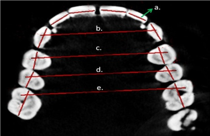
Figure 1. Tooth size and arch size measurements: (a) Mesiodistal width of the tooth; (b) inter-canine width measured as the distance between the two canine cusp tips; (c,d) inter-premolar width measured as the distance between the buccal cusp tips of the right and left premolars; (e) inter-molar width measured as the distance between the mesiobuccal cusp tips of the right and left first molars.
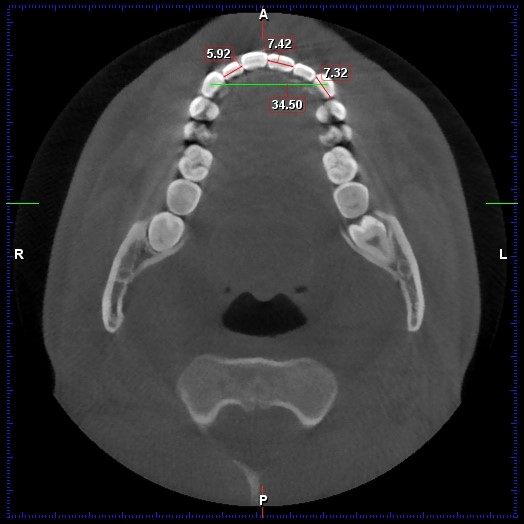
Figure 2. Measuring the mesiodistal width of the teeth and arch size.
Results
The mesiodistal tooth width and arch size of patients were measured. From 70 patients that was evaluated in this study, 36 (51.4%) were females and 34 (48.6%) were males. The mean age of participants was 25.69 ± 5.59 years (range 16 to 36 years). The mean age was 25.11 ± 5.25 years in females and 26.26 ± 5.81 years in males. Due to wide dispersion of age, no statistical analysis could be performed on age of participants in this study (P=0.386).
Table 1 presents the mesiodistal width of different teeth. As shown, the largest mean width was recorded for tooth #46 (mean value of 10.48 ± 0.71 mm), and the smallest mean width was recorded for tooth #31 (mean value of 5.01 ± 0.45 mm). The maximum mesiodistal width was 12.11 mm in tooth #46, and the minimum mesiodistal width was 4 mm in tooth #31.
| Tooth number | Number | Minimum | Maximum | Mean and std. deviation |
| 16 | 70 | 6.80 | 10.50 | 9.42 ± 0.7 |
| 15 | 70 | 5.03 | 7.22 | 6.02 ± 0.51 |
| 14 | 70 | 5.18 | 7.13 | 6.14 ± 0.49 |
| 13 | 70 | 6.18 | 8.76 | 7.05 ± 0.5 |
| 12 | 70 | 4.70 | 7.41 | 6.17 ± 0.58 |
| 11 | 70 | 6.49 | 9.53 | 8.09 ± 0.61 |
| 21 | 70 | 6.77 | 9.66 | 8.12 ± 0.57 |
| 22 | 70 | 4.33 | 7.87 | 6.13 ± 0.63 |
| 23 | 70 | 6.00 | 8.36 | 6.99 ± 0.53 |
| 24 | 70 | 5.18 | 7.30 | 6.14 ± 0.47 |
| 25 | 70 | 5.03 | 7.42 | 6.11 ± 0.48 |
| 26 | 70 | 7.34 | 10.48 | 9.45 ± 0.59 |
| 36 | 70 | 7.25 | 11.90 | 10.27 ± 1.08 |
| 35 | 70 | 5.91 | 9.97 | 6.84 ± 0.76 |
| 34 | 70 | 5.53 | 8.84 | 6.43 ± 0.58 |
| 33 | 70 | 4.76 | 7.41 | 6.26 ± 0.49 |
| 32 | 70 | 4.58 | 6.48 | 5.58 ± 0.44 |
| 31 | 70 | 4.00 | 6.07 | 5.01 ± 0.45 |
| 41 | 70 | 4.18 | 6.07 | 5.08 ± 0.44 |
| 42 | 70 | 4.43 | 6.74 | 5.57 ± 0.45 |
| 43 | 70 | 5.52 | 7.65 | 6.29 ± 0.59 |
| 44 | 70 | 5.34 | 8.49 | 6.54 ± 0.59 |
| 45 | 70 | 5.92 | 10.06 | 6.82 ± 0.74 |
| 46 | 70 | 7.92 | 12.11 | 10.48 ± 0.71 |
Table 2 presents the arch size according to the inter-canine, inter-first premolar, inter-second premolar, and inter-first molar widths. As shown, the mean arch size was the largest at the inter-first molar width in the maxilla (50.33 ± 3.34 mm), and the smallest at the inter-canine width in the mandible (26.75 ± 1.66 mm). The largest inter-first molar width in the maxilla was 55.5 mm while the smallest inter-canine width in the mandible was 23.47 mm.
| Arch width | Number | Minimum | Maximum | Mean and std. deviation |
| Maxillary inter-canine | 70 | 28.90 | 38.40 | 33.72 ± 1.91 |
| Mandibular inter-canine | 70 | 23.47 | 31.59 | 26.75 ± 1.66 |
| Maxillary inter-first premolars | 70 | 33.95 | 45.05 | 40.55 ± 2.36 |
| Mandibular inter-first premolars | 70 | 30.39 | 39.12 | 35.21 ± 1.94 |
| Maxillary inter-second premolars | 70 | 39.94 | 50.53 | 45.71 ± 2.65 |
| Mandibular inter-second premolars | 70 | 37.23 | 47.18 | 41.26 ± 2.31 |
| Maxillary inter-first molars | 70 | 42.90 | 55.50 | 50.33 ± 3.34 |
| Mandibular inter-first molars | 70 | 41.13 | 54.51 | 46.67 ± 3.11 |
The Kolmogorov-Smirnov test showed normal distribution of all data except for the mesiodistal width of tooth #26. Thus, independent t-test was used to compare the normally distributed data between males and females, while the non-parametric Mann-Whitney U test was applied to compare non-normally distributed data between males and females.
Table 3 presents the mean arch size in males and females. The largest width was recorded at inter-first molars at the maxilla in both females (mean value of 49.02 ± 3.72 mm) and males (51.83 ± 2.04 mm).
| Arch width | Gender | Mean and std. deviation | P-value |
| Maxillary inter-canine | Female | 33.27 ± 1.72 | 0.054 |
| Male | 34.18 ± 2.04 | ||
| Mandibular inter-canine | Female | 26.74 ± 1.75 | 0.927 |
| Male | 26.77 ± 1.58 | ||
| Maxillary inter-first premolars | Female | 39.77 ± 2.46 | 0.003 |
| Male | 41.5 ± 1.89 | ||
| Mandibular inter-first premolars | Female | 34.79 ± 1.99 | 0.082 |
| Male | 35.62 ± 1.82 | ||
| Maxillary inter-second premolars | Female | 44.79 ± 3.12 | 0.008 |
| Male | 46.61 ± 1.73 | ||
| Mandibular inter-second premolars | Female | 40.25 ± 2.13 | <0.001 |
| Male | 42.3 ± 2.02 | ||
| Maxillary inter-first molars | Female | 49.02 ± 3.72 | 0.001 |
| Male | 51.83 ± 2.04 | ||
| Mandibular inter-first molars | Female | 45.15 ± 2.68 | 0.001 |
| Male | 48.01 ± 2.86 |
Independent t-test and Mann Whitney U test showed a significant difference in mesiodistal width of teeth #13, 23, 36, 33, 41, and 44 between males and females (P<0.05). The mesiodistal width of tooth #41 in females was larger than that in males. In teeth #13, 23, 36, and 33, the mesiodistal width was larger in males than females. Also, significant differences were noted between males and females in maxillary inter-first premolars, maxillary and mandibular inter-second premolars, and maxillary and mandibular inter-first molars widths (P<0.05).
Comparison of mesiodistal width of the teeth in the maxilla and mandible between the right and left quadrants by paired t-test showed no significant difference in this regard between the right and left quadrants neither in the maxilla (P=0.178) nor in the mandible (P=0.287). Thus, the mean mesiodistal width of the teeth in the right and left sides was calculated and compared between males and females (Table 4). As shown, significant differences were noted in the mesiodistal width of maxillary canine (P=0.001) and mandibular canine (P=0.035) and first premolar (P=0.025) between males and females such that the mesiodistal width of the abovementioned teeth in females was smaller than that in males.
| Mesiodistal width | Gender | Mean and std. deviation | P-value | |
| Maxilla | Tooth #1 | Females | 8.11 ± 0.45 | 0.94 |
| Males | 8.10 ± 0.63 | |||
| Tooth #2 | Females | 6.18 ± 0.51 | 0.517 | |
| Males | 6.09 ± 0.63 | |||
| Tooth #3 | Females | 6.83 ± 0.37 | 0.001 | |
| Males | 7.20 ± 0.50 | |||
| Tooth #4 | Females | 6.07 ± 0.42 | 0.137 | |
| Males | 6.24 ± 0.47 | |||
| Tooth #5 | Females | 5.99 ± 0.41 | 0.197 | |
| Males | 6.15 ± 0.48 | |||
| Tooth #6 | Females | 9.45 ± 0.49 | 0.271 | |
| Males | 9.64 ± 0.78 | |||
|
Mandible |
Tooth #1 | Females | 5.13 ± 0.38 | 0.053 |
| Males | 4.94 ± 0.43 | |||
| Tooth #2 | Females | 5.60 ± 0.35 | 0.684 | |
| Males | 5.56 ± 0.46 | |||
| Tooth #3 | Females | 6.11 ± 0.48 | 0.035 | |
| Males | 6.40 ± 0.60 | |||
| Tooth #4 | Females | 6.35 ± 0.52 | 0.026 | |
| Males | 6.64 ± 0.50 | |||
| Tooth #5 | Females | 6.71 ± 0.41 | 0.104 | |
| Males | 7.01 ± 0.90 | |||
| Tooth #6 | Females | 10.17 ± 0.72 | 0.058 | |
| Males | 10.60 ± 0.77 |
Considering the normal distribution of data, the Pearson’s correlation test was used to analyze the correlation of mesiodistal width of the teeth and arch size in the maxilla and mandible. The results showed 70% correlation between mesiodistal width of the teeth and maxillary arch size, which was statistically significant (P<0.001). Also, the correlation between mesiodistal width of the teeth and mandibular arch size was 73%, which was statistically significant as well (P<0.001). The correlation between the maxillary and mandibular arch sizes was 89%, which was also statistically significant (P<0.001).
Discussion
Tooth size and arch size are influenced by a number of factors such as age, gender, and race. Assessment of tooth size and arch size is important in orthodontic treatment and forensic medicine [2,9]. Teeth can greatly help in age estimation, and gender and race determination for identification of burned or decomposed corpse. Studies on sexual dimorphism can provide valuable anthropological and epidemiological information [10]. Racial differences in tooth dimensions are an important topic in this respect; however, the findings of studies on each race are only generalizable to the respective racial population. Thus, assessment of tooth dimensions and their ratios is imperative in different races [11].
Considering the lack of comprehensive studies regarding tooth size measurements and the controversial results in this respect, as well as the significance of this topic for identification purposes in forensic medicine, this study aimed to assess the mesiodistal tooth width and arch size in an Iranian population using CBCT.
Khursheed et al. [1], in 2014 evaluated tooth dimensions and arch size of 32 males and 21 females using CBCT, and showed that tooth size was the same in the right and left quadrants except for right second premolar, which was slightly larger than the left second premolar. The greatest difference in males was noted in maxillary lateral incisors and mandibular second premolars and lateral incisors while in females, maximum difference was noted in maxillary canines and mandibular central incisors. The size of mandibular and maxillary canine teeth showed the highest sexual dimorphism. For dental arch dimensions, the greatest difference was noted in inter-canine width in the maxilla in males, and inter-canine and inter-molar width in the maxilla in females. No significant difference was noted in males or females of different age groups [1]. In the present study, since the difference in mesiodistal width of the same teeth in the right and left quadrants was not significant, the mean of right and left-side measurements was used for the purpose of comparison; the results showed a significant difference in mesiodistal width of maxillary canine teeth and mandibular canine and first premolar teeth between males and females, such that all the above-mentioned teeth were smaller in females than males. Also, the arch width at the site of maxillary inter-first premolars, maxillary and mandibular inter-second premolars, and maxillary and mandibular inter-first molars were significantly different between males and females, and the measured widths were smaller in females than males. Moreover, the maximum mean inter-arch width in both males and females was recorded at the site of maxillary inter-first molars. In the present study, no significant difference was found with respect to age. Differences in the results of studies can be due to differences in study populations and sample size. Kaushal et al. [12], in 2004 showed that the mean distance between the crowns of mandibular canine teeth in the right and left sides was significantly different between males and females. They also showed a significant difference in mesiodistal width of mandibular canine teeth between males and females. In the present study, the inter-canine width was not significantly different between males and females neither in the maxilla nor in the mandible. Regarding the mesiodistal width of the teeth, the mesiodistal width of maxillary canine and mandibular canine and first premolar teeth was significantly different between males and females. Controversy in the results may be due to the fact that Kaushal et al., only evaluated the mandibular canine teeth as reference while all maxillary and mandibular teeth were evaluated in the present study. Also, this difference may be due to different methods of measurements, study populations, and races. Khursheed et al. [4], in 2019 measured tooth dimensions in adults of Saudi-Arabian, Jordanian, and Egyptian populations by using CBCT. They measured tooth dimensions from the second molar of one side to the second molar of the other side in the maxilla and mandible. The results revealed a significant difference in tooth dimensions in 9 out of 14 groups in the maxilla and mandible. They found no significant difference in tooth dimensions among the study populations. In another study, Salam et al. [13], in 2022 evaluated dental arch dimensions in an Egyptian population, and compared males and females in this respect. They found significant differences in inter-canine width and arch length both in the maxilla and mandible between males and females.
Tarazona et al. [14], in 2013 compared tooth dimensions by CBCT and digital radiography and found significant differences in dimensions of maxillary right first premolar, maxillary left first molar, mandibular right first premolar, and mandibular right second premolar measured on CBCT scans and digital radiographs. They indicated that digital CBCT models obtained from plaster casts were precise and reliable. Elsande et al. [15], in 2010 assessed the accuracy of CBCT panoramic-like images to measure the mesiodistal root angulation, and found significant differences between the coordinate measuring machine and CBCT in measurement of mesiodistal root angulation of 16 out of 28 teeth. In comparison with previous studies that used conventional panoramic radiography, the mesiodistal angles of teeth measured on CBCT scans were closer to the actual values. Bakkannavar et al. [16], in 2014 evaluated gender determination according to the mandibular canine index in an Egyptian population. They demonstrated that the mean mandibular canine index was significantly different between males and females in both the right and left sides, and had a prediction accuracy of approximately 73% for gender determination. However, this index was not significantly different between males and females for the maxillary canine tooth neither in the right nor in the left side. In general, gender determination by maxillary canine index had low statistical significance in their study.
Presence of differences and significant dimorphisms in teeth can be due to relatively high amounts of dentin in males compared with females. Agnihotri et al. [17], attributed the difference in mesiodistal width of teeth in males and females to genetics, because the chromosome Y determines the tooth size and chromosome X determines the enamel thickness. Also, variations in the results conducted in different countries may be due to ethnic and racial differences in the study populations.
Conclusion
Teeth are the most durable body parts which may remain sound for long periods of time after decomposition of bones. CBCT images can help in gender determination based on measurement of mesiodistal width of teeth and arch width. The present study evaluated the mesiodistal width of teeth and arch width on CBCT scans of patients presenting to the Oral and Maxillofacial Radiology Department of School of Dentistry, Tabriz University of Medical Sciences, showed that the mesiodistal width of teeth and arch width in an Iranian population had some differences with those of Indian and Malaysian populations. The differences in mesiodistal width of teeth and arch width between males and females were also evaluated. Comparison of mesiodistal width of teeth in the right and left quadrants revealed no significant difference in the maxilla or mandible. Also, comparison of mesiodistal width of teeth between males and females showed significant differences in mesiodistal width of maxillary canine and mandibular canine and first premolar teeth, and they were smaller in females. Also, the arch width at maxillary inter-first premolars, maxillary and mandibular inter-second premolars, and maxillary and mandibular inter-first molars was significantly smaller in females than males.
References
2. Burris BG, Harris EF. Maxillary arch size and shape in American blacks and whites. The Angle Orthodontist. 2000;70(4):297-302.
3. Kaur S, Chattopadhyay PK. Sexual dimorphism of incisors: a study of the Jat Sikhs. Legal Medicine. 2003;5:S261-S2.
4. Alam MK, Alzarea BK, Ganji KK, Kundi I, Patil S. 3D CBCT human adult odontometrics: Comparative assessment in Saudi, Jordan and Egypt population. The Saudi Dental Journal. 2019;31(3):336-42.
5. Gröndahl HG, Huumonen S. Radiographic manifestations of periapical inflammatory lesions. Endodontic Topics. 2004;8(1):55-67.
6. Pinsky H, Dyda S, Pinsky R, Misch K, Sarment D. Accuracy of three-dimensional measurements using cone-beam CT. Dentomaxillofacial Radiology. 2006;35(6):410-6.
7. Valladares Neto J, Estrela C, Bueno MR, Guedes OA, Porto OCL, Pécora JD. Mandibular condyle dimensional changes in subjects from 3 to 20 years of age using Cone-Beam Computed Tomography: A preliminary study. Dental Press Journal of Orthodontics. 2010;15(5):172-81.
8. Shahid F, Alam MK, Khamis MF, Kato I, Kubo K, Maeda H. A New Anterior and Posterior Maxillary Expansion Index in Orthodontics via Digital Dental Models. Journal of Hard Tissue Biology. 2015;24(3):241-8.
9. Khamis MF, Taylor JA, Malik SN, Townsend GC. Odontometric sex variation in Malaysians with application to sex prediction. Forensic Science International. 2014;234:183. e1-. e7.
10. Camps F. Identification by the Skeletal Structures," Gradwohl's Legal Medicine. Year Book Medical Publishers Inc Chicago. 1976:110-1.
11. Hasanreisoglu U, Berksun S, Aras K, Arslan I. An analysis of maxillary anterior teeth: facial and dental proportions. The Journal of Prosthetic Dentistry. 2005;94(6):530-8.
12. Kaushal S, Patnaik V, Sood V, Agnihotri G. Sex determination in north Indians using mandibular canine index. 2004.
13. Salam E, El-feky H, Khalifa A. Assessment of arch length prediction based on CBCT measurements of inter-canine width in Egyptian population sample. Egyptian Dental Journal. 2022;68(1):433-43.
14. Tarazona B, Llamas J, Cibrian R, Gandia J, Paredes V. A comparison between dental measurements taken from CBCT models and those taken from a digital method. The European Journal of Orthodontics. 2011;35(1):1-6.
15. Van Elslande D, Heo G, Flores-Mir C, Carey J, Major PW. Accuracy of mesiodistal root angulation projected by cone-beam computed tomographic panoramic-like images. American Journal of Orthodontics and Dentofacial Orthopedics. 2010;137(4):S94-S9.
16. Ayoub F, Rizk A, Yehya M, Cassia A, Chartouni S, Atiyeh F, et al. Sexual dimorphism of mandibular angle in a Lebanese sample. Journal of Forensic and Legal Medicine. 2009;16(3):121-4.
17. Agnihotri G, Sikri V. Crown and cusp dimensions of the maxillary first molar: a study of sexual dimorphism in Indian Jat Sikhs. Dental Anthropology. 2010;23:1-6.